. 2025 Jul 16;21(7):e1013353.
doi: 10.1371/journal.ppat.1013353. eCollection 2025 Jul.

Spatio-genetically coordinated TPR domain-containing proteins modulate c-di-GMP signaling in Vibrio vulnificus

Affiliations

Spatio-genetically coordinated TPR domain-containing proteins modulate c-di-GMP signaling in Vibrio vulnificus

Shobnom Mustaree et al. PLoS Pathog. .

Abstract

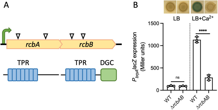
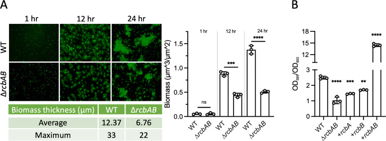
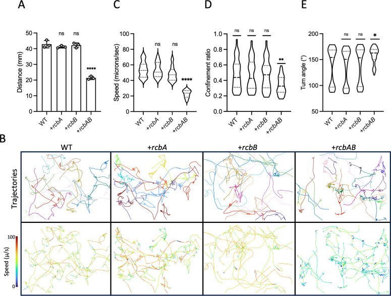
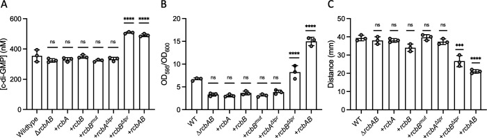
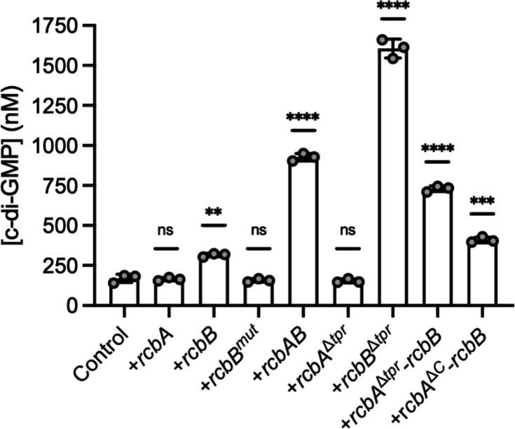
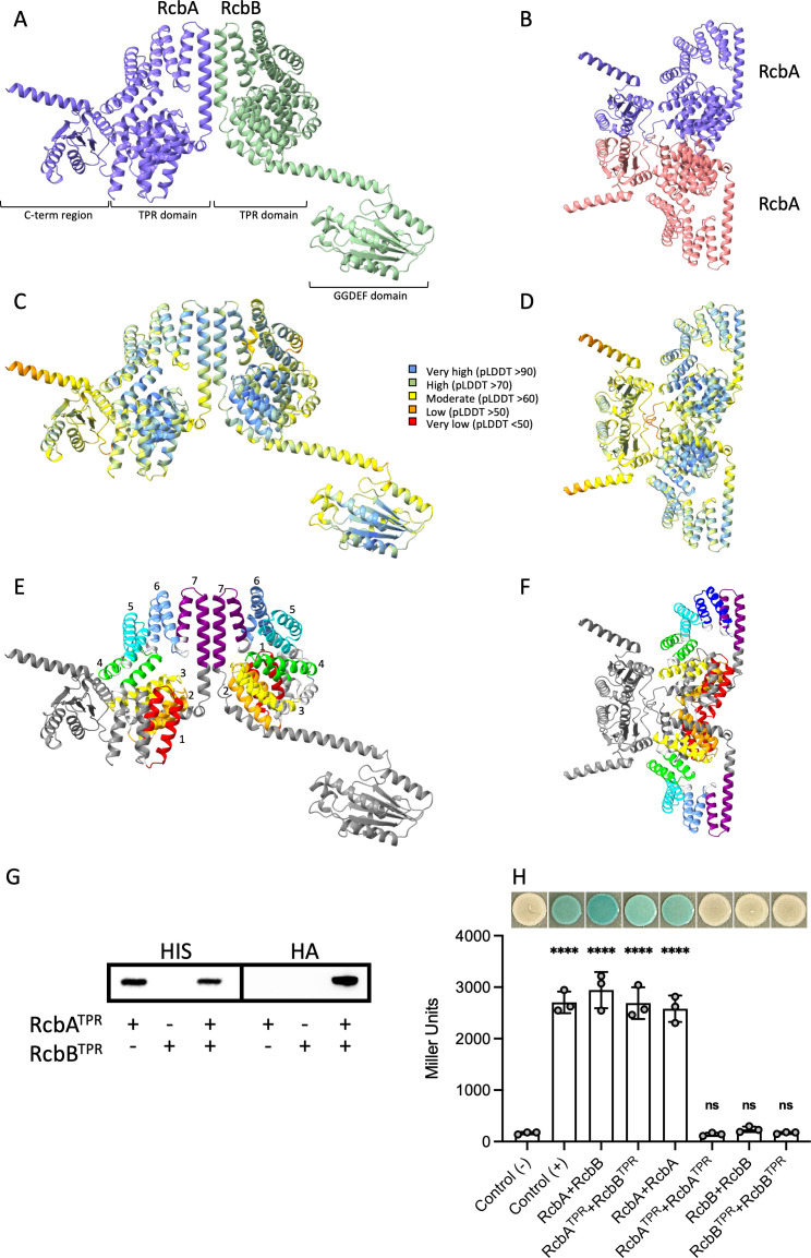
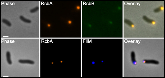
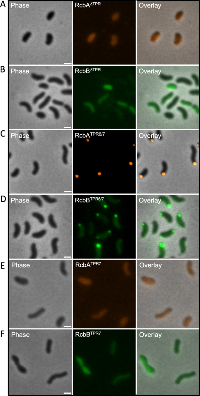
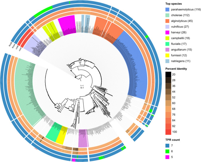
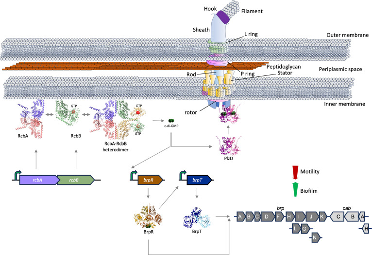
Vibrio species, which include several pathogens, are autochthonous to estuarine and warm coastal marine environments, where biofilm formation bolsters their ecological persistence and transmission. Here, we identify a bicistronic operon, rcbAB, whose products synergistically inhibit motility and promote biofilm maturation post-attachment by modulating intracellular c-di-GMP levels in the human and animal pathogen V. vulnificus. RcbA contains an N-terminal tetratricopeptide repeat (TPR) domain and a structured C-terminal region of unknown function, while RcbB possesses an N-terminal TPR domain and a C-terminal GGDEF domain characteristic of diguanylate cyclases. The TPR domain of RcbB represses its diguanylate cyclase activity, while RcbA's TPR domain and C-terminal region co-operatively de-repress it. Localization of both proteins to the flagellar pole is TPR-dependent but not co-dependent, although RcbA anchors RcbB to the pole in the absence of polar landmarks such as HubP and flagella. The conservation of rcbAB across diverse bacterial taxa substantiates its fundamental importance in bacterial biology. This work demonstrates how spatio-genetically coordinated TPR domain-containing proteins modulate c-di-GMP signaling, contributing to our understanding of biofilm formation in Vibrio species and potentially other bacteria. It also reveals the first evidence of inter-protein interaction via the TPR domains of both partners, challenging the conventional paradigm in which only one bears the domain.


Conflict of interest statement

The authors have declared that no competing interests exist.

Figures

Fig 1
Fig 1. The rcbAB operon regulates calcium-induced brp exopolysaccharide expression.
A, genetic organization of rcbAB. White arrows indicate Tn insertions identified in independent mutants and the bent green arrow represents a putative promoter. The domain structure of the encoded proteins is below. A tetratricopeptide (TPR) domain containing 7 repeats (blue) is present in both proteins, along with a diguanylate cyclase (DGC) domain (green) in RcbB. B, representative images of growth of wildtype V. vulnificus (WT) and ∆rcbAB mutant (Tn) strains bearing a PbrpAlacZ reporter on LB without or supplemented with 15 mM CaCl2 (LB + Ca2+). A plot of the corresponding PbrpAlacZ expression levels is below. Bars indicate the respective mean values and error bars represent the standard deviation of triplicate assays. Statistically significant differences between samples (****p < 0.0001; ns, no significant difference) were determined by unpaired Student’s t-test (two-tailed).
Fig 2
Fig 2. RcbA and RcbB co-operatively promote biofilm development post-attachment.
A, representative fluorescence images of biofilms formed by wildtype (WT) and ∆rcbAB strains under constant flow in IO20YP after 1, 12 and 24 hours. The average and maximum biomass thickness is shown below and a plot of the biomass for the same strains is on the right. B, biofilm formation (OD595/OD600) by WT and ∆rcbAB strains expressing (+) rcbA, rcbB or both genes. Bars indicate the respective mean values and error bars represent the standard deviation of triplicate assays. Statistically significant differences were determined by an unpaired Student’s t-test (two-tailed) in A and relative to WT cells by one-way ANOVA with a Tukey’s multiple comparisons post-hoc test in B (**p < 0.01, ***p < 0.001, ****p < 0.0001; ns, no significant difference). Expression was induced with 1 µM IPTG.
Fig 3
Fig 3. RcbA and RcbB co-operatively inhibit V. vulnificus swimming motility.
A, plot of the swimming motility zones in soft agar by wildtype (WT) cells harboring the empty plasmid (pC2X6HT) or expressing (+) rcbA, rcbB or rcbAB, respectively. Bars indicate the respective mean values and error bars represent the standard deviation of triplicate assays. B, representative images of motility traces for the same samples in A. Different colored lines in the traces on top represent trajectories for individual cells and the corresponding traces colored by swimming speed are below. C-E, violin plots of the swimming speed, confinement ratio (net displacement/total distance traveled), and turn angles for the cells in B. Dashed horizontal lines denote the median, first and third quartiles for the data range from 100 tracks for each strain. Statistically significant differences (*p < 0.05, **p < 0.01, ****p < 0.0001; ns, no significant difference) relative to WT cells were determined by one-way ANOVA with Dunnett’s multiple comparisons post hoc test. Expression was induced with 10 µM IPTG.
Fig 4
Fig 4. RcbA and RcbB co-operatively regulate c-di-GMP-dependent phenotypes.
Plot of cellular c-di-GMP levels (A), biofilm formation (B) and motility in soft agar (C) of wildtype V. vulnificus (WT) and ∆rcbAB cells that carry the empty expression plasmid or express (+) rcbA, rcbB, rcbBmut (GGAEF active site mutant), rcbAΔtpr (an RcbA TPR deletion mutant), rcbBΔtpr (an RcbB TPR deletion mutant) or the rcbAB operon. Bars show the respective mean values for each strain and error bars represent the standard deviation of triplicate assays. Statistically significant differences relative to wildtype were determined by one-way ANOVA with a Dunnett’s multiple comparisons post-hoc test (**p < 0.01, ***p < 0.001, ****p < 0.0001; ns, no significant difference). Expression was induced with 10 µM IPTG in A, 1 µM in B and 10 µM in C.
Fig 5
Fig 5. RcbA and RcbB co-operatively regulate c-di-GMP production and the TPR domain of RcbB suppresses its DGC activity.
Plot of intracellular c-di-GMP levels in wildtype E. coli MG1655 cells that carry the empty expression plasmid (control) or express (+) rcbA, rcbB, rcbBmut (GGAEF active site mutant), rcbAB, rcbAΔtpr (an RcbA TPR deletion mutant), rcbBΔtpr (an RcbB TPR deletion mutant), rcbAΔC-rcbB (in which the C-terminus of RcbA is deleted), or rcbAΔtpr-rcbB. (in which the TPR domain of RcbA is deleted). Bars show the respective mean values for each strain and error bars represent the standard deviation of triplicate assays. Statistically significant differences relative to the respective controls were determined by one-way ANOVA with a Dunnett’s multiple comparisons post-hoc test (**p < 0.01, ****p < 0.0001; ns, no significant difference). Expression was induced with 10 µM IPTG.
Fig 6
Fig 6. RcbA and RcbB interact via their TPR domains.
AlphaFold 3 predicted structure of RcbA and RcbB complexes. Shown are a RcbA-RcbB heterodimer (A, C and E) and RcbA homodimer (B, D and F) colored by chain (A and B), per-atom confidence estimate (pLDDT; C and D) and rainbow for the TPR repeats (E and F). The N-terminal TPR domains of RcbA and RcbB are predicted to contain 7 TPR repeats each (numbered 1-7 and colored red, orange, yellow, green, cyan, light blue and purple, respectively). The C-terminus of RcbA and GGDEF domain of RcbB are colored grey. G, western blot of pull-downs using Ni-coated beads from extracts of E. coli producing either the HIS-tagged TPR domain of RcbA (RcbATPR) or the HA-tagged TPR domain of RcbB (RcbBTPR) alone or together. Anti-HIS antibody (HIS) was used to detect RcbATPR. The blot was stripped for subsequent detection of RcbBTPR with anti-HA antibody (HA). Expression was induced with 50 µM IPTG. H, Bacterial Adenylate Cyclase Two-Hybrid (BACTH) assays of protein-protein interactions. RcbA, RcbB, RcbATPR and RcbBTPR were fused to the C-terminus of Bordetella pertussis adenylate cyclase subunits T18 or T25. The plot shows β-galactosidase activity (Miller units) for the indicated protein combinations. Leucine zipper fusions to T18 and T25 served as a positive control (+), while empty plasmids were used as the negative control (-). Bars indicate the respective mean values and error bars represent the standard deviation of triplicate assays. Statistically significant differences (****p < 0.0001; ns, no significant difference) relative to the negative control were determined by one-way ANOVA with a Dunnett’s multiple comparisons post-hoc test. Representative images of the respective strains are shown above the plot.
Fig 7
Fig 7. RcbA and RcbB localize to the flagellar pole.
Representative phase, fluorescence and overlay images of ∆rcbAB cells expressing rcbA-mRuby3 and rcbB-mNeonGreen (top panels) and ∆fliMrcbA cells expressing rcbA-mRuby3 and fliM-bfp (bottom panels). Scale bars = 1 μm. Expression of rcbA and rcbB was induced with 10 µM IPTG and fliM was induced with 0.05% L-arabinose.
Fig 8
Fig 8. TPR6 and TPR7 of RcbA and RcbB are necessary for polar localization.
From left to right, representative phase, fluorescence and overlay images of RcbA∆TPR-mRuby3 signal in ∆rcbA cells (A), RcbB∆TPR-mNeonGreen signal in ∆rcbB cells (B), RcbATPR6/7-mRuby3 signal in the ∆rcbA cells (C), RcbBTPR6/7-mNeonGreen signal in ∆rcbB cells (D), RcbATPR7-mRuby3 signal in the ∆rcbA cells (E) and RcbBTPR7-mNeonGreen signal in ∆rcbB cells (F). Scale bar = 1 μm. Expression was induced with 10 µM IPTG.
Fig 9
Fig 9. Polar localization of RcbA and RcbB is not co-dependent.
Representative phase, fluorescence and overlay images of ∆rcbAB cells expressing rcbA-mRuby3 (top panels) or rcbB-mNeonGreen (bottom panels). Scale bars = 1 μm. Expression was induced with 10 µM IPTG.
Fig 10
Fig 10. RcbA anchors RcbB to the pole in the absence of the polar landmark protein HubP.
From left to right, representative phase, fluorescence and overlay images of RcbA-mRuby3 (A), RcbB-mNeonGreen (B), both (C), RcbA∆TPR-mRuby3 and RcbB-mNeonGreen (D) or RcbATPR6/7-mRuby3 and RcbBTPR6/7-mNeonGreen signal (E) in ∆hubPrcbAB cells. Scale bar = 1 μm. Expression was induced with 10 µM IPTG.
Fig 11
Fig 11. Phylogenetic distribution of rcbAB among Vibrio species.
From inside-out, Maximum Likelihood tree (rooted with V. vulnificus) based on 133 common core genes from 617 complete Vibrio genome assemblies, with branch lengths representing substitutions per site. Clades are color-coded for the ten most frequently represented Vibrio species, with the number of genomes harboring rcbAB indicated in parentheses. Taxa names are labeled on the tree. The inner two color-gradient tracks show percent identity to rcbA and rcbB from V. vunificus ATCC27562 of orthologs from the respective strain, while the outer two tracks indicate the number of tetratricopeptide repeats (TPR count) identified in the corresponding proteins.
Fig 12
Fig 12. Model for subcellular localization and interaction of RcbA and RcbB in regulating bacterial biofilm formation and motility.
Expression of the bicistronic operon produces RcbA (purple) and RcbB (green), which can independently localize to the flagellar pole. RcbA can self-assemble into homomultimers (purple/red), while RcbB cannot; however, RcbA may facilitate the formation of higher-order oligomers involving RcbB (green/gold) that are functionally important, since DGCs typically function as protein dimers. The N-terminal TPR domain of RcbB strongly inhibits its C-terminal DGC activity, keeping it in an autoinhibited state. RcbA anchors RcbB to the flagellar pole for optimal phenotypic effect and relieves RcbB’s autoinhibition. Elevated c-di-GMP production increases expression of brpR (brown), which encodes a transcriptional activator and potential c-di-GMP effector. BrpR in turn upregulates expression of the regulator encoded by brpT (blue). This cascade induces the synthesis of biofilm matrix components such as BRP exopolysaccharides (EPS) and calcium-binding proteins (dark and light grey, respectively), promoting biofilm development. Additionally, c-di-GMP may be sensed by effector proteins such as PlzD, which localizes to the flagellar pole and reduces the swimming speed and turn frequency of motile cells.

Similar articles

References

    1. Sampaio A, Silva V, Poeta P, Aonofriesei F. Vibrio spp.: Life Strategies, Ecology, and Risks in a Changing Environment. Diversity. 2022;14(2):97.
    1. Grimes DJ. The Vibrios: Scavengers, Symbionts, and Pathogens from the Sea. Microb Ecol. 2020;80(3):501–6. doi: 10.1007/s00248-020-01524-7 - DOI - PubMed
    1. Yildiz FH, Visick KL. Vibrio biofilms: so much the same yet so different. Trends Microbiol. 2009;17(3):109–18. doi: 10.1016/j.tim.2008.12.004 - DOI - PMC - PubMed
    1. Baker-Austin C, Oliver JD, Alam M, Ali A, Waldor MK, Qadri F, et al. Vibrio spp. infections. Nat Rev Dis Primers. 2018;4(1):1–19. - PubMed
    1. Thompson F, Iida T, Swings J. Biodiversity of vibrios. Microbiol Mol Biol Rev. 2004;68(3):403–31. - PMC - PubMed

MeSH terms

LinkOut - more resources