. 2025 Jul 10;104(10):105524.
doi: 10.1016/j.psj.2025.105524. Online ahead of print.

Development of an ELISPOT assay for numerating IFN-γ-secreting T cells in chicken using novel monoclonal antibodies

Affiliations

Development of an ELISPOT assay for numerating IFN-γ-secreting T cells in chicken using novel monoclonal antibodies

Zhou Zhou et al. Poult Sci. .

Abstract

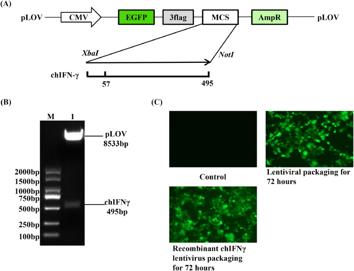
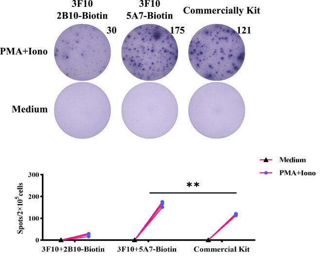
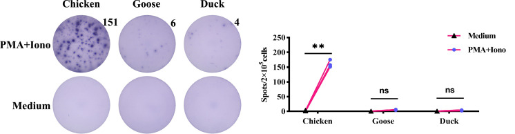
Cellular immune responses play critical roles in the control of pathogenic infection. The measurement of antigen-specific IFN-γ-secreting T cells via an enzyme-linked immunospot assay (ELISPOT) is a valuable method for the evaluation of cellular immune responses. However, in chicken, few of monoclonal antibodies (mAbs) against chicken IFN-γ (chIFN-γ) are suitable for this application. In this study, three anti-chIFN-γ mAbs (2B10, 3F10, 5A7) were generated by immunization with pcDNA-chIFN-γ plasmid and recombinant Hela cell line stably expressing chIFN-γ (Hela-chIFN-γ). Indirect immunofluorescence assay showed that these mAbs specifically recognized eukaryotically-expressed chIFN-γ in DF-1 cells and natural chIFN-γ secreted by mitogen-activated chicken splenocytes. Furthermore, using 3F10 as a capture antibody and biotinylated 5A7 as a detection antibody, an ELISPOT assay was established for numerating IFN-γ-secreting T cells of chicken. This chIFN-γ ELISPOT assay had higher reactivity than a commercially available kit and showed no cross-reactivity with activated lymphocytes from geese and ducks. This assay was further applied to detect the frequency of MDV antigen-specific IFN-γ-secreting T cells in the spleen of CVI988-immunized chickens. Collectively, we developed and validated an ELISPOT assay for detecting IFN-γ-secreting T cells in chickens using novel anti-chIFN-γ mAbs. Our study provides an important immunological tool for in-depth analysis of cellular immune response in chicken after infection or vaccination.

Keywords: Chicken interferon-γ; ELISPOT; IFN-γ-secreting T cells; Monoclonal antibody.


Conflict of interest statement

Disclosures We declare that we have no financial and personal relationships with other people or organizations that can inappropriately influence our work, and there is no professional or other personal interest of any nature or kind in any product, service and/or company that could be construed as influencing the content of this paper.

Figures

Fig 1
Fig. 1
Eukaryotic recombinant chIFN-γ protein was expressed and identified in Hale cells. The pcDNA3.1-chIFN-γ plasmid was transfected into Hale cells, and anti-His tag was used mAb to analyze the recombinant chIFN-γ protein through IFA and Western blot. (A) IFA showed that recombinant chIFN-γ was expressed in pcDNA3.1-chIFN-γ-transfected (upper panel) but not pcDNA3.1-transfected (lower panel) Hale cells. (B)The recombinant chIFN-γ was detected by Western blot in the cell lysate of pcDNA3.1-chIFN-γ-transfected Hale cells (lane 3) rather than pcDNA3.1-transfected Hale cells (lane 2) and Hale cells (lane 1).
Fig 2
Fig. 2
Recombinant chIFN-γ lentivirus was packaged in 293T cells. The expression plasmid (pLOV-chIFN-γ), packaging plasmid (psPAX2), and envelope plasmid (pMD2.G) were co-transfected into 293T cells to package recombinant chIFN-γ lentivirus. (A) Plasmid profile of lentiviral packaging system. The full-length chIFN-γ gene was cloned into pLOV-EGFP-3Flag vectors. (B) Identification of recombinant plasmid pLOV-chIFN-γ by enzyme digestion. M, DNA ladder; lane 1, plasmid enzyme digest product. (C) The packaging of the recombinant lentivirus. The 293T cells were transfected with either pLOV-EGFP-3Flag or pLOV-chIFN-γ plasmids, in combination with psPAX2 and pMD2.G plasmids, respectively. The green fluorescence from the packaged recombinant lentivirus was observed 72 h after transfection, with untransfected Hela cells used as controls.
Fig 3
Fig. 3
Establishment and characterization of a recombinant Hela cell line stably expressing rchIFN-γ. Hela cells were infected with recombinant chIFN-γ lentivirus (MOI=50) and selected with puromycin (2 μg/mL) to establish a recombinant Hela cell line stably expressing rchIFN-γ (Hela-chIFN-γ). Hela-chIFN-γ cells were identified by RT-PCR, IFA and Western blot. (A) Puromycin selection of Hela cells infected with recombinant chIFN-γ lentivirus. Green fluorescence expression in puromycin selected Hela cells at 7, 15, 30, 45, 60 days post-infection. (B) Agarose gel electrophoresis of amplicons obtained from Hela-chIFN-γ using RT-PCR tests. M, DNA ladder; lane 2, amplicons obtained from Hela-chIFN-γ; lane 1 and 3, negative amplification control from Hela and Hela-EGFP. (C) IFA showed that rchIFN-γ was expressed in Hela-chIFN-γ cells (upper panel) but not Hela-EGFP cells (lower panel). Cells were stained with the AF594 secondary antibody, and recombinant chIFN-γ was visualized by red fluorescence. (D) Recombinant chIFN-γ was detected by Western blot in the cell lysate of Hela-chIFN-γ (lane 3) rather than Hela and Hela-EGFP cells (lane 1 and 2).
Fig 4
Fig. 4
Screening and characterization of mAbs against chIFN-γ protein. Using hybridoma supernatant as the primary antibody, the reactivity of the mAbs with the eukaryotic recombinant chIFN-γ expressed in DF-1 cells and natural chIFN-γ expressed in mitogen-activated chicken splenocytes was identified by IFA and Western blot. (A, B) The green fluorescence observed in the IFA showed that all anit-chIFN-γ mAbs (2B10, 3F10 and 5A7) specifically recognize recombinant chIFN-γ expressed in DF-1 cells. Additionally, these mAbs also recognized natural chIFN-γ expressed by mitogen-activated chicken splenocytes. (C) Western blot was performed on natural chIFN-γ in splenocytes with anit-chIFN-γ mAbs (2B10, 3F10 and 5A7) and HRP-conjugated rabbit anti-mouse IgG antibody.
Fig 5
Fig. 5
The chIFN-γ ELISPOT analysis of the frequency of IFN-γ-secreting cells in splenocytes stimulated with PMA and Ionomycin. ELISPOT analyses were conducted using antibody pairs 3F10 (capture) with biotinylated 5A7 (detection), 3F10 (capture) with biotinylated 2A10 (detection), and commercial antibody pair. The ELISPOT assay employs a concentration of 5 µg/mL for the capture antibody and 0.5 µg/mL for the detection antibody. Splenocytes were stimulated for 48 h with medium or PMA (50 ng/mL) and Ionomycin (500 ng/mL). Spot counts represent the number of chIFN-γ-secreting cells and approximately 200,000 splenocytes are contained in each representative plot (upper panel). The sensitivity of the antibody pair 3F10 (capture) and biotinylated 5A7 (detection) are superior to those of commercially available kits (lower panel). Asterisks indicate p-value: ** p≤0.01.
Fig 6
Fig. 6
Cross-reactivity of the chIFN-γ ELISPOT assay with ducks and geese. ELISPOT analyses were conducted using antibody pairs 3F10 (capture, 5 µg/mL) and biotinylated 5A7 (detection, 0.5 µg/mL). Splenocytes from chicken, duck, and goose were a stimulated for 48 h with medium or PMA (50 ng/mL) and Ionomycin (500 ng/mL). Spot counts represent the number of IFN-γ-secreting cells in approximately 20 0,000 splenocytes. Asterisks indicate p-value: ** p≤0.01.
Fig 7
Fig. 7
The chIFN-γ ELISPOT assay for the frequency of antigen-specific IFN-γ-producing T cells in chickens following CVI988 immunization. ELISPOT analyses were conducted using antibody pairs 3F10 (capture, 5 µg/mL) and biotinylated 5A7 (detection, 0.5 µg/mL). The SMCs from chickens following CVI988 immunization were a stimulated for 48 h with antigens of MDV or medium. Unimmunized chickens served as controls. Spot counts represent the number of IFN-γ-secreting cells in approximately 2 × 10⁶ SMCs. Asterisks indicate p-value: ** p≤0.01.

References

    1. Andersen S.H., Vervelde L., Sutton K., Norup L.R., Wattrang E., Juul-Madsen H.R., Dalgaard T.S. Quantification and phenotypic characterisation of peripheral IFN-γ producing leucocytes in chickens vaccinated against Newcastle disease. Vet. Immunol. Immunopathol. 2017;193-194:18–28. - PMC - PubMed
    1. Ariaans M.P., van de Haar P.M., Lowenthal J.W., van Eden W., Hensen E.J., Vervelde L. ELISPOT and intracellular cytokine staining: novel assays for quantifying T cell responses in the chicken. Dev. Comp. Immunol. 2008;32:1398–1404. - PubMed
    1. Axelsson B. Detection of cytokine-secreting cells by enzyme-linked immunospot (ELISpot) Methods Mol. Biol. 2022;2386:61–79. - PubMed
    1. Boodhoo N., Behboudi S. Differential virus-specific IFN-gamma producing T cell responses to Marek's disease virus in chickens with B19 and B21 MHC haplotypes. Front. Immunol. 2021;12 - PMC - PubMed
    1. Boodhoo N., Behboudi S. Marek's disease virus-specific T cells proliferate, express antiviral cytokines but have impaired degranulation response. Front. Immunol. 2022;13 - PMC - PubMed