. 2025 Jul 16;15(1):25797.
doi: 10.1038/s41598-025-10244-2.

A conserved motif within the NSP2 of SARS-CoV-2 is required for processing of the distal NSP1/NSP2 junction by NSP3

Affiliations

A conserved motif within the NSP2 of SARS-CoV-2 is required for processing of the distal NSP1/NSP2 junction by NSP3

Thea Kristensen et al. Sci Rep. .

Abstract

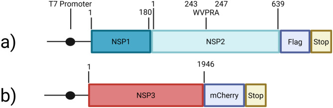
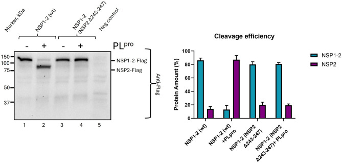
In 2019, the severe acute respiratory syndrome coronavirus 2 virus (SARS-CoV-2) started to spread globally and caused the COVID-19 pandemic. SARS-CoV-2, like other members of the Coronaviridae, has a single-stranded, positive sense RNA genome about 30 kb in length, which is translated to generate 16 non-structural proteins (NSPs); a set of sub-genomic mRNAs encode the structural and accessory proteins. The ORF1a precursor includes NSP1-11 and is processed by virus-encoded proteases to produce the mature proteins. We recently identified a short, highly conserved motif (YCPRP) within the structural protein precursor of foot-and-mouth disease virus (FMDV), a member of the Picornaviridae. This motif is conserved among picornaviruses and is found as (W/F/Y)-x-P-R-(P/A). The motif has a major influence on the processing of the FMDV capsid precursor (P1-2A) by the viral protease 3Cpro. We have now identified a similar motif (WVPRA) within the NSP2 of SARS-CoV-2. Interestingly, this motif is required for the efficient processing of the NSP1-NSP2 junction by the SARS-CoV-2 protease PLpro (NSP3) and a single amino acid substitution within the motif can abrogate cleavage of this junction. We hypothesise that this motif acts, within NSP1-NSP2, to enable this precursor to fold correctly and allow efficient processing of the NSP1/NSP2 junction.

Keywords: Coronavirus; Intramolecular chaperone; NSP2; Proteolytic processing; SARS-CoV-2.


Conflict of interest statement

Declarations. Competing interests: The authors declare no competing interests.

Figures

Fig. 1
Fig. 1
Schematic representation of the SARS-CoV-2 genome organization. The RNA genome is translated from two overlapping reading frames, ORF1a and ORF1b, to generate two different polyproteins, pp1a and pp1ab, the latter is generated following a programmed ribosomal frameshift that occurs within the overlap between ORF1a and ORF1b. The two polyproteins are cleaved by the virus-encoded proteases PLpro (NSP3) and 3CLpro (NSP5) into a total of 16 non-structural proteins (NSPs). The sites cleaved by the PLpro (marked with red arrows) and 3CLpro (marked with yellow arrows) are indicated. Downstream of the ORF1b, the RNA genome encodes 10 more proteins, which are translated from sub-genomic mRNAs, including the structural proteins (spike (S), envelope (E), membrane (M) and nucleocapsid (N)) and several accessory proteins. The conserved motif (WVPRA) is found within NSP2 of pp1a and is marked on the lower part of the figure. Created in BioRender. Kristensen, T. (2025) https://BioRender.com/s80a354.
Fig. 2
Fig. 2
Alignment of partial NSP2 amino acid sequences containing the conserved motif from 9 different betacoronaviruses. The alignment was performed using ClustalW in Geneious 9.0.5. The entire ORF1a was included in the alignment but just a short region of the NSP2 sequence is shown here. The amino acids are numbered based on the sequence of the SARS-CoV-2 NSP2. The conserved motif is highlighted in different colors depending on their amino acid properties, i.e. W and F have similar properties (aromatic and non-polar) and thus have a similar color. V, L, I and A have similar properties to each other (aliphatic and non-polar) so also have a similar color. Only the P residue (which has distinct properties as an imino acid) is totally conserved. The R-V/I change is rather non-conservative. A consensus sequence from the alignment is shown in the bottom of the Figure where x represents any amino acid. The sequences used were as follows: SARS-CoV-2 (Beta/subB) (GenBank accession number QWC81719.1), SARS-CoV (Beta/subB) (RefSeq accession number YP_009944365.1), Bat coronavirus 279/2005(Beta/SubB) (GenBank accession number GCA_031121315.1), Bat SARS coronavirus HKU3(Beta/SubB) (GenBank accession number QND76018.1), MERS-CoV (Beta/SubB) (GenBank accession number AVN89452.1), Murine hepatitis virus(Beta/subA) (GenBank accession number AWB14623.1), Human coronavirus OC43(Beta/subA) (GenBank accession number GCA_003972325.1), Equine coronavirus(Beta/subA) (GenBank accession number UGN73921.1) and Bovine coronavirus(Beta/subA) (GenBank accession number UZT75406.1).
Fig. 3
Fig. 3
Overview of the coding capacity of plasmids used in the transient expression assays. (a) The coding sequence for the SARS-CoV-2 NSP1-NSP2 plus a C-terminal Flag-tag and a stop codon were inserted into the pGEM-3Z plasmid, containing the T7 promoter upstream of the insert. The lengths of both NSP1 and NSP2 (in terms of the number of amino acid residues) are indicated, and the location of the conserved motif (WVPRA) is marked. This plasmid is referred to as the wt. (b) The NSP3-mCherry construct, encoding a functional PLpro, was a gift from Bruno Antonny (see Materials and Methods), a T7 promoter sequence was inserted into this plasmid, upstream of the coding sequence for NSP3, as described in Materials & Methods. Created in BioRender. Kristensen, T. (2025) https://BioRender.com/d9lyyyv.
Fig. 4
Fig. 4
A short, conserved motif within SARS-CoV-2 NSP2 is required for processing of the NSP1/NSP2 junction by the PLpro (NSP3). Cell lysates from BHK cells infected with vTF7-3 and transfected with plasmids that express the SARS-CoV-2 NSP1-NSP2-Flag (wt or with a small deletion, removing the conserved motif) either alone (odd numbered lanes) or with the plasmid encoding the SARS-CoV-2 PLpro (even numbered lanes) were analyzed by immunoblotting using rabbit anti-Flag antibodies, followed by anti-rabbit HRP-conjugated secondary antibodies and a chemiluminescence detection kit. Molecular mass markers (kDa) are indicated on the left. A negative control (no DNA) was included (lane 5). The uncut membrane is shown in Supplementary Fig. S1. This experiment has been repeated three times in total with separate transfections and western blots and resulted in very similar results. The additional two experiments are shown in Supplementary Figs. S2 and S3. Band intensities for each experiment were quantified using pixel count in ImageJ. Each lane was set to 100%, and the relative percentage of each of the two bands was calculated. The mean values of the triplicates were plotted, and the bar diagram with error bars representing the standard deviation (SD) are shown on the right side of the figure.
Fig. 5
Fig. 5
Identification of amino acid residues within the conserved WVPRA motif within the SARS-CoV-2 NSP2 that are required for efficient processing of the NSP1/NSP2 junction by PLpro (NSP3). Cell lysates from BHK cells infected with vTF7-3 and transfected with plasmids that express the SARS-CoV-2 NSP1-NSP2-Flag (wt or with single amino acid substitutions within the conserved WVPRA motif) either alone (odd numbered lanes) or with the plasmid encoding the SARS-CoV-2 PLpro (even numbered lanes) were analyzed by immunoblotting as in Fig. 4. Molecular mass markers (kDa) are indicated on the left. A negative control was included (lane 9). The NSP1-NSP2-Flag and the NSP2-Flag products are indicated on the right side of the western blot picture and were detected using anti-Flag-antibodies. Panel (a) shows the wt and various substitutions of the NSP2 W243 and a negative control. Panel (b) shows the wt, the NSP2 R246A, the NSP2 P247A, NSP2 P247G and a negative control. The uncut membranes are shown in Supplementary Figs. S4 and S7. This experiment has been repeated three times in total with separate transfections and western blots and resulted in very similar results. The additional experiments are shown in Supplementary Figs. S5, S6, S8 and S9. Band intensities for each experiment were quantified using pixel count in ImageJ. Each lane was set to 100%, and the relative percentage of each of the two bands was calculated. The mean values of the triplicates were plotted, and the bar diagram with error bars representing the standard deviation (SD) are shown on the right side of panels a and b.

Similar articles

References

    1. Wu, D., Wu, T., Liu, Q. & Yang, Z. The SARS-CoV-2 outbreak: What we know. Int. J. Infect. Dis.94, 44–48 (2020). - PMC - PubMed
    1. Naqvi, A. A. T. et al. Insights into SARS-CoV-2 genome, structure, evolution, pathogenesis and therapies: Structural genomics approach. BBA Mol. Basis Dis.1866, 1–17 (2020). - PMC - PubMed
    1. Beyerstedt, S., Casaro, E. B. & Rangel, É. B. COVID-19: Angiotensin-converting enzyme 2 (ACE2) expression and tissue susceptibility to SARS-CoV-2 infection. Eur. J. Clin. Microbiol. Infect. Dis.40, 905–919 (2021). - PMC - PubMed
    1. Wu, A. et al. Genome composition and divergence of the novel coronavirus (2019-nCoV) originating in China. Cell Host Microbe27, 325–328 (2020). - PMC - PubMed
    1. Murugan, C. et al. COVID-19: A review of newly formed viral clades, pathophysiology, therapeutic strategies and current vaccination tasks. Int. J. Biol. Macromol.193, 1165–1200 (2021). - PMC - PubMed

MeSH terms

Substances

LinkOut - more resources