. 2025 Aug;44(16):4611-4630.
doi: 10.1038/s44318-025-00493-2. Epub 2025 Jul 16.

The proteostatic landscape of healthy human oocytes

Affiliations

The proteostatic landscape of healthy human oocytes

Gabriele Zaffagnini et al. EMBO J. 2025 Aug.

Abstract

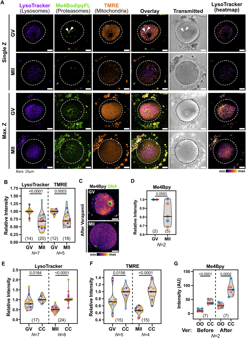
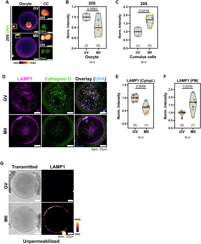
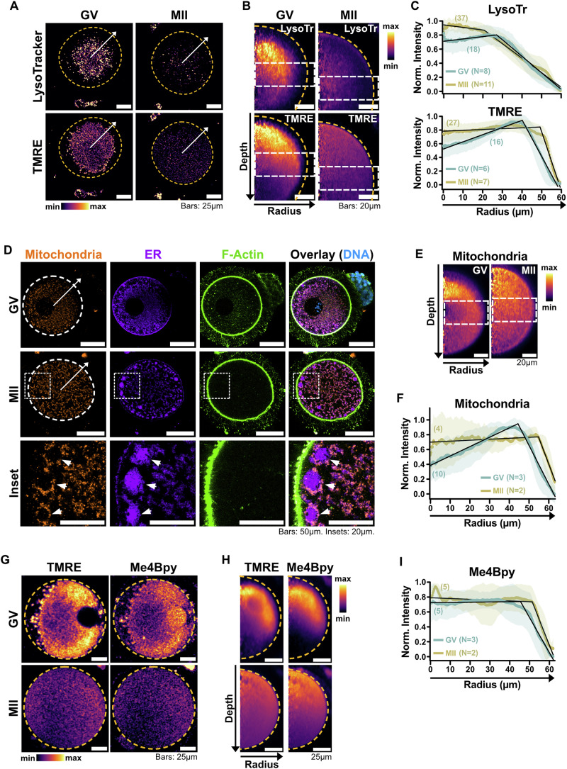
Oocytes, female germ cells that develop into eggs, are among the longest-lived cells in the animal body. Recent studies on mouse oocytes highlight unique adaptations in protein homeostasis (proteostasis) within these cells. However, the mechanisms of proteostasis in human oocytes remain virtually unstudied. We present the first large-scale study of proteostatic activity in human oocytes using over 100 freshly donated oocytes from 21 healthy women aged 19-34 years. We analysed the activity and distribution of lysosomes, proteasomes, and mitochondria in both immature and mature oocytes. Notably, human oocytes exhibit nearly twofold lower proteolytic activity than surrounding somatic cells, with further decreases as oocytes mature. Oocyte maturation is also coupled with lysosomal exocytosis and a decrease in mitochondrial membrane potential. We propose that reduced organelle activity preserves key cellular components critical for early embryonic development during the prolonged maturation of human oocytes. Our findings highlight the distinctive biology of human oocytes and the need to investigate human-specific reproductive biology to address challenges in female fertility.

Keywords: Female Fertility; Human Oocytes; Lysosomes; Mitochondria; Proteostasis.


Conflict of interest statement

Disclosure and competing interests statement. The authors declare no competing interests.

Figures

Figure 1
Figure 1. Degradative and mitochondrial activity decreases during oocyte maturation in humans.
(A) Representative live confocal images of human immature (GV) and mature (MII) oocytes labelled with LysoTracker Deep Red, Me4Bpy and TMRE to mark the activity of lysosomes, proteasomes, and mitochondria, respectively. Single confocal slices at the equatorial plane and maximal Z projections are shown. Dashed lines indicate the oocyte perimeter. Notice the clustered appearance of mitochondria and lysosomes in GVs, that leave an organelle-free area below the cell cortex. Lysosomes were also centrally distributed in MIIs. Arrowheads denote autofluorescent refractile bodies in the cytoplasm of oocytes. (B) Comparison of the average Lysotracker (lysosomes) and TMRE (mitochondria) intensity in GV and MII oocytes. Data points represent individual oocytes and are colour-coded by donor. Colour-coding is consistent across all figures (see Table EV1). Only donors with at least one GV and one MII were considered. N numbers indicate the total donors considered for each quantification. For each donor, data were normalized to the median of GVs. Numbers in parentheses indicate the total oocytes analysed per condition. Data averaged by donor are shown in Fig. EV2A. p values: unpaired t tests with Welch’s correction. (C) Live Confocal images of GV and MII oocytes treated with Verapamil and labelled with Me4Bpy to mark proteasomal activity (shown as a heatmap). Scale bars: 25 µm. (D) Quantification of the average Me4Bpy intensity in Verapamil-treated GV and MII oocytes from N = 2 donors. For each donor, data were normalized to GVs. Data averaged by donor are shown in Fig. EV2G. p value: unpaired t test with Welch’s correction. (E, F) Comparison of Lysotracker (E) and TMRE (F) intensities between oocytes and the corresponding cumulus cells (CC). For each donor, data were normalized to the median of CCs. A pairwise comparison between each oocyte and the corresponding cumulus cells is shown in Fig. EV2H,I, respectively. p values: unpaired t tests with Welch’s correction. (G) Quantification of Me4Bpy intensity in the same oocytes (OO) (2 GVs and 5 MIIs from N = 2 donors) and cumulus cells (CC) before and after incubation with Verapamil (Ver). Comparison between GV and MII oocytes is shown in Fig. 1D. p values: unpaired t tests with Welch’s correction. Source data are available online for this figure.
Figure 2
Figure 2. The abundance of degradative organelles decreases during oocyte maturation.
(A) Confocal images of oocytes and cumulus cells (CC) immunolabelled with an antibody against 20S core proteasome (displayed as a heatmap). Scale bars: 25 µm. Insets: 10 µm. (B, C) Comparison of the average 20S intensity in immunolabelled oocytes (B) and cumulus cells (C) from N = 2 donors. Data points represent individual oocytes and are colour-coded by donor. For each donor, data were normalized to the median of GVs. The number of oocytes quantified per condition is indicated into parentheses. A pairwise comparison averaged by donor is shown in Fig. EV3A,C, respectively. p values: unpaired t tests with Welch’s correction. (D) Representative confocal images of GV and MII oocytes immunolabelled for the lysosomal proteins LAMP1 and Cathepsin D. (E, F) Quantification of the mean intracellular (E) or plasma membrane (F) LAMP1 intensity in GV and MII oocytes shown in (D). Data points represent individual oocytes from N = 3 donors and are colour-coded by donor. For each donor, data were normalized to the median of GVs. The number of oocytes quantified per condition is indicated into parentheses. A pairwise comparison averaged by donor is shown in Fig. EV3D,E, respectively. p values: unpaired t tests with Welch’s correction. (G) Confocal images of non-permeabilized GV and MII oocytes immunolabelled with anti-LAMP1 (heatmap). Scale bars: 25 µm. Source data are available online for this figure.
Figure 3
Figure 3. Human oocytes contain aggregated proteins in few enlarged lysosomes.
(A) Representative confocal images of oocytes from multiple donors labelled with anti-LAMP1 and with or without Proteostat. Notice the higher overall background of both LAMP1 and Proteostat in oocytes from Donor 5, highlighting variability between different donors. (B) Box-and-whiskers plot of the number of LAMP1- and Proteostat-positive refractile bodies in human MII oocytes from different donors. Data points represent individual oocytes colour-coded by donor. The horizontal line indicates the median, the box represents the interquartile range (IQR), and the whiskers denote the minimum and maximum, respectively. Numbers in parentheses indicate the total amount of oocytes quantified from N = 3 donors. Source data are available online for this figure.
Figure 4
Figure 4. Organelles are asymmetrically distributed in human oocytes.
(A) Representative equatorial confocal planes of GV and MII oocytes labelled with LysoTracker and TMRE. Dashed circles indicate the oocyte boundaries. Arrows indicate the direction of the radius used for the radial reslicing shown in (B, C). (B) Radial reslicing of the oocytes shown in (A) along the depicted radiuses. Average projections of 360 radiuses spaced by 1° are shown. Orange dashed lines indicate the oocyte boundaries. White dashed boxes indicate the equatorial regions quantified in (C). (C) Radial distribution of LysoTracker (top) and TMRE (bottom) intensity in GV and MII oocytes, quantified as shown in (A, B). (D) Representative confocal images of GV and MII oocytes immunolabelled with anti-Calnexin (CANX) and anti-ATP5A to highlight the endoplasmic reticulum (ER) and mitochondria, respectively. The actin cytoskeleton was labelled with Phalloidin. Arrows indicate the direction of radiuses used for radial quantification shown in (F). Arrowheads indicate subcortical clusters of ER membranes surrounded by mitochondria in MIIs. (E) Radial reslicing of the oocytes shown in (D) along the depicted radiuses. Average projections of 360 radiuses spaced by 1° are shown. Dashed boxes indicate the equatorial regions quantified in (F). (F) Radial distribution of immunolabelled mitochondria in fixed oocytes, quantified as shown in (D, E). (G) Live confocal images of oocytes labelled with TMRE and Me4Bpy after Verapamil treatment. Orange dashed circles indicate the oocyte boundaries. See Fig. EV5A for additional pictures of the same oocytes for Lysotracker staining. (H) Radial reslice of the oocytes shown in (G). Orange dashed lines indicate the oocyte boundaries. (I) Radial distribution of Me4Bpy intensity in GV and MII oocytes. For (C, F), (I), numbers in parentheses indicate the total number of oocytes quantified per condition from multiple donors. N indicates the number of donors considered per condition. Mean and SD are shown. Data were normalized to the maximal and minimal intensities of each curve as 1 and 0, respectively. Black lines represent segmented models, confidence intervals (CIs) for GVs and MIIs do not overlap across conditions: Breakpoints of each model are as follows: Lysotracker GVs (27,012 µm; 95% CI 26,402–27,623 µm), Lysotracker MIIs (20,682; 95% CI 19,773–21,590 µm), TMRE GVs (40,118 µm; 95% CI 39,870–40,366 µm), TMRE MII (49,154 µm; 95% CI 48,820–49,489 µm), Mitochondria GV (44,377 µm; 95% CI 44,141–44,613 µm), Mitochondria MII (54,723 µm; 95% CI 54,481–54,965 µm), Me4Bpy GV (45,391 µm; 95% CI: 45,042–45,740 µm), Me4Bpy MII (51,336 µm; 95% CI 50,866–51,807 µm). Source data are available online for this figure.
Figure EV1
Figure EV1. Morphometric analysis of the retrieved oocytes and validation of LysoTracker, TMRE and Me4Bpy labelling in human cumulus-oocyte complexes.
(A) Quantification of the oocyte diameter from live-cell imaging experiments. Data points represent individual oocytes and are colour-coded by donor (see Table EV1). The horizontal lines indicate the median of each distribution, boxes represent the interquartile ranges (IQR), and whiskers denote the minima and maxima, respectively. Numbers in parentheses indicate the total amount of oocytes quantified per condition from N = 9 and N = 12 donors, respectively. p value: unpaired t test with Welch’s correction. (B) Representative confocal images of the chromatin configurations in GVs isolated from N = 7 donors and labelled with Hoechst 33342. Classification was performed according to Combelles et al (2002). The number of oocytes in each class is indicated. (C) Relative proportion of the chromatin configuration classes among the analysed GVs. The original data from Combelles et al (2002) are reported for comparison. Number into parentheses indicate the total amount of oocytes scored per study. The p value was calculated with a Fisher’s exact test on the raw oocyte counts. (D) Representative live confocal images of Zona pellucida-attached cumulus cells unlabelled or labelled with LysoTracker Deep Red, Me4Bpy and TMRE to highlight active lysosomes, proteasomes, and mitochondria, respectively. (E) Representative live epifluorescence images of the same oocyte before and after CCCP treatment to dissipate the mitochondrial membrane potential. Mitochondria were labelled with the membrane potential-sensitive dye TMRE before CCCP treatment. (F) Live confocal images of GV-stage oocytes treated with or without Bafilomycin A1 and MG-132 to inhibit lysosomes and proteasomes, respectively. Oocytes were labelled in presence of Verapamil with LysoTracker, Me4Bpy and TMRE to highlight active lysosomes, proteasomes and mitochondria, respectively. DNA was counterstained with Hoechst 33342. (G) Representative live confocal images of Zona-attached cumulus cells treated with or without Bafilomycin A1 and MG-132 to inhibit lysosomes and proteasomes, respectively. Cells were labelled with LysoTracker Deep Red, Me4Bpy and TMRE to highlight active lysosomes, proteasomes and mitochondria, respectively. Notice that treated cells were still alive, as indicated by the retention of TMRE signal. Source data are available online for this figure.
Figure EV2
Figure EV2. Characterization of LysoTracker, TMRE, and Me4Bpy in oocytes and cumulus cells before and after Verapamil treatment.
(A) Comparison of the average Lysotracker (lysosomes) and TMRE (mitochondria) intensity in GV and MII oocytes as shown in Fig. 1B. Data from multiple oocytes were averaged by donor and normalized to the GV value for each donor. N numbers indicate the total donors considered for each quantification. p values: ratio-paired t tests. (B) Representative live confocal images of oocytes labelled with or without Me4Bpy. Arrowheads indicate autofluorescent refractile bodies. (C) Comparison of LysoTracker, TMRE and Me4Bpy intensity in cumulus cells attached to GV- and MII-stage oocytes from multiple donors. Data were averaged by donor and normalized, for each donor and dye, to the GV value. Only donors with at least one GV and one MII were considered. p values: ratio-paired t tests. (D) Representative live confocal images of the same oocytes labelled with LysoTracker, TMRE and Me4Bpy and imaged before and after incubation with Verapamil. DNA was counterstained with Hoechst 33342. (E, F) Quantification of LysoTracker, TMRE and Me4Bpy intensities in the same oocytes (E) and cumulus cells (F) before and after verapamil addition. Data points represent individual COCs from N = 2 donors. For each COC, data were normalized to the value before verapamil. p values: ratio-paired t tests. (G) Quantification of the average Me4Bpy intensity in Verapamil-treated GV and MII oocytes from N = 2 donors, as shown in Fig. 1D. Data from multiple oocytes were averaged by donor and normalized to the GV value for each donor. p value: ratio-paired t test. (H, I) Pairwise comparison of Lysotracker (H) and TMRE (I) intensities between oocytes and the corresponding cumulus cells (CC) as shown in Fig. 1E,F. Data points represent individual Cumulus Oocyte Complexes (COCs) and are colour-coded by donor. For each COC, data were normalized to the CC value. p values: ratio-paired t tests. (J) Pairwise quantification of Me4Bpy intensity in the same oocytes (OO) and cumulus cells (CC) before and after incubation with Verapamil (Ver) as shown in Fig. 1G. Data points represent individual COCs and were normalized to the OO value before Verapamil for each COC. p values: ratio-paired t tests. The comparison between oocyte intensities before and after Verapamil treatment is shown in Fig. EV2E. (K) Quantification of the LysoTracker, TMRE and Me4Bpy intensity ratio between each oocyte (OO) and its corresponding cumulus cells (CC) after verapamil addition.
Figure EV3
Figure EV3. The reduction in lysosome levels in MIIs is accompanied by lysosomal exocytosis.
(A, C) Comparison of the average 20S intensity in immunolabelled oocytes (A) and cumulus cells (C) from N = 2 donors. Data from multiple oocytes were averaged by donor and normalized to the GV value for each donor. p values: ratio-paired t tests. (B) Relative change in lysosomal (indigo) and proteasomal (green) proteins between GVs and MIIs, as detected by single-cell proteomics of human oocytes. Data were retrieved from (Galatidou et al, 2024). Proteins were selected based on GO Cellular Component (CC) terms matching “lysosome” and “proteasome”, respectively. The dashed line indicates p = 0.05. (D, E) Quantification of the mean intracellular (D) or plasma membrane (E) LAMP1 intensity in GV and MII oocytes shown in Fig. 2D–F. For each donor (N = 3), data from multiple oocytes were averaged and normalized to the GV value. p values: ratio-paired t tests. (F) Representative confocal images of non-permeabilized MII oocytes labelled with or without anti-LAMP1 H3A4. LAMP1 intensity is displayed as a heatmap.
Figure EV4
Figure EV4. Proteostat intensity does not change between GVs and MIIs.
Quantification of mean proteostat intensity in refractile bodies in GV and MII oocytes from N = 2 donors. Only donors with at least 1 GV and 1 MII were considered. Data points represent individual oocytes and were normalized to the median of GVs for each donor. Numbers in parentheses indicate the number of oocytes quantified per condition. p value: unpaired t test with Welch’s correction.
Figure EV5
Figure EV5. Lysosomes are clustered in both human GVs and MIIs.
(A) Live confocal images of GV and MII oocytes labelled with LysoTracker and TMRE after Verapamil incubation. Intensities are displayed as heatmaps. Additional images of the same oocytes are shown in Fig. 4G. (B) Confocal images of GV and MII oocytes from multiple donors immunolabelled with anti-LAMP1 (shown as heatmap). Arrows indicate radiuses used for the radial reslicing shown in (C, D). Examples of oocytes from a third donor are shown in Fig. 2D. (C) Radial reslicing of the oocytes shown in (B) along the depicted radiuses. Average projections of 360 radiuses spaced by 1° are shown. Dashed boxes indicate the equatorial regions quantified in (D). (D) Radial distribution of LAMP1 signal in the cytoplasm of fixed oocytes. Mean and SD are shown. Numbers in parentheses indicate the total amount of oocytes quantified per condition from multiple donors. N indicates the number of donors considered per condition. Data were normalized to the min and max of each curve as 0 and 1, respectively. Note that the lower levels of LAMP1 antibody staining on MIIs cause a low signal-to-noise ratio, increasing the deviation on its average curve. Radius length does not include plasma membrane in these measurements. Black lines represent segmented models. Breakpoints of each model are as follows: GVs (29,260; 95% CI 28,836–29,684 µm), MIIs (5308 µm; 95% CI 4573–6043 µm).

References

    1. Allen EA, Baehrecke EH (2020) Autophagy in animal development. Cell Death Differ 27:903–918 - PMC - PubMed
    1. AL-Zubaidi U, Liu J, Cinar O, Robker RL, Adhikari D, Carroll J (2019) The spatio-temporal dynamics of mitochondrial membrane potential during oocyte maturation. Mol Hum Reprod 25:695–705 - PMC - PubMed
    1. Berkers CR, Leeuwen FWB, van, Groothuis TA, Peperzak V, Tilburg EW van, Borst J, Neefjes JJ, Ovaa H (2007) Profiling proteasome activity in tissue with fluorescent probes. Mol Pharm 4:739–748 - PubMed
    1. Bomba-Warczak EK, Velez KM, Zhou LT, Guillermier C, Edassery S, Steinhauser ML, Savas JN, Duncan FE (2024) Exceptional longevity of mammalian ovarian and oocyte macromolecules throughout the reproductive lifespan. eLife 13:RP93172 - PMC - PubMed
    1. Chazotte B (2011) Labeling lysosomes in live cells with LysoTracker. Cold Spring Harb Protoc 2011:pdb.prot5571 - PubMed

Substances

LinkOut - more resources