. 2025 Jul 16;20(1):661.
doi: 10.1186/s13018-025-06067-6.

Metformin facilitates osteogenic differentiation of bone marrow stromal cells through AMPK-dependent autophagy: an investigation into the healing of osteoporotic fractures in murine models

Affiliations

Metformin facilitates osteogenic differentiation of bone marrow stromal cells through AMPK-dependent autophagy: an investigation into the healing of osteoporotic fractures in murine models

Daoguang Du et al. J Orthop Surg Res. .

Abstract

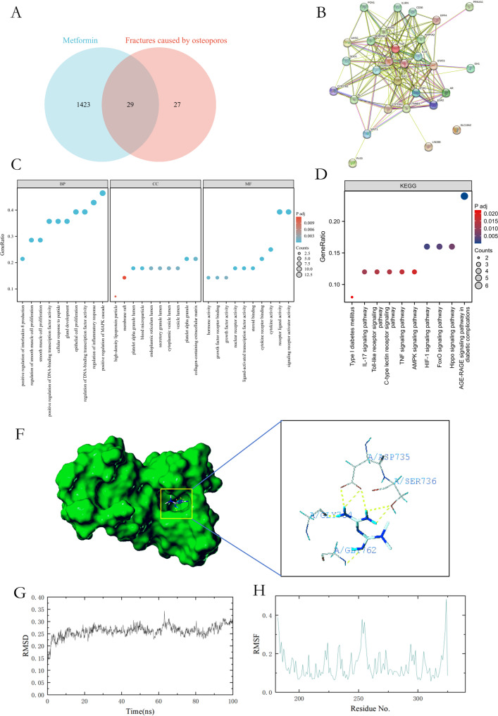
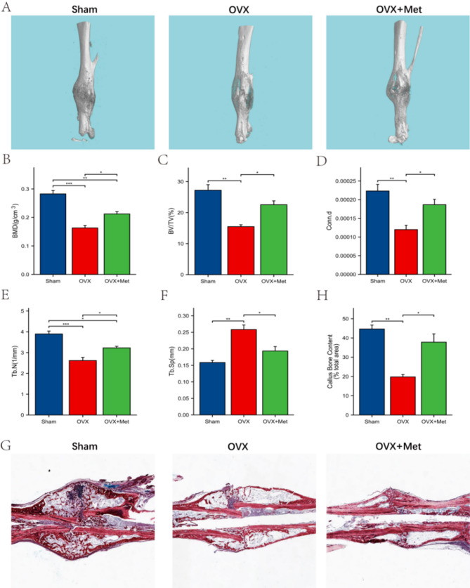
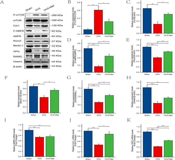
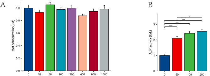
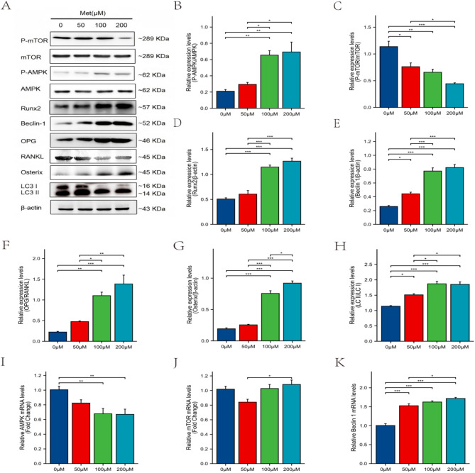
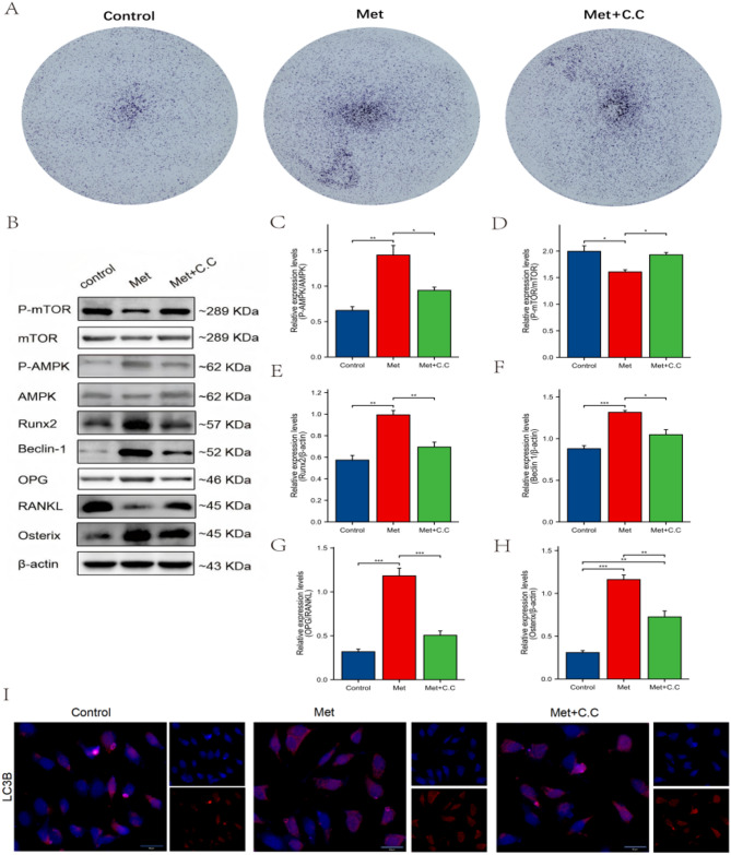
Osteoporosis is a widespread metabolic bone disorder characterized by a reduction in bone density and structural deterioration, leading to an increased susceptibility to fractures. This study investigates the role of metformin (Met) in promoting the osteogenic differentiation of bone marrow stromal cells (BMSCs) through the activation of AMP-activated protein kinase (AMPK) and explores its potential application in the treatment of osteoporotic fractures. We conducted a series of in vivo and in vitro experiments to elucidate the mechanisms underlying the effects of metformin. In a murine model of osteoporosis, metformin treatment significantly enhanced tibia fracture healing, as evidenced by increased bone mineral density (BMD), bone volume fraction (BV/TV), trabecular number (Tb.N), and decreased trabecular separation (Tb.Sp), as observed through micro-computed tomography (Micro-CT) and histological analyses. Immunoblot and real-time PCR demonstrated that metformin upregulated collagen type I (Col-I), a key osteogenic marker, and osteoprotegerin (OPG), an inhibitor of osteoclast differentiation that contributes to bone homeostasis via activated the AMPK signaling pathway. In vitro, metformin enhanced the osteogenic differentiation of BMSCs, as indicated by elevated alkaline phosphatase (ALP) activity. Western blot and PCR analyses further revealed that metformin increased the expression of AMPK, phosphorylated AMPK (p-AMPK), mammalian target of rapamycin (mTOR), phosphorylated mTOR (p-mTOR), Beclin-1, and LC3-II/LC3-I, suggesting enhanced autophagy. The application of the AMPK inhibitor Compound C attenuated these effects, confirming the role of AMPK-mediated autophagy in metformin-induced osteogenesis. These findings suggest that metformin, through the activation of AMPK and subsequent enhancement of autophagy, promotes the osteogenic differentiation of BMSCs and accelerates the healing of osteoporotic fractures. Future research should focus on optimizing metformin dosage and administration routes and evaluating the long-term safety and efficacy of this therapeutic approach for patients with osteoporotic fractures.

Supplementary Information: The online version contains supplementary material available at 10.1186/s13018-025-06067-6.

Keywords: AMPK; Autophagy; Metformin; Osteoblast differentiation; Osteoporotic fracture.


Conflict of interest statement

Declarations. Competing interests: The authors declare no competing interests.

Figures

Fig. 1
Fig. 1
Network pharmacology analysis illustrating the relationship between metformin and osteoporosis-induced fractures. (A) Venn diagram showing the intersection of metformin-related molecules and osteoporosis-related fracture molecules. (B) PPI network analysis of the intersecting molecules identified in the Venn diagram. (C) GO analysis of the intersecting molecules, highlighting the biological processes, cellular components, and molecular functions involved. (D) KEGG pathway analysis of the intersecting molecules, illustrating the relevant signaling pathways. (E) Molecular docking model of metformin and AMPK. (F-G) MD analysis of Metformin with AMPK
Fig. 2
Fig. 2
Effect of metformin on tibia fracture healing in osteoporosis mice. (A) MicroCT reconstructions; (B) Bone mineral density (BMD); (C) Bone volume fraction BV/TV; (D) trabecular link density Conn.d; (E) trabecular number Tb.N; (F): trabecular separation Tb.sp; (G) H&E staining. (H) Callus Bone Content. Data are presented as the mean ± SD, n = 3. *P < 0.05. **P < 0.01, ***P < 0.001
Fig. 3
Fig. 3
Effect of metformin on the expression of AMPK signaling pathway in osteoporosis mice. (A-H) WB method detected the protein expression level of p-mTOR, mTOR, Col-I, p-AMPK, AMPK, Beclin-1, OPG, RANKL, Osterix; (I-K) Realtime-PCR detection the expression of AMPK, Col-I and OPG. Data are presented as the mean ± SD, n = 3. *P < 0.05. **P < 0.01, ***P < 0.001
Fig. 4
Fig. 4
Effect of metformin on the cell viability and osteogenic differentiation of mice BMSCs. (A) CCK8; (B) ALP activity. Data are presented as the mean ± SD, n = 3. *P < 0.05. **P < 0.01, ***P < 0.001
Fig. 5
Fig. 5
Effects of metformin on AMPK signaling pathway and autophagy in osteogenic differentiation of BMSCs. (A) Representative Western blot images of p-mTOR, mTOR, p-AMPK, AMPK, Runx2, Beclin-1, OPG, RANKL, Osterix, and LC3 (II/I) in BMSCs treated with different concentrations of metformin. (B-H) Quantification of protein expression levels of p-mTOR (B), p-AMPK (C), Runx2 (D), Beclin-1 (E), OPG (F), RANKL (G), and LC3-II/I ratio (H). (I-K) Relative mRNA expression levels of AMPK (I), Beclin-1 (J), and LC3 (K) assessed by real-time PCR. Data are presented as the mean ± SD, n = 3. *P < 0.05. **P < 0.01, ***P < 0.001
Fig. 6
Fig. 6
Effects of AMPK Inhibitor on metformin-Induced autophagy and osteogenic differentiation of BMSCs. (A) ALP staining; (B) Representative Western blot images of AMPK, p-AMPK, mTOR, p-mTOR, Runx2, Beclin-1, OPG, RANKL and Osterix; (C-G) Quantification of protein expression levels of p-AMPK/AMPK (C), p-mTOR/mTOR (D), Runx2 (E), Beclin-1 (F), OPG/RANKL (G), (H), and Osterix; (I) Immunofluorescence staining of LC3B. Quantitively data are presented as the mean ± SD, n = 3. *P < 0.05. **P < 0.01, ***P < 0.001

Similar articles

References

    1. Hou C, Wang X, Jiang W, Bian Z, Zhu L, Li M. Peptide 11R–VIVIT promotes fracture healing in osteoporotic rats. Int J Mol Med. 2021;48(2):162. - PMC - PubMed
    1. Eastell R, O’Neill TW, Hofbauer LC, et al. Postmenopausal osteoporosis. Nat Reviews Disease Primers. 2016;2:16069. - PubMed
    1. Li Q, Zhang W, Deng J, et al. Ameliorative effects of extracellular vesicles derived from mesenchymal stem cells on apoptosis and differentiation of osteoblasts treated with CoCl(2). Cell Reprogram. 2023;25:99–108. - PubMed
    1. Zheng L, Shen X, Xie Y, Lian H, Yan S, Wang S. Metformin promotes osteogenic differentiation and prevents hyperglycaemia-induced osteoporosis by suppressing PPARgamma expression. Acta Biochim Biophys Sin (Shanghai). 2023;55:394–403. - PMC - PubMed
    1. Zhang W, Hou W, Chen M, et al. Upregulation of parkin accelerates osteoblastic differentiation of bone marrow-derived mesenchymal stem cells and bone regeneration by enhancing autophagy and beta-catenin signaling. Front Cell Dev Biol. 2020;8:576104. - PMC - PubMed

LinkOut - more resources