. 2025 Jul 16;20(1):84.
doi: 10.1186/s13062-025-00664-5.

Extracellular vesicle-enriched secretome of adipose-derived stem cells upregulates clusterin to alleviate doxorubicin-induced apoptosis in cardiomyocytes

Affiliations

Extracellular vesicle-enriched secretome of adipose-derived stem cells upregulates clusterin to alleviate doxorubicin-induced apoptosis in cardiomyocytes

Wan-Tseng Hsu et al. Biol Direct. .

Abstract

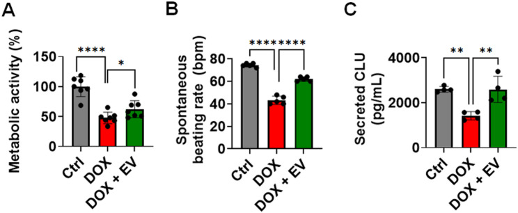
Doxorubicin (DOX) is a potent chemotherapeutic widely used against various cancers, but its clinical application is limited by DOX-induced cardiotoxicity (DIC). This study explored the cardioprotective potential of extracellular vesicle-enriched secretome derived from adipose stem cells (EVSASC) in mitigating DOX-induced apoptosis in cardiomyocytes. Adipose-derived stem cells were cultured, and their conditioned medium and extraceullular vesicles were isolated and characterized according to the Minimal Information for Studies of Extracellular Vesicles 2023 guidelines. HL-1 cardiomyocytes were pretreated with EVSASC before exposure to 1 µM DOX. Cell viability was assessed via the cell counting kit-8 assay, while apoptosis markers and survival mediators were evaluated through Western blotting. RNA sequencing identified differentially expressed genes, including clusterin (Clu), which was further quantified using an enzyme-linked immunosorbent assay. The functional role of clusterin was validated through siRNA-mediated knockdown. EVSASC significantly improved cell viability in DOX-exposed cardiomyocytes and reduced the cleaved caspase-3 to procaspase-3 ratio. Clusterin expression was highest in EVSASC-treated cells, and its knockdown markedly increased caspase-3 cleavage, confirming its pivotal role in cardioprotection. Moreover, EVSASC enhanced the phosphorylation of AKT, Bcl2-associated agonist of cell death, and glycogen synthase kinase-3β, implicating PI3K/AKT pathway activation in clusterin upregulation and anti-apoptotic effects. These findings demonstrate that EVSASC mitigates DOX-induced apoptosis in cardiomyocytes through clusterin upregulation and PI3K/AKT pathway activation. Clusterin is identified as a potential biomarker for evaluating EVSASC efficacy. While EVSASC shows promise as a cardioprotective strategy against DIC, further studies are needed to optimize its therapeutic safety by addressing potential oncogenic risks.


Conflict of interest statement

Declarations. Ethics approval and consent to participate: All animal experiments were performed following approved protocols by the Institutional Animal Care and Use Committee (IACUC) of National Taiwan University and were reported in line with the ARRIVE guidelines 2.0. (1) Title of the approved project: Mechanistic exploration of candidate oxysterols on breast tumorigenesis and tumorigenic microenvironment; (2) Name of the institutional approval committee: National Taiwan University College of Medicine and College of Public Health Institutional Animal Care and Use Committee; (3) Approval number: 20201104; (4) Date of approval: November 28, 2022. Consent for publication: Not applicable. Competing interests: The authors declare no competing interests.

Figures

Fig. 1
Fig. 1
Persistent doxorubicin-induced cardiomyocyte damage and apoptosis despite dexrazoxane interventions (A) Cardiomyocytes were treated with 1 µM doxorubicin (DOX) for 24 h and subsequently cultured in a fresh medium without DOX for an additional 24 h. Cell viability was assessed using a CCK-8 assay. The results demonstrate a significant reduction in cell viability following DOX treatment (*p < 0.05 vs. 0 µM), which persists even after the removal of the drug, indicating irreversible viability loss. (B) Treatment with dexrazoxane (DRZ) at 50 µM restored cell viability to levels comparable to control, whereas concentrations above 100 µM appeared to have diminished protective effects. (C) Apoptotic cells were detected using annexin V staining. Representative images show increased annexin V-positive cardiomyocytes after 24 h of DOX treatment and continued apoptosis 24 h post-drug removal. Quantitative analysis of annexin V-positive cells confirmed significant apoptosis induced by DOX, which remains elevated after the removal of the drug. (D) Western blotting of cleaved caspase-3 in cardiomyocyte lysates. Cropped gels and blots are displayed, with full-length blots provided in Supplementary Fig. 1. The results demonstrate increased levels of cleaved caspase-3 following DOX treatment, which remain elevated even after drug removal, suggesting persistent activation of the apoptotic pathway. *p < 0.05; **p < 0.01; ***p < 0.005; ****p < 0.0001
Fig. 2
Fig. 2
ASCs decreased doxorubicin-induced apoptosis and caspase-3 activation through paracrine effects in a Transwell coculture system (A) Differentiation of ASCs into adipocytes and osteocytes was confirmed using Oil Red O staining and Alizarin Red staining. Scale bar, 100 μm. (B) Comparing surface markers of ASCs in MesenPro medium and Complete Claycomb medium. (C) Schematic diagram of the Transwell coculture experimental timeline. HL-1 cardiomyocytes were seeded into the lower wells of a 6-well plate, and ASCs were seeded into the upper Transwell inserts. Both cell types were maintained in a Complete Claycomb medium (containing 10% FBS) throughout the experiment. After a 4-hour coculture adaptation period, DOX was added to induce cardiotoxicity. ASCs were retained in the upper chamber during DOX exposure to maintain continuous paracrine signaling and simulate coexposure conditions. Scale bar, 100 μm. (D) Caspase-3 activation was assessed in cardiomyocytes treated with 1 µM DOX alone or in coculture with ASCs using Western blotting. Cropped gels and blots are displayed, with full-length blots provided in Supplementary Fig. 2. (E) Quantitative analysis of annexin V-positive cells and cleaved caspase-3 bands normalized to GAPDH confirmed the statistically significant reduction in apoptosis and caspase-3 activation in the presence of ASCs compared to DOX treatment alone. *p < 0.05; **p < 0.01; ***p < 0.005
Fig. 3
Fig. 3
CM attenuated DOX-induced apoptosis in cardiomyocytes (A) The schematic illustrates the timeline of cell seeding, coculture, and treatment phases. CM was prepared by culturing ASCs in DMEM-HG supplemented with 10% standard FBS for 72 h, followed by filtration. All DOX-only and control groups were cultured in DMEM-HG medium containing 10% FBS to confirm that CM-specific effects were not due to FBS. (B) Adipose-derived stem cells (ASCs) secrete a range of pro-angiogenic and cardioprotective factors, including hepatocyte growth factor (HGF), granulocyte colony–stimulating factor (G-CSF), insulin-like growth factor 1 (IGF-I), platelet-derived growth factor (PDGF), placental growth factor (PIGF), vascular endothelial growth factor (VEGF), stem cell factor (SCF), epidermal growth factor (EGF), transforming growth factor (TGF-β1), fibroblast growth factor-2 (FGF-2), angiopoietin-1 (Ang1), interleukin (IL)-6, and thrombopoietin (TPO). (C) HL-1 cardiomyocytes were pretreated with CM derived from either mitomycin C-treated ASCs (CM MMC−treated ASC) or untreated ASCs (CM proliferative ASC) for 4 h prior to doxorubicin (DOX, 1 µM) exposure, and CM was replenished during the 24-hour DOX treatment. Cell viability was assessed using the CCK-8 assay. (D) The impacts of ASC-derived CM on DIC, as determined by annexin V and propidium iodide (PI) staining, highlight its role in reducing cardiomyocyte apoptosis. *p < 0.05; ***p < 0.005; ****p < 0.0001
Fig. 4
Fig. 4
EV fulfilled with Minimal Information for Studies of Extracellular Vesicles 2023 guidelines interacts with cardiomyocytes (A) Schematic representation of the isolation of extracellular vesicles (EVs) using the combination of qEV size exclusion columns (SEC) and an automatic fraction collector, followed by characterization and tracking of EVs in HL-1 cardiomyocytes. The first six fractions (F1-F6) after the buffer were collected, with each fraction being 0.7 mL. (B) Simultaneous measurement of size and concentration using tunable resistive pulse sensing (TRPS). (C) Statistical analysis of particle diameters. (D) Concurrent measurement of both size and zeta potential via TRPS. (E) Transmission electron microscopy (TEM) images. (F) Detection of internal markers; cropped gels and blots are shown, with full-length blots available in Supplementary Fig. 4. (G) Profiling of the exosomal marker CD63 within EVs and Dulbecco’s Phosphate-Buffered Saline (DPBS). (H) Profiles of growth factors present in EVs. (I) Interaction and uptake of EVs by HL-1 cardiomyocytes, emphasizing their proximity to mitochondria. Two co-staining protocols were utilized: the first employed Hoechst for nuclei, PlasMem for cell membranes, and ExoSparkler for exosomal membranes; the second used Hoechst for nuclei, MitoBright for mitochondria, and ExoSparkler for exosomal membranes. Colocalization of EVs with mitochondrial markers suggests their involvement in modulating mitochondrial function and oxidative stress in cardiomyocytes, providing insights into potential therapeutic mechanisms against DIC. Scale bar, 100 μm. Magnified view of the white-boxed region. Scale bar, 10 μm. HCI = High-Content Imaging System. SEC = size exclusion chromatography
Fig. 5
Fig. 5
EV restored the viability of HL-1 cardiomyocytes post-DOX exposure (A) Interaction and uptake of extracellular vesicles (EVs) by HL-1 cardiomyocytes 1 h after DOX exposure, compared to control cells, demonstrating initial EV internalization. (B) Interaction and uptake of EVs by HL-1 cardiomyocytes 20 h after DOX exposure, compared to control cells, indicating sustained EV presence and cellular uptake over time. (C) Impacts of EVs and their vehicle, PBS, on control cells and DOX-treated cells, providing a baseline for EV interaction in the absence of DOX-induced stress. (D) Effects of EV treatment on cardiomyocyte survival at the end of the 24-hour DOX exposure period and 24 h after DOX removal, highlighting the sustained cardioprotective effects of EVs in mitigating DOX-induced cytotoxicity. HCI = High-Content Imaging (HCI) System. * p < 0.05; ** p < 0.01; **** p < 0.0001. Scale bar, 100 μm. Magnified view of the white-boxed region. Scale bar, 10 μm
Fig. 6
Fig. 6
Clusterin played a critical role in CM and EVs against DOX-induced apoptosis (A) Venn diagram of differentially expressed genes (DEGs) across three experimental groups. A total of 621 genes uniquely modulated by CM in response to DIC are highlighted within the green circle. The accompanying volcano plot illustrates the statistical significance and magnitude of changes in the expression of these 621 genes, facilitating the rapid identification of significant DEGs. (B) Heat maps of protein-coding DEGs with TPM values above 5 in the CM-treated group are clustered hierarchically to reveal expression patterns across samples. (C) KEGG pathway analysis highlights representative pathways featuring the top 7 gene ratios. Gene ratio (x-axis) is the percentage of the number of genes present in this GO term over the total number of genes in this category. (D) The top 5 terms from the functional analysis of biological processes based on false discovery rate (FDR) in the CM-treated group are presented. (E) The quantification of clusterin (CLU) levels in HL-1 cardiomyocyte lysates and supernatants shows an upregulation following CM or EV treatment. (F) A schematic representation explains the siRNA-mediated knockdown of clusterin, outlining the experimental workflow, including quantification of clusterin in HL-1 cardiomyocyte lysates (intracellular CLU) and supernatant (secreted CLU) post-siRNA treatment, confirming effective knockdown. (G) Metabolic activity assessments reveal comparable protective effects of CM and EVs, as demonstrated by CCK-8 assays. (H) A representative Western blot shows caspase-3 activation, illustrating the protective effect of CM and EV treatment. Cropped gels and blots are shown, with the full-length blots available in Supplementary Fig. 6. Densitometric analysis of Western blot data provides statistical validation. * p < 0.05; **p < 0.01; ***p < 0.005; ****p < 0.0001. si-CLU = siRNA targeting clusterin; si-Ctrl = negative control siRNA; TR = Lipofectamine RNAiMAX Transfection Reagent. None = no siRNA or TR treatment
Fig. 7
Fig. 7
CM activated AKT and reduced mitochondrial superoxide production (A) Representative Western blot images illustrating the levels of caspase-3 activation alongside the phosphorylation of AKT, BAD, and GSK3β in response to CM treatment, highlighting the regulatory effects of CM on AKT-BAD and AKT-GSK3β signaling. Cropped gels and blots are shown, with full-length blots available in Supplementary Fig. 7. Densitometric analysis of the Western blot results provides quantitative validation of the observed protein expression changes. (B) Representative high-content images (HCI) show co-staining for Hoechst (nuclei), MitoBright (mitochondria), and mtSOX (mitochondrial superoxide), along with merged panels that illustrate cellular localization and oxidative stress response. Enlarged images from the boxed regions highlight the colocalization of mitochondria and oxidative stress markers, providing deeper insight into the intracellular effects of CM treatment. Scale bar, 100 μm. Magnified view of the white-boxed region. Scale bar, 10 μm. (C) A representative histogram of flow cytometry analysis displays the fluorescence of mtSOX superoxide. (D) Quantitative analysis of mtSOX fluorescence intensity is conducted using both HCI and flow cytometry to assess oxidative stress levels in cardiomyocytes across different treatment conditions. (E) Oxygen consumption rate (OCR) measurement by using the Seahorse XFe96 extracellular flux analyzer at baseline and after the addition of oligomycin, carbonyl cyanide-4-(trifluoromethoxy) phenylhydrazone (FCCP), and rotenone + antimycin A. Mitochondrial function, focusing on basal and maximal respiration in HL-1 cardiomyocytes pretreated with DRZ, CM, or EV, was compared to groups treated only with DOX. (F) Control experiments verified that treatment with LY294002 alone, ranging from 5 to 20 µM, did not induce significant cytotoxicity. (G) HL-1 cells were pretreated with LY294002 (20 µM) for 2 h before the addition of CM and/or DOX. After pretreatment, cells were exposed to DOX (1 µM) and CM for 24 h. Caspase-3 cleavage was subsequently assessed by Western blotting to evaluate apoptosis. The experimental groups included: (1) Control (Ctrl), (2) DOX-only, (3) DOX + CM, (4) DOX + CM + LY294002, and Ctrl + LY294002. *p < 0.05; **p < 0.01; ***p < 0.005; ****p < 0.0001
Fig. 8
Fig. 8
EVs restored metabolic activity and clusterin expression in induced pluripotent stem cell-derived human cardiomyocytes exposed to DOX treatment (A) Metabolic activity of cardiomyocytes was evaluated using the CCK-8 assay. (B) Spontaneous beating rates of human cardiomyocytes were recorded using an inverted phase-contrast microscope equipped with a high-resolution digital camera. (C) Clusterin expression levels were quantified via ELISA. *p < 0.05; **p < 0.01; ****p < 0.0001
Fig. 9
Fig. 9
Schematic representation of the EV-enriched secretome from ASCs, which modulates mitochondrial ROS, reduces apoptosis, and increases clusterin in cardiomyocytes treated with DOX

Similar articles

Cited by

References

    1. Siegel RL, Miller KD, Fuchs HE, et al. Cancer statistics, 2021. CA Cancer J Clin. 2021;71:7–33. - PubMed
    1. Rivankar S. An overview of doxorubicin formulations in cancer therapy. J Cancer Res Ther. 2014;10:853–8. - PubMed
    1. Thorn CF, Oshiro C, Marsh S, et al. Doxorubicin pathways: pharmacodynamics and adverse effects. Pharmacogenet Genomics. 2011;21:440–6. - PMC - PubMed
    1. Yang F, Teves SS, Kemp CJ, et al. Doxorubicin, DNA torsion, and chromatin dynamics. Biochim Biophys Acta. 2014;1845:84–9. - PMC - PubMed
    1. Octavia Y, Tocchetti CG, Gabrielson KL, et al. Doxorubicin-induced cardiomyopathy: from molecular mechanisms to therapeutic strategies. J Mol Cell Cardiol. 2012;52:1213–25. - PubMed