. 2025 Jul 16;23(1):518.
doi: 10.1186/s12951-025-03555-z.

Efficacy and safety of Fe-curcumin coordination polymer nanodots to prevent corneal neovascularization in alkali burn models

Affiliations

Efficacy and safety of Fe-curcumin coordination polymer nanodots to prevent corneal neovascularization in alkali burn models

Yan Huo et al. J Nanobiotechnology. .

Abstract

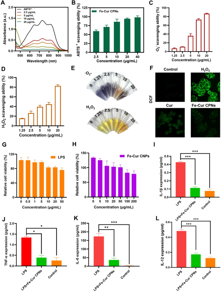
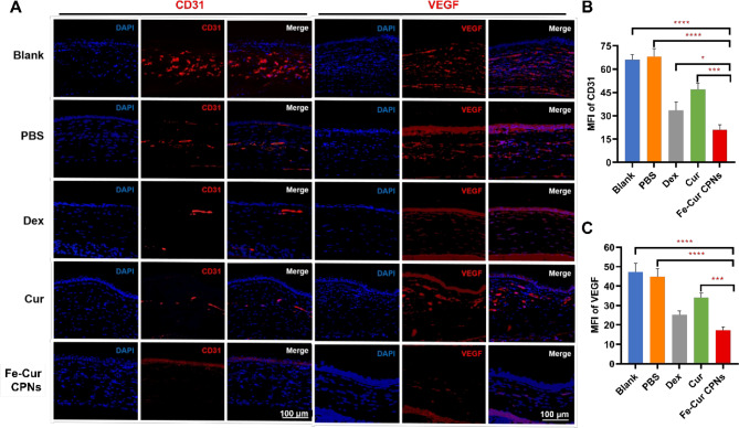
Alkali burns pose a significant risk of corneal injury, leading to potential blindness. During the progression of alkali burns, heightened oxidation levels can induce corneal damage, resulting in diminished clarity and vision loss. In this study, we chose metallic iron in conjunction with a small molecule, curcumin, to synthesize a curcumin-iron coordinated nanocomposite aimed at enhancing the bioaccessibility and targeting capabilities of curcumin. It could be found that Fe-curcumin coordination polymer nanodots (Fe-Cur CPNs) were comparably effective in suppressing corneal neovascularization, and they exhibited notable advantages in promoting corneal epithelial repair with minimal adverse effects. Additionally, Fe-Cur CPNs inhibited the activation of the nuclear factor-κ-gene binding (NF-κB) signaling pathway by scavenging reactive oxygen species (ROS), thus mitigating corneal neovascularization, which might represent a potential mechanism underlying the therapeutic effect of the Fe-Cur CPNs in alkali burn treatment. Moreover, treatment with the Fe-Cur CPNs did not result in any signs of cytotoxicity, hematological toxicity, or internal organ damage, further confirming the safety profile of this therapeutic agent. In conclusion, Fe-Cur CPNs present a novel, safe, and efficacious approach for addressing corneal alkali burns.

Keywords: Corneal alkali burn; Fe-curcumin coordination polymer nanodots (Fe-Cur CPNs); Neovascularization; Reactive oxygen species; Toxicity.


Conflict of interest statement

Declarations. Ethics approval and consent to participate: All animal experiments were conducted according to the Principles of Labora tory Animal Care of the People’s Republic of China and the Guidelines for the Care and Use of Laboratory Animals of Peking University First Hospital, China (Ethical Approval No: J202175). Consent for publication: All authors consent to publication. Competing interests: The authors declare no competing interests.

Figures

Scheme 1
Scheme 1
A schematic representation illustrates the synthesis of Fe-Cur CPNs and their targeted therapeutic delivery for corneal alkali burns. Fe-Cur CPNs were synthesized via a self-assembly method using curcumin as the organic ligand and iron ions as the coordinating metal. Upon administration, Fe-Cur CPNs overcome ocular delivery barriers, demonstrating enhanced solubility and bioavailability, to specifically target alkali burn lesions. These nanodots exhibit multifaceted therapeutic effects, including potent anti-angiogenic activity through inhibition of VEGF-mediated signaling, robust antioxidant properties via scavenging ROS, and significant anti-inflammatory effects by suppressing the NF-κB signaling pathway. Together, these mechanisms effectively mitigate corneal neovascularization, reduce inflammation, and promote epithelial repair with excellent biocompatibility and minimal toxicity, presenting a promising therapeutic strategy for oxidative stress-induced ocular injuries
Fig. 1
Fig. 1
Physicochemical characterization, cytocompatibility, and antioxidant properties of Fe-Cur CPNs. (A) TEM images of Fe-Cur CPNs. (B) Particle size distribution of Fe-Cur CPNs. (C) Hydrodynamic diameter of Fe-Cur CPNs measured by DLS. (D, E) Cytocompatibility assay of Fe-Cur CPNs at various concentrations. (F) Zeta potential of Fe-Cur CPNs. (G) FTIR spectra of PVP, Fe-Cur and Fe-Cur-PVP conjugates. (H) High-resolution XPS spectra of Fe 2p in Fe-Cur CPNs. (I) DLS profiles of Fe-Cur CPNs after incubation in PBS for 1 and 7 days, demonstrating excellent colloidal stability. (J) Photographs of Fe-Cur CPNs suspensions in PBS and culture medium at Day 1 and Day 7, showing no significant color change or precipitation. (K) EDS elemental analysis of Fe-Cur CPNs
Fig. 2
Fig. 2
Antioxidant and anti-inflammatory evaluations of Fe-Cur CPNs. (A) UV–vis spectroscopy was used to assess the ABTS•+ radical scavenging activity of Fe-Cur CPNs. (B) ABTS assay was performed to quantify the radical scavenging ability of Fe-Cur CPNs. (C) NBT reduction assay was used to evaluate superoxide anion (·O2⁻) scavenging activity. (D) H2O2 scavenging was evaluated via the titanium sulfate assay. (E) Colorimetric NBT and H2O2 assays were used to visually assess ROS scavenging based on observable color changes. (F) DCF-DA fluorescent probe staining was used to detect intracellular ROS levels. (G) CCK-8 assay was performed to evaluate the effect of LPS on HCECs viability. (H) CCK-8 assay was used to assess the cytotoxicity of Fe-Cur CPNs on HCECs. (IL) ELISA was used to measure the expression levels of IL-1β (I), TNF-α (J), IL-6 (K), and IL-12 (L)
Fig. 3
Fig. 3
Fe-Cur CPNs inhibit corneal neovascularization. (A) Timeline of examinations following a corneal alkali burn. (B) Corneal neovascularization was assessed at 1, 4, 7, and 14 days post-burn. (C) The quantified corneal neovascularization within 14 days. (D) Pathohistological and histological changes in corneal and limbal neovascularization in the rat cornea for each group on day 7 post-burn injury. (E) Statistical analysis results of CoNV density for each group on day 7 post-burn injury
Fig. 4
Fig. 4
Evaluation of Epithelial Repair and Corneal Permeability of Fe–Cur CPNs. (A) Observation of corneal epithelial sodium fluorescein-stained areas was conducted in each group on days 0, 1, 2, 3, 4, and 7 following corneal alkali burns. (B) Semi-quantitative analysis of the percentage area of corneal epithelial defects in each group at various time points following corneal alkali burns. (C) Permeability of Fe-Cur CPNs from the bulbar conjunctiva to the cornea, observed on day 7 post-alkali burn
Fig. 5
Fig. 5
Immunofluorescence. (A) Immunofluorescence plots of CD31 and VEGF in corneas of each group. (B, C) Quantitative analysis of staining intensity across the groups
Fig. 6
Fig. 6
Immunohistochemical staining. (A) Immunohistochemical staining for TNF-α and NF-κB in the corneas of each group. (B, C) Quantitative analysis of staining intensity across the groups. (D) Corneal MDA levels measured in rats from all groups on day 7 after alkali burn
Fig. 7
Fig. 7
Biosafety evaluation. (A) HE staining of major organs was conducted in different groups of rats 7 days after the corneal alkali burn. (B) Biochemical parameters were measured in orbital venous blood from different groups of rats 7 days after the alkali burn

References

    1. Bizrah M, Yusuf A, Ahmad S. An update on chemical eye burns. Eye. 2019;33:1362–77. - PMC - PubMed
    1. Dua HS, Ting DSJ, Al Saadi A, Said DG. Chemical eye injury: pathophysiology, assessment and management. Eye. 2020;34:2001–19. - PMC - PubMed
    1. Saccu G, Menchise V, Giordano C, Delli Castelli D, Dastrù W, Pellicano R, Tolosano E, Van Pham P, Altruda F, Fagoonee S. Regenerative approaches and future trends for the treatment of corneal burn injuries. J Clin Med. 2021;10. - PMC - PubMed
    1. Feng Y, Yang X, Huang J, Shen M, Wang L, Chen X, Yuan Y, Dong C, Ma X, Yuan F. Pharmacological Inhibition of glutaminase 1 attenuates alkali-induced corneal neovascularization by modulating macrophages. Oxid Med Cell Longev. 2022;2022:1106313. - PMC - PubMed
    1. Luo LJ, Lai JY, Chou SF, Hsueh YJ, Ma DH. Development of gelatin/ascorbic acid cryogels for potential use in corneal stromal tissue engineering. Acta Biomater. 2018;65:123–36. - PubMed

MeSH terms

LinkOut - more resources