. 2025 Jul 16;22(1):76.
doi: 10.1186/s12987-025-00690-5.

Imatinib prevents blood-spinal cord barrier disruption by inhibiting PDGFR-mediated JMJD3 expression and activation after spinal cord injury

Affiliations

Imatinib prevents blood-spinal cord barrier disruption by inhibiting PDGFR-mediated JMJD3 expression and activation after spinal cord injury

Chan Sol Park et al. Fluids Barriers CNS. .

Abstract

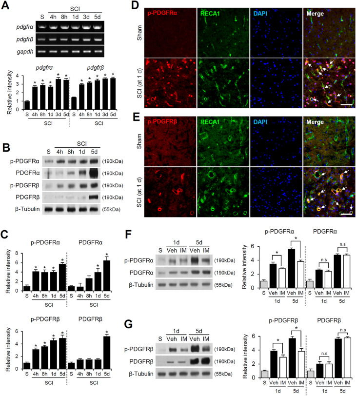
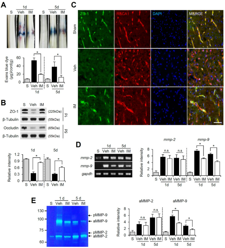
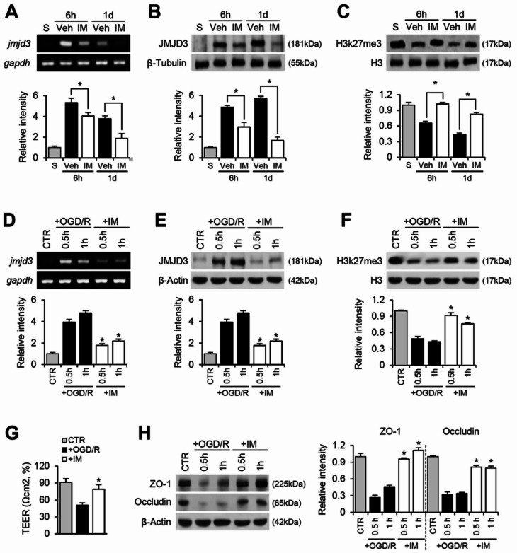
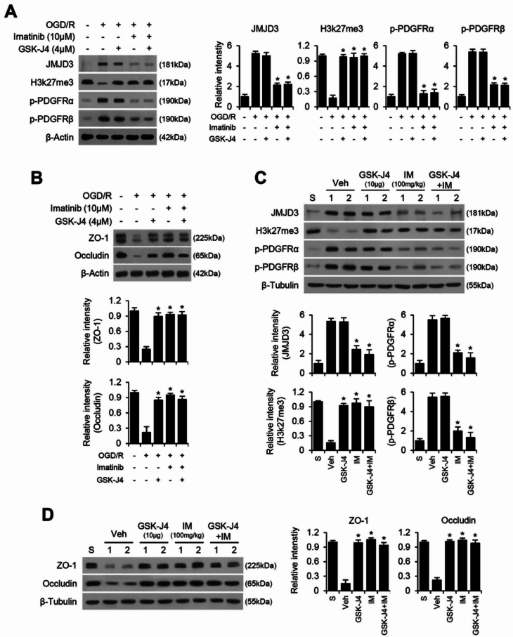
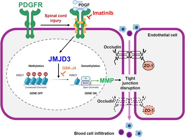
Background: After a spinal cord injury (SCI), disruption of the blood-spinal cord barrier (BSCB) leads to secondary injuries, including inflammatory responses and apoptotic cell death, ultimately causing permanent neurological deficits. Imatinib, a tyrosine kinase inhibitor, has been reported to enhance BSCB integrity and improve functional recovery after SCI. However, the mechanism by which imatinib regulates BSCB integrity remains unclear. Recent studies have identified the histone H3K27me3 demethylase JMJD3 as a key mediator of BSCB disruption, with high expression observed in blood vessels after SCI. In this study, we investigated whether imatinib regulates JMJD3 expression and activation through PDGFR signaling, thereby mitigating BSCB disruption following SCI.

Methods: Imatinib (100 mg/kg) was administered intraperitoneally to rats subjected to a contusion injury at the T9 level of the spinal cord and was continued daily for 14 days.

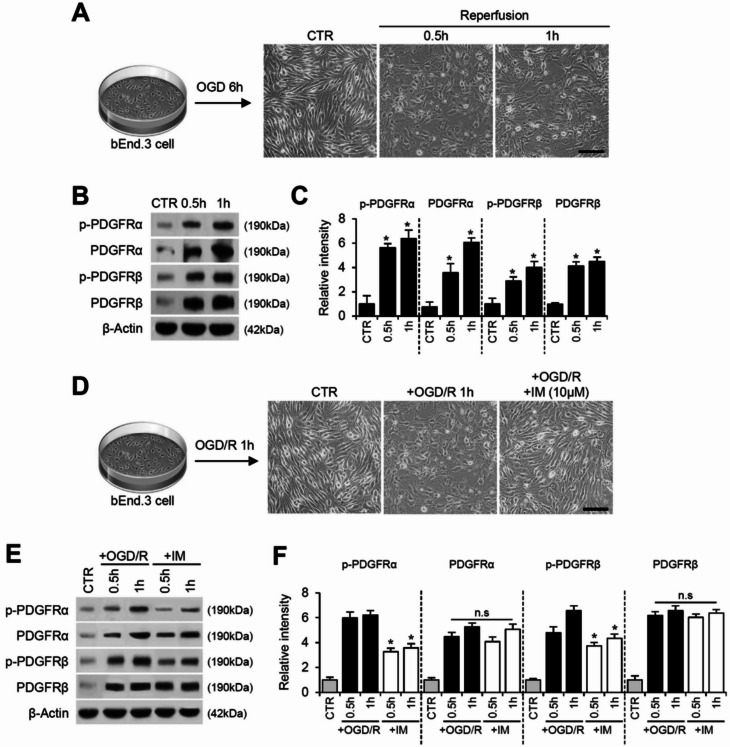
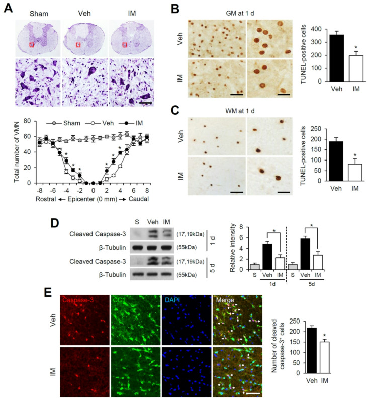
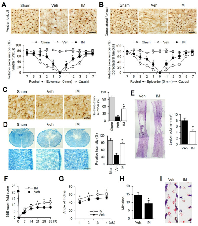
Results: Our results indicate that imatinib inhibited the phosphorylation of PDGFRα and PDGFRβ, both tyrosine kinase receptors, without affecting their expression levels. Additionally, imatinib reduced JMJD3 and MMP-9 expression and activation in blood vessels, thereby decreasing macrophage infiltration after SCI. In an oxygen-glucose deprivation (OGD)-induced bEnd.3 cell model, phosphorylated PDGFRα and PDGFRβ, along with JMJD3 expression and activation, were significantly upregulated but were effectively inhibited by imatinib treatment. Furthermore, imatinib suppressed secondary damage, including cell death, blood cell infiltration (e.g., neutrophils and macrophages), inflammation, axonal and myelin loss, and lesion volume. These effects collectively resulted in significant improvements in functional recovery after SCI.

Conclusion: Based on these findings, we propose that imatinib exerts a neuroprotective effect, in part by inhibiting PDGFR-mediated JMJD3 expression and activation following SCI.

Keywords: Blood-spinal cord barrier; Imatinib; JMJD3; MMP; PDGFR; Spinal cord injury.


Conflict of interest statement

Declarations. Ethics approval and consent to participate: All animal experiments were conducted in accordance with the guidelines set by the Animal Care Committee of Kyung Hee University (permission number: KHSASP-23-320). Consent for publication: Not applicable. Competing interests: The authors declare no competing interests.

Figures

Fig. 1
Fig. 1
Imatinib inhibits the activation of PDGFRα and β, which are upregulated in vascular endothelial cells after spinal cord injury. (A) RT-PCR and densitometric analysis of PDGFRα and β after SCI (mean ± SD, n = 5). *p < 0.05 vs. Sham (one-way ANOVA). (B) Western blot of p-PDGFRα, PDGFRα, p-PDGFRβ, PDGFRβ and β-Tubulin after SCI. (C) Densitometric analysis of Western blot (mean ± SD, n = 5). *p < 0.05 vs. Sham (one-way ANOVA). (D) Double immunofluorescence staining of p-PDGFRα with RECA1(blood vessel) in injured spinal cord at 1 d after SCI. Scale bar, 50 μm. (E) Double immunofluorescence staining of p-PDGFRβ with RECA1 in injured spinal cord at 1 d after SCI. Scale bar, 50 μm (n = 3). (F-G) Western blot and densitometric analysis for p-PDGFRα, PDGFRα, p-PDGFRβ, PDGFRβ and β-Tubulin at 1 d and 5 d after SCI. (mean ± SD, n = 5). *p < 0.05 vs. Vehicle (one-way ANOVA)
Fig. 2
Fig. 2
Imatinib also inhibits PDGFRα and β activation in OGD/R-induced bEnd.3 cells. (A) Schematic illustration of experimental protocol. Cells were exposed to OGD for 6 h, followed by reperfusion for 0.5–1 h. Photographs show morphological change of bEnd.3 cells. scale bar, 50 μm. (B) Western blot for p-PDGFRα, PDGFRα, p-PDGFRβ, PDGFRβ and β-actin. (C) Densitometric analysis of Western blot (mean ± SD, n = 5). *p < 0.05 vs. CTR (one-way ANOVA). (D) Cells were exposed to OGD for 6 h, followed by reperfusion for 1 h. Photographs show morphological change of bEnd.3 cells. scale bar, 50 μm. (E) Western blot for p-PDGFRα, PDGFRα, p-PDGFRβ, PDGFRβ and β-actin in OGD/R-induced bEnd.3 cells treated with imatinib. (F) Densitometric analysis of western blot (mean ± SD, n = 5). *p < 0.05 vs. +OGD/R (one-way ANOVA)
Fig. 3
Fig. 3
Imatinib prevents BSCB disruption and tight junction breakdown by inhibiting MMP-9 expression and activation after SCI. (A) Representative spinal cord showing Evans blue dye permeabilized into moderately injured spinal cord at 1 d and 5 d after SCI. Quantification of the amount of Evans blue at 1 d and 5 d after injury (mean ± SEM, n = 5). *p < 0.05 vs. Vehicle (unpaired t-test). (B) Western blot for ZO-1 (at 1 d) and occludin (at 5 d) after SCI and densitometric analysis of western blot (mean ± SD, n = 5). (C) Double immunofluorescence staining of ZO-1 with RECA1(blood vessel) in injured spinal cord at 1 d after SCI. Scale bar, 50 μm. (D) RT-PCR and densitometirc analysis of mmp-2 and − 9 mRNA (mean ± SD, n = 5). (E) Gelatin zymography and densitometric analysis of zymography (mean ± SD, n = 5). *p < 0.05 vs. Vehicle (one-way ANOVA). pMMP-2, pro MMP-2; aMMP-2, active MMP-2; pMMP-9, pro MMP-9; aMMP-9, active MMP-9
Fig. 4
Fig. 4
Imatinib alleviates tight junction breakdown by inhibiting JMJD3 expression and activation after SCI and in OGD/R-induced bEnd.3 cells. (A) RT-PCR for jmjd3 after SCI and densitometric analysis of RT-PCR (mean ± SD, n = 5). Western blot for (B) JMJD3 and (C) H3k27me3 after SCI. Densitometric analysis of Western blot (mean ± SD, n = 5). *p < 0.05 vs. Vehicle (one-way ANOVA). (D) RT-PCR for jmjd3 and densitometric of RT-PCR (mean ± SD, n = 5). (E) Western blot for JMJD3 and densitometric analysis (mean ± SD, n = 5). (F) Western blot for H3k27me3/H3 and densitometric analysis (mean ± SD, n = 5). (G) TEER (mean ± SD, n = 5). (H) Western blot for ZO-1 and occludin. Densitometric analysis of Western blot (mean ± SD, n = 5). *p < 0.05 vs. +OGD/R (one-way ANOVA)
Fig. 5
Fig. 5
PDGFR-JMJD3 axis mediates tight junction breakdown after SCI and OGD/R injury. Western blot and densitometric analysis for (A) JMJD3, H3k27me3, p-PDGFRα, p-PDGFRβ, (B) ZO-1 and occludin in imatinib and/or GSK-J4 treated End.3 cells subjected to 1 h of OGD/R injury (mean ± SD, n = 5). *p < 0.05 vs. +OGD/R (one-way ANOVA). Western blot and densitometric analysis for (C) JMJD3, H3k27me3, p-PDGFRα, p-PDGFRβ, (D) ZO-1 and occludin in injured spinal cord at 1 d after SCI (mean ± SD, n = 5). *p < 0.05 vs. Vehicle (one-way ANOVA)
Fig. 6
Fig. 6
Imatinib inhibits neutrophil and macrophage infiltration and reduces chemokines and cytokines expression after SCI. After SCI, blood infiltration was assessed by measuring the fluorescent intensity of MPO or ED-1 immunoreactive area at 1 and 5 d or Western blot for ED-1 at 5 d after injury as described in the Materials & methods section. (A) Representative photographs at 1500 and 2000 μm rostral to lesion epicenter showed MPO-labeled neutrophils and ED-1-labeled macrophages in the dorsal column of injured spinal tissues (cross section) injected with and without imatinib. Scale bar, 50 μm. (B) Relative fluorescent intensity of MPO- and ED-1-positive cells (mean ± SD, n = 5). *p < 0.05 vs. Vehicle (unpaired t-test). (C) Western blot and densitometric analysis of ED-1 at 5 d after injury (mean ± SD, n = 5). (D) RT-PCR and densitometric analysis for cytokines (tnf-α at 2 h, il-1β and il-6 at 6 h, cox-2 and inos at 1 d) (mean ± SD, n = 5). (E) RT-PCR and densitometric analysis for chemokines (mcp-1, mip-1α and mip-1β at 6 h, gro-α and mip-2α at 2 h) (mean ± SD, n = 5). (F) Western blot and densitometric analysis of COX-2 and iNOS at 1 d after SCI (mean ± SD, n = 5). *p < 0.05 vs. Vehicle (one-way ANOVA)
Fig. 7
Fig. 7
Imatinib inhibits neuronal and oligodendrocyte cell death after SCI. (A) Representative Cresyl violet staining showing ventral horn of spinal cord at 3 mm rostral from lesion site at 1 d. Scale bar, 50 μm. The spatial pattern of the number of VMN (mean ± SD, n = 3). *p < 0.05 vs. Vehicle (unpaired t-test). (B) Representative TUNEL staining in the GM of the spinal cord at 2 mm rostral from lesion site at 1 d and quantitative analysis of TUNEL-positive cells at 1 d (mean ± SD, n = 3). *p < 0.05 vs. Vehicle (unpaired t-test). Right panels show high-power views. Scale bars, 20 μm. (C) Representative TUNEL staining in the WM at 7 mm rostral from lesion site at 5 d and quantitative analysis of TUNEL-positive cells (mean ± SD, n = 3). *p < 0.05 vs. Vehicle (unpaired t-test). Right panels show high-power views. Scale bars, 20 μm. (D) Western blot and densitometirc analysis of cleaved caspase-3 at 1 d and 5 d after injury (mean ± SD, n = 5). *p < 0.05 vs. Vehicle (one-way ANOVA). (E) Immunohistochemical analysis of cleaved caspase-3 and CC1. Double labeling shows that oligodendrocytes in the WM were positive for cleaved caspse-3 after SCI (arrow). Scale bar, 50 μm. Quantitative analysis of cleaved caspase-3-positive cells (mean ± SD, n = 3). *p < 0.05 vs. Vehicle (unpaired t-test) (n = 3)
Fig. 8
Fig. 8
Imatinib attenuates axon and myelin loss and improves functional recovery after SCI. (A) Representative photographs of NF200-positive axons in ventral funiculus of spinal cord. Scale bars, 50 μm. Quantitative analysis of NF200-positive axons (mean ± SD, n = 5). *p < 0.05 vs. Vehicle (unpaired t-test). (B) Representative photographs of NF200-positive axons in dorsolateral funiculus of spinal cord and quantitative analysis (mean ± SD, n = 5). *p < 0.05 vs. Vehicle (unpaired t-test). (C) Representative photographs of 5-HT-positive axon in ventral horn areas in Sect. 3 mm caudal to the lesion site. Scale bars, 50 μm. Quantitative analysis of 5-HT (mean ± SD, n = 5). *p < 0.05 vs. Vehicle (unpaired t-test). (D) Transverse sections (lateral funiculus) selected from 2 mm rostral to the lesion site were processed for Luxol fast blue staining. Note that the extent of myelin loss was attenuated by imatinib treatment compared to the vehicle control. Scale bar, 100 μm. Quantitative analysis of Luxol fast blue intensity (mean ± SD, n = 5). *p < 0.05 vs. Vehicle (unpaired t-test). (E) Representative spinal cord tissues (1.2 mm from the dorsal surface) showing cavitation in the lesion site and quantitative analysis at 35 d after injury. Scale bar, 1 mm. Data are presented as mean ± SD (n = 5). *p < 0.05 vs. Vehicle (unpaired t-test). (F) BBB scores, (G) Inclined plane test, (H) Grid walk test, and (I) Foot print analysis of vehicle and imatinib-treated groups after SCI. Behavior data are presented as mean ± SEM (n = 10). *p < 0.05 vs. Vehicle (one-way repeated measured ANOVA)
Fig. 9
Fig. 9
Schematic diagram

Similar articles

References

    1. Jain A, Kim YT, McKeon RJ, et al. In situ gelling hydrogels for conformal repair of spinal cord defects, and local delivery of BDNF after spinal cord injury. Biomaterials. 2006;27:497–504. 10.1016/j.biomaterials.2005.07.008. - PubMed
    1. Saunders LL, Selassie A, Cao Y, et al. Epidemiology of pediatric traumatic spinal cord injury in a Population-Based cohort, 1998–2012. Top Spinal Cord Inj Rehabil. 2015;21:325–32. 10.1310/sci2104-325. - PMC - PubMed
    1. Chen G, Zhou Z, Sha W, et al. A novel CX3CR1 inhibitor AZD8797 facilitates early recovery of rat acute spinal cord injury by inhibiting inflammation and apoptosis. Int J Mol Med. 2020;45:1373–84. 10.3892/ijmm.2020.4509. - PMC - PubMed
    1. Lopez-Serrano C, Santos-Nogueira E, Francos-Quijorna I, et al. Lysophosphatidic acid receptor type 2 activation contributes to secondary damage after spinal cord injury in mice. Brain Behav Immun. 2019;76:258–67. 10.1016/j.bbi.2018.12.007. - PMC - PubMed
    1. Echeverry S, Shi XQ, Rivest S, et al. Peripheral nerve injury alters blood-spinal cord barrier functional and molecular integrity through a selective inflammatory pathway. J Neurosci. 2011;31:10819–28. 10.1523/JNEUROSCI.1642-11.2011. - PMC - PubMed

MeSH terms

Substances

LinkOut - more resources