

High-fat diet activates pyroptosis of retinal pigment epithelial cells in aged TgAPPswePS1 transgenic mice
- PMID: 40671142
- PMCID: PMC12269102
- DOI: 10.1186/s40001-025-02898-5
High-fat diet activates pyroptosis of retinal pigment epithelial cells in aged TgAPPswePS1 transgenic mice
Erratum in
-
Correction: High-fat diet activates pyroptosis of retinal pigment epithelial cells in aged TgAPPswePS1 transgenic mice.Eur J Med Res. 2025 Aug 8;30(1):724. doi: 10.1186/s40001-025-02979-5. Eur J Med Res. 2025. PMID: 40775372 Free PMC article. No abstract available.
Abstract
Purpose: This study assesses the impact of high-fat diet on retinal pigment epithelium (RPE) of aged TgAPPswePS1 transgenic mice, focusing on the involvement of RPE cell pyroptosis.
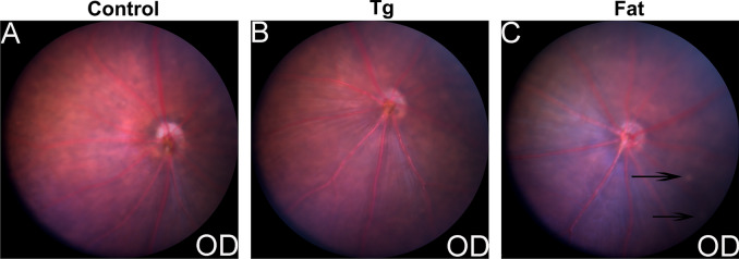
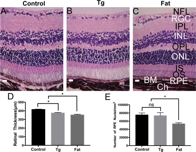
Methods: Twenty-four TgAPPswePS1 transgenic mice (18 months) was randomly divided into Tg group (n = 12) and Fat group (n = 12). Mice in Fat group were fed with high-fat diet consisting of 81.85% standard chow, supplemented with 1% cholesterol, 015% cholic acid, and 17% hydrogenated vegetable oil. Another 12 wild-type C57BL/6J mice were serve as control group. The fundus was examined through Micron IV. The eyes of mice were removed for paraffin-embedding and sectioning. HE staining was carried out to observe the structure of retina and measure retinal thickness. The expressions of amyloid-beta (Aβ), NOD-like receptor thermal protein domain associated protein 3 (NLRP3), Caspase-1, gasdermin D (GSDMD), IL-1β and IL-18 in RPE, as well as the number of RPE were detected.
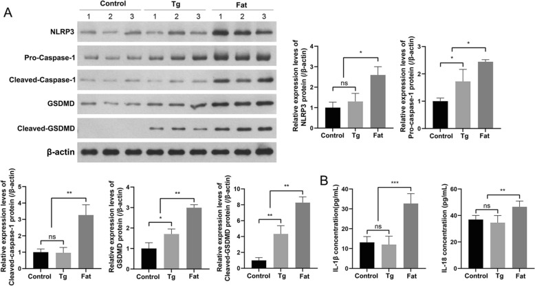
Results: The RPE in Fat group showed obvious Aβ accumulation (p < 0.0001). Compared with the Tg group, the thickness of retina in the Fat group was significantly reduced (t = 5, p = 0.0075), and the number of RPE was statistically decreased (t = 4.243, p = 0.0132). In addition, the expressions of pyroptosis-related proteins NLRP3, Caspase-1, GSDMD, IL-1β and IL-18 in RPE were significantly increased in Fat group (p < 0.05).
Conclusions: High-fat diet leads to Aβ accumulation in the RPE of aged TgAPPswePS1 transgenic mice, causes RPE damage, in which RPE cell pyroptosis may play a crucial role.
Keywords: Amyloid-beta; Pyroptosis; Retinal pigment epithelium; TgAPPswePS1 transgenic mice.
© 2025. The Author(s).
Conflict of interest statement
Declarations. Ethics approval and consent to participate: This study approved by the Laboratory Animal Ethics Committee of Wenzhou Medical University (approval number: xmsq2023-0837, date: 2023.06.30). Competing interests: The authors declare no competing interests.
Figures


Similar articles
-
[Role and mechanism of microRNA-145-5p in hypoxia-induced pyroptosis of human alveolar epithelial cells].Zhonghua Wei Zhong Bing Ji Jiu Yi Xue. 2025 Apr;37(4):354-360. doi: 10.3760/cma.j.cn121430-20240217-00134. Zhonghua Wei Zhong Bing Ji Jiu Yi Xue. 2025. PMID: 40814708 Chinese.
-
HMGB1-mediated pyroptosis promotes inflammation and contributes to skeletal muscle atrophy induced by cigarette smoke.Am J Physiol Cell Physiol. 2025 Jul 1;329(1):C325-C340. doi: 10.1152/ajpcell.01014.2024. Epub 2025 May 30. Am J Physiol Cell Physiol. 2025. PMID: 40445389
-
AIM2 activation mediated by RIPK1/3-dependent mitochondrial DNA release drives Aβ1-40-Induced retinal pigment epithelium injury.Cell Commun Signal. 2025 Jun 21;23(1):301. doi: 10.1186/s12964-025-02294-w. Cell Commun Signal. 2025. PMID: 40544279 Free PMC article.
-
Adiponectin alleviates inflammatory response in metabolic dysfunction-associated steatohepatitis by inhibiting NLRP3 inflammasome-mediated hepatocyte pyroptosis.Hepatobiliary Pancreat Dis Int. 2025 Aug;24(4):433-443. doi: 10.1016/j.hbpd.2025.04.004. Epub 2025 Apr 21. Hepatobiliary Pancreat Dis Int. 2025. PMID: 40307114
-
[Moxibustion promotes endometrial repair in rats with thin endometrium by inhibiting the NLRP3/pyroptosis axis via upregulating miR-223-3p].Nan Fang Yi Ke Da Xue Xue Bao. 2025 Jul 20;45(7):1380-1388. doi: 10.12122/j.issn.1673-4254.2025.07.04. Nan Fang Yi Ke Da Xue Xue Bao. 2025. PMID: 40673300 Free PMC article. Chinese.
Cited by
-
Correction: High-fat diet activates pyroptosis of retinal pigment epithelial cells in aged TgAPPswePS1 transgenic mice.Eur J Med Res. 2025 Aug 8;30(1):724. doi: 10.1186/s40001-025-02979-5. Eur J Med Res. 2025. PMID: 40775372 Free PMC article. No abstract available.
References
-
- Bai Y, Pan Y and Liu X. Mechanistic insights into gasdermin-mediated pyroptosis. Nat Rev Mol Cell Biol. 2025; 0:501–521. - PubMed
MeSH terms
Substances
Grants and funding
LinkOut - more resources
Full Text Sources
Medical
Miscellaneous
