. 2025 Aug;34(8):e70209.
doi: 10.1002/pro.70209.

Genetic mutations disrupt the coordinated mode of tyrosinase's intra-melanosomal domain

Affiliations

Genetic mutations disrupt the coordinated mode of tyrosinase's intra-melanosomal domain

Sarah Toay et al. Protein Sci. 2025 Aug.

Abstract

Oculocutaneous albinism type 1 is a genetic disorder caused by the disruption of tyrosinase activity in the melanogenesis pathway. The tyrosinase's intramelanosomal domain can be subdivided into the catalytic and Cys-rich subdomains, integral for protein stability and catalytic activity. To understand the movement in the tyrosinase intra-melanosomal subdomains and their link to its catalytic activity, we perform essential dynamics on homology models for tyrosinase and the mutant variants R217Q, R402Q, and R217Q/R402Q. Dimensional reduction techniques, such as principal component analysis (PCA), are fundamental to systematically comprehending collective movements in protein structure. The alpha-carbon atomic coordinates for all residues across a 100-ns molecular dynamics trajectory were input into the PCA function, and the results were analyzed alongside correlated movements and free energy profiles for each protein structure. The PCA-identified coordinated movement underlying the stable conformations of wild-type tyrosinase arises within the H9 and H10 helices, which are proximal to the flexible tunnel system and the interface of the catalytic and Cys-rich subdomains. In contrast, genetic mutations R217Q and R217Q/R402Q disrupt the coordinated movement of the tyrosinase intra-melanosomal domain, indicating a cause of mutant variant instability.

Keywords: PCA; coordinated protein motions; molecular dynamics; oculocutaneous albinism type 1; tyrosinase.


Figures

FIGURE 1
FIGURE 1
Protein sequence and ribbon structural model of the intra‐melanosomal domain of tyrosinase. Amino acid sequence and secondary structure presentation of the tyrosinase intra‐melanosomal domain produced using the PDBSum generate server (a) and alpha‐helical structure (b). Alpha‐helices H7, H9, H10, and H19 form a bundle, helping to maintain the tyrosinase catalytic site.
FIGURE 2
FIGURE 2
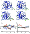
RMSF colored tyrosinase structures and changes over time for solvent‐accessible surface area and radius of gyration. The RMSF for tyrosinase (a), R217Q (b), R402Q (c), and R217Q/R402Q (d) are colored with a rainbow scale with larger values in red and smaller values in blue. The concatenated trajectories for SASA (e) and Rg (f) are colored by protein: wild‐type Tyr (black), R217Q (red), R402Q (green), and R217Q/R402Q (blue) proteins.
FIGURE 3
FIGURE 3
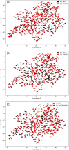
K‐means score plots for principal components. Presented are PC1 and PC2 of WT (a), R217Q (b), R402Q (c), and R217Q/R402Q (d).
FIGURE 4
FIGURE 4
Top 10% loadings projected onto reference PDB structures. Visualization of top 10% of loadings (blue) for WT (a–c, Clusters 1–3), R217Q (d–f, Clusters 1–3), R402Q (g–i, Clusters 1–3), and R217Q/R402Q (j–l, Clusters 1–3) mutant variants are presented by cluster.
FIGURE 5
FIGURE 5
Dynamical cross‐correlation matrices represent the movement relationships between amino acid residues. The matrices are shown for Tyr (a), R217Q (b), R402Q (c), and R217Q/R402Q (d) mutant variants.
FIGURE 6
FIGURE 6
The superposition of porcupine plots of each mutant variant and wild‐type tyrosinase. Porcupine plots are shown for R217Q (a), R402Q (b), and R217Q/R402Q (c) mutant variants. The approximate active site location is denoted with a gold star on (c).
FIGURE 7
FIGURE 7
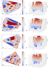
Tyrosinases Free Energy Landscapes. The landscapes are represented for wild‐type tyrosinase (a, b), R217Q (c, d), R402Q (e, f), and R217Q/R402Q (g, h). Mutant variants are shown in both 2D and 3D plots, respectively.
FIGURE 8
FIGURE 8
Tunnel formation correlates with flexible Tyr regions. Tunnels are shown in wild‐type Tyr (a) and R217Q/R402Q mutant variant (b). The N‐terminus (residues Y7‐R115) and C‐terminus (residues A381‐S424, R434‐Y449), between which the tunnel forms in the R217Q/R402Q mutant variant, are shown in red and blue, respectively (c). The surface rendition of the tunnel opening shows residues lining the tunnel (d, red).
FIGURE 9
FIGURE 9
Potential tyrosinase binding pockets for chaperone molecules. Shifting regions containing residues Y7‐R115 and A381‐S424, R434‐Y449 are shown in blue and red, respectively. Three potential binding pockets (a) and Afegostat are highlighted in light/dark green (b), dark/light blue (c), and pink/magenta (d). All potential clusters are also shown (d).

Update of

Similar articles

References

    1. Abouzied AS, Alqarni S, Younes KM, Alanazi SM, Alrsheed DM, Alhathal RK, et al. Structural and free energy landscape analysis for the discovery of antiviral compounds targeting the cap‐binding domain of influenza polymerase pb2. Sci Rep. 2024;14(1):25441. - PMC - PubMed
    1. Abulaiti D, Tuerxun N, Wang H, Ma L, Zhao F, Liu Y, et al. Fungal secondary metabolites as a potential inhibitor of t315i‐ bcr::Abl1 mutant in chronic myeloid leukemia by molecular docking, molecular dynamics simulation and binding free energy exploration approaches. J Genet Eng Biotechnol. 2024;22(4):100444. - PMC - PubMed
    1. Amadei A, Linssen AB, Berendsen HJ. Essential dynamics of proteins. Proteins. 1993;17(4):412–425. - PubMed
    1. Arnold GE, Ornstein RL. Molecular dynamics study of time‐correlated protein domain motions and molecular flexibility: cytochrome p450bm‐3. Biophys J. 1997;73(3):1147–1159. - PMC - PubMed
    1. Chen WC, Tseng TS, Hsiao NW, Lin YL, Wen ZH, Tsai CC, et al. Discovery of highly potent tyrosinase inhibitor, t1, with significant anti‐melanogenesis ability by zebrafish in vivo assay and computational molecular modeling. Sci Rep. 2015;5:7995. - PMC - PubMed

MeSH terms

Substances

Supplementary concepts

LinkOut - more resources