. 2025 Jul 8;53(13):gkaf646.
doi: 10.1093/nar/gkaf646.

A human high-fidelity DNA polymerase holoenzyme has a wide range of lesion bypass activities

Affiliations

A human high-fidelity DNA polymerase holoenzyme has a wide range of lesion bypass activities

Rachel L Dannenberg et al. Nucleic Acids Res. .

Abstract

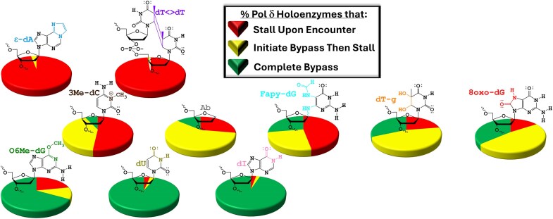
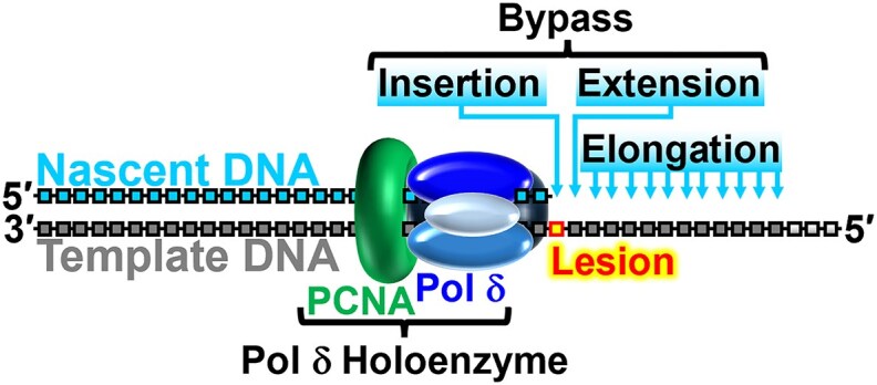
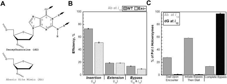
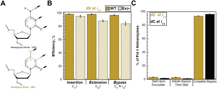
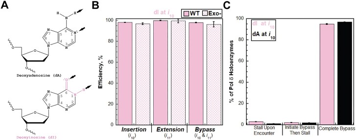
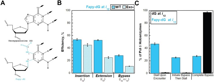
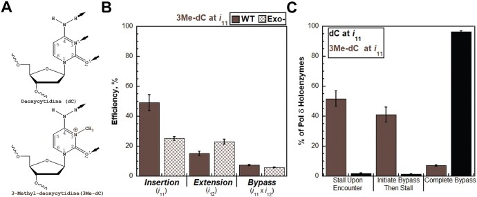
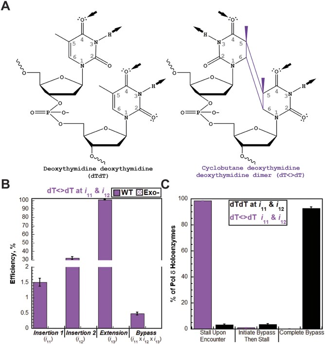
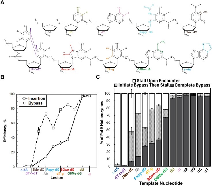
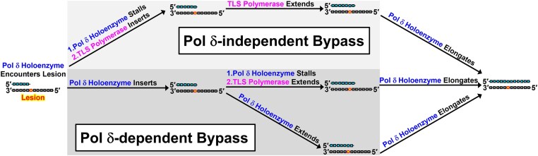
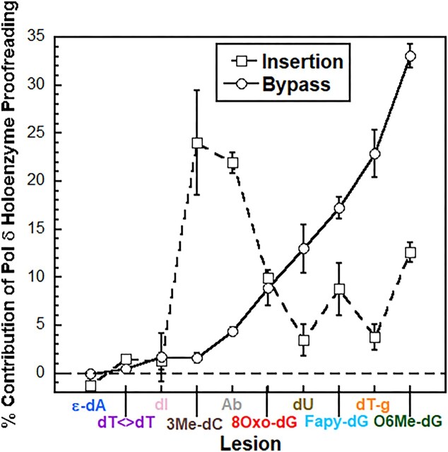
During replication, lagging strand lesions are initially encountered by high-fidelity DNA polymerase (pol) holoenzymes comprised of pol δ and the PCNA sliding clamp. To proceed unhindered, pol δ holoenzymes must bypass lesions without stalling. This entails dNMP incorporation opposite the lesion (insertion) and the 5' template deoxynucleotide (extension). Historically, it was viewed that high-fidelity pol holoenzymes stall upon encountering lesions, activating DNA damage tolerance pathways that are ultimately responsible for lesion bypass. Our recent study of four prominent lesions revealed that human pol δ holoenzymes support insertion and/or bypass for multiple lesions and the extent of these activities depends on the lesion and pol δ proofreading. In the present study, we expand these analyses to other prominent lesions. Collectively, analyses of 10 lesions from both studies reveal that the insertion and bypass efficiencies of pol δ holoenzymes each span a complete range (0%-100%). Consequently, the fates of pol δ holoenzymes upon encountering lesions are quite diverse. Furthermore, pol δ proofreading promoted holoenzyme progression at 7 of the 10 lesions and did not deter progression at any. Altogether, the results significantly alter our understanding of the replicative capacity of high-fidelity pol holoenzymes and their functional role(s) in lesion bypass.


Conflict of interest statement

None declared.

Figures

Graphical Abstract
Graphical Abstract
Figure 1.
Figure 1.
A progressing pol δ holoenzyme encountering a lesion within a lagging strand template.
Figure 2.
Figure 2.
Progressing pol δ holoenzymes encountering an Ab lesion. (A) Structure of Ab. Atomic positions of the nitrogenous base are indicated in gray. Canonical base pairing sites within the nucleobase are indicated by black arrows. Arrows pointed toward and away from the nucleobase indicate hydrogen-bond acceptors and donors, respectively. An Ab is generated from dG (top) by cleavage of the N-glycosidic bond connecting position 9 of the nitrogenous base to the deoxyribose sugar. Shown is an Ab mimic (bottom). (B) Efficiency of replicating an Ab. The efficiencies of dNMP incorporation opposite an Ab (i.e. insertion), 1 nt downstream of an Ab (i.e. extension), and bypass for WT and exonuclease-deficient (Exo−) pol δ holoenzymes are plotted as percentages. The respective dNMP incorporation step(s) (i) for each efficiency is indicated below. (C) Fates of pol δ holoenzymes after encountering an Ab. The distribution of pol δ holoenzyme fates observed after an Ab is encountered at i12 (Ab at i12) is plotted in gray. For comparison, the distribution of pol δ holoenzyme fates observed after a native dG is encountered at i12 (dG at i12) is plotted in black.
Figure 3.
Figure 3.
Progressing pol δ holoenzymes encountering a dU lesion. (A) Structure of dU. Atomic positions of the nitrogenous bases are indicated in gray. Canonical base pairing sites within the nucleobase are indicated by black arrows. Arrows pointed toward and away from the nucleobase indicate hydrogen-bond acceptors and donors, respectively. A dU (bottom) is generated from dC (top) by deamination of position 4. (B) Efficiency of replicating a dU. The efficiencies of dNMP incorporation opposite a dU (i.e. insertion), 1 nt downstream of a dU (i.e. extension), and bypass for WT and exonuclease-deficient (Exo−) pol δ holoenzymes are plotted as percentages. The respective dNMP incorporation step(s) (i) for each efficiency is indicated below. (C). Fates of pol δ holoenzymes after encountering a dU. The distribution of pol δ holoenzyme fates observed after a dU is encountered at i11 (dU at i11) is plotted in olive. For comparison, the distribution of pol δ holoenzyme fates observed after a native dC is encountered at i11 (dC at i11) is plotted in black.
Figure 4.
Figure 4.
Progressing pol δ holoenzymes encountering a dI lesion. (A) Structure of dI. Atomic positions of the nitrogenous bases are indicated in gray. Canonical base pairing sites within the nucleobase are indicated by black arrows. Arrows pointed toward and away from the nucleobase indicate hydrogen-bond acceptors and donors, respectively. A dI (bottom) is generated from a dA (top) by deamination of position 6. (B) Efficiency of replicating a dI. The efficiencies of dNMP incorporation opposite a dI (i.e. insertion), 1 nt downstream of a dI (i.e. extension), and bypass for WT and exonuclease-deficient (Exo−) pol δ holoenzymes are plotted as percentages. The respective dNMP incorporation step(s) (i) for each efficiency is indicated below. (C) Fates of pol δ holoenzymes after encountering a dI. The distribution of pol δ holoenzyme fates observed after a dI is encountered at i10 (dI at i10) is plotted in pink. For comparison, the distribution of pol δ holoenzyme fates observed after a native dA is encountered at i10 (dA at i10) is plotted in black.
Figure 5.
Figure 5.
Progressing pol δ holoenzymes encountering a Fapy-dG lesion. (A) Structure of Fapy-dG. Atomic positions of the nitrogenous bases are indicated in gray. Canonical base pairing sites within the nucleobase are indicated by black arrows. Arrows pointed toward and away from the nucleobase indicate hydrogen-bond acceptors and donors, respectively. A Fapy-dG (bottom) is generated from a dG (top) by β-fragmentation of the imidazole ring at position 6. The N-glycosidic bond of Fapy-dG is displayed as a wave to highlight the anomers of this lesion where the bond is either in the α- or β-position. (B) Efficiency of replicating a Fapy-dG. The efficiencies of dNMP incorporation opposite a Fapy-dG (i.e. insertion), 1 nt downstream of a Fapy-dG (i.e. extension), and bypass for WT and exonuclease-deficient (Exo−) pol δ holoenzymes are plotted as percentages. The respective dNMP incorporation step(s) (i) for each efficiency is indicated below. (C) Fates of pol δ holoenzymes after encountering a Fapy-dG. The distribution of pol δ holoenzyme fates observed after a Fapy-dG is encountered at i12 (Fapy-dG at i10) is plotted in cyan. For comparison, the distribution of pol δ holoenzyme fates observed after a native dG is encountered at i12 (dG at i12) is plotted in black.
Figure 6.
Figure 6.
Progressing pol δ holoenzymes encountering a 3Me-dC lesion. (A) Structure of 3Me-dC. Atomic positions of the nitrogenous bases are indicated in gray. Canonical base pairing sites within the nucleobase are indicated by black arrows. Arrows pointed toward and away from the nucleobase indicate hydrogen-bond acceptors and donors, respectively. A 3Me-dC (bottom) is generated from a dC (top) by methylation of position 3. (B) Efficiency of replicating a 3Me-dC. The efficiencies of dNMP incorporation opposite a 3Me-dC (i.e. insertion), 1 nt downstream of a 3Me-dC (i.e. extension), and bypass for WT and exonuclease-deficient (Exo−) pol δ holoenzymes are plotted as percentages. The respective dNMP incorporation step(s) (i) for each efficiency is indicated below. (C) Fates of pol δ holoenzymes after encountering a 3Me-dC. The distribution of pol δ holoenzyme fates observed after a 3Me-dC is encountered at i11 (3Me-dC at i11) is plotted in brown. For comparison, the distribution of pol δ pol δ holoenzyme fates observed after a native dC is encountered at i11 (dC at i11) is plotted in black.
Figure 7.
Figure 7.
Progressing pol δ holoenzymes encountering a dT<>dT lesion. (A) Structure of dT<>dT. Atomic positions of the nitrogenous bases are indicated in gray. Canonical base pairing sites within the nucleobases are indicated by black arrows. Arrows pointed toward and away from the nucleobase indicate hydrogen-bond acceptors and donors, respectively. A dT<>dT (right) is generated from a native dTdT sequence (left) by crosslinking the 5 and 6 positions of adjacent deoxythymidines via covalent carbon–carbon bonds. (B) Efficiency of replicating a dT<>dT. The efficiencies of dNMP incorporation opposite the 5′ deoxynucleotide in a dT<>dT (i.e. insertion 1), opposite the 3′ deoxynucleotide in a dT<>dT (i.e. insertion 2), 1 nt downstream of a dT<>dT (i.e. extension), and bypass for WT and exonuclease-deficient (Exo−) pol δ holoenzymes are plotted as percentages. Insertion 1, insertion 2, extension, and bypass were not observed for Exo− pol δ holoenzymes. The respective dNMP incorporation step(s) (i) for each efficiency is indicated below. (C) Fates of progressing pol δ holoenzymes after encountering a dT<>dT. The distribution of pol δ holoenzyme fates observed after a dT<>dT is encountered at i11 and i12 (dT<>dT at i11 & i12) is plotted in purple. For comparison, the distribution of pol δ holoenzyme fates observed after a native dTdT sequence is encountered at i11 and i12 (dTdT at i11 & i12) is plotted in black.
Figure 8.
Figure 8.
Progressing pol δ holoenzymes encountering DNA lesions. (A) Structures of the DNA lesions analyzed in our previous and present studies. For each, the covalent modifications are uniquely color-coded. (B) Efficiency of replicating and bypassing DNA lesions. The efficiencies of insertion and bypass for DNA lesions analyzed in our previous and present studies are plotted. For dT<>dT, the efficiency of dNMP incorporation opposite the 5′ nucleotide within the CPD (i.e. insertion 1) is plotted. (C) Distributions of pol δ holoenzyme fates observed after damaged or native template nucleotides are encountered. For each, the % distribution of pol δ holoenzymes that stall upon encounter, initiate bypass then stall, and complete bypass is displayed as a stacked column where the sum of the percentages for all events is equal to 100%. The data for Ab, dU, dI, Fapy-dG, 3Me-dC, and dT<>dT lesions and dG, dC, and dA native template nucleotides are from Figs 2C, 3C, 4C, 5C, 6C, and 7C. The data for ϵ-dA, dT-g, O6Me-dG, and 8Oxo-dG lesions and a native T template nucleotide are from our previous study [3].
Figure 9.
Figure 9.
3Me-dC, Fapy-dG, Ab, dT-g, and 8Oxo-dG lesions in lagging strand templates are bypassed by pol δ-independent and -dependent mechanisms. Only P/T DNA is shown for simplicity. Nascent (primer) and template DNA strands are depicted in cyan and gray, respectively. (Top) Pol δ-independent lesion bypass. (Bottom) Pol δ-dependent lesion bypass mechanisms.
Figure 10.
Figure 10.
Proofreading by pol δ holoenzymes and lesion bypass. The % contribution of proofreading by pol δ holoenzymes to insertion opposite lesions (i.e. initiation of bypass) and (complete) bypass is plotted. The data for Ab, dU, dI, Fapy-dG, 3Me-dC, and dT<>dT lesions are calculated from Figs 2B, 3B, 4B, 5B, 6B, and 7B. The data for ϵ-dA, dT-g, O6Me-dG, and 8Oxo-dG lesions are from our previous study [3]. Negative values, which are not observed within experimental error, indicate that proofreading by pol δ holoenzymes restricts insertion and/or bypass; positive values indicate that proofreading by pol δ holoenzymes promotes insertion and/or bypass.

Update of

Similar articles

References

    1. Lee M, Wang X, Zhang S et al. Regulation and modulation of human DNA polymerase delta activity and function. Genes (Basel). 2017; 8:190. 10.3390/genes8070190. - DOI - PMC - PubMed
    1. Hedglin M, Kumar R, Benkovic SJ Replication clamps and clamp loaders. Cold Spring Harb Perspect Biol. 2013; 5:a010165–83. 10.1101/cshperspect.a010165. - DOI - PMC - PubMed
    1. Dannenberg RL, Cardina JA, Pytko KG et al. Tracking of progressing human DNA polymerase delta holoenzymes reveals distributions of DNA lesion bypass activities. DNA Repair (Amst). 2022; 50:20401–6. 10.1093/nar/gkac745. - DOI - PMC - PubMed
    1. Berdis AJ Mechanisms of DNA polymerases. Chem Rev. 2009; 109:2862–79. 10.1021/cr800530b. - DOI - PubMed
    1. Hedglin M, Benkovic SJ Eukaryotic translesion DNA synthesis on the leading and lagging strands: unique detours around the same obstacle. Chem Rev. 2017; 117:7857–77. 10.1021/acs.chemrev.7b00046. - DOI - PMC - PubMed

LinkOut - more resources