. 2025 Dec;17(1):2531210.
doi: 10.1080/19490976.2025.2531210. Epub 2025 Jul 17.

Ginger-derived vesicle-like nanoparticles loaded with curcumin to alleviate ionizing radiation-induced intestinal damage via gut microbiota regulation

Affiliations

Ginger-derived vesicle-like nanoparticles loaded with curcumin to alleviate ionizing radiation-induced intestinal damage via gut microbiota regulation

Xinrui Zhang et al. Gut Microbes. 2025 Dec.

Abstract

Emerging insights have been approached that gut microbiota act as a critical regulator for ionizing radiation (IR)-induced damage. Herein, an available strategy has been explored to shape gut microbiota for radioprotection by loading curcumin (Cur) into ginger-derived vesicle-like nanoparticles (GDNs). Engineered biomimetic nanovesicles (GDN-Cur) exhibited superb stability in the gastrointestinal tract, thereby significantly enhancing the oral bioavailability of Cur. Consequently, the intrinsic antioxidative, anti-inflammatory, and anti-apoptotic properties of GDNs and Cur granted this nanosystem exceptional protective effect against IR-induced injuries, especially in mitigating intestinal damage. Particularly, the dysbacteriosis triggered by IR could be counteracted through the oral administration of GDN-Cur, resulting in gut microbiota regulation-mediated syndrome mitigation. Furthermore, elevated abundances of Akkermansia muciniphila (A. muciniphila), a bacterial strain of Akkermansia taxa responsive to GDN-Cur, especially their supernatants, were associated with post-radiation protection of intestinal function. This beneficial effect was attributed to the identified radioprotective metabolites secreted by A. muciniphila, such as tanespimycin (17-AAG), which was demonstrated to deactivate AKT/NF-κB signaling pathway. These findings reveal the impact of plant products on radioprotective microbes and metabolites to target host processes and alleviate IR-induced intestinal damage, shedding light on new insights in the development of novel radioprotectants.

Keywords: Ionizing radiation-induced intestinal damage; curcumin; ginger-derived vesicle-like nanoparticles; gut microbiota; metabolites; radioprotection.


Conflict of interest statement

No potential conflict of interest was reported by the author(s).

Figures

Scheme 1.
Scheme 1.
Schematic illustration of GDNs loaded with Cur to alleviate IRID via gut microbiota regulation.
Figure 1.
Figure 1.
Preparation and characterization of GDN-Cur. (a) Schematic representation of the GDN-Cur development process. (b) TEM images, (c) AFM images, (d) size distribution determined by DLS analysis, and (e) zeta potential of the nanovesicles. (f) Predicted lipid species and (g) protein composition of GDNs. (h) CLSM images of PKH67-labeled GDN-Cur. (i) Size changes and (j) percentage of released Cur from GDN-Cur at different pHs over time. (k) Tissue fluorescence observed on CLSM in mice following oral administration of PKH67-labeled GDNs at 4 h. (l) Cur fluorescence in the gastrointestinal tract and main organs analyzed by in vivo imaging system in mice at various times post oral administration of GDN-Cur or Cur. (m) Quantitative analysis of fluorescence intensity across the entire gastrointestinal tract. ***p<0.001.
Figure 2.
Figure 2.
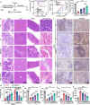
The effect of GDNs loaded with Cur on IR-induced whole-body injury. (a) Illustration of oral administration and radiation on mice. (b) The survival curve (n=20 in each group), (c) clinical score monitoring assessed by body parameters, (d) average dietary intake of feedstuff during the observation period (10 mice per cage), and (e) H&E staining of tissues in mice pretreated with GDN-Cur following by IR (blue arrow: necrosis and shedding of crypt epithelial cells, vacuolar degeneration of hepatocytes, trabeculae splenicae, bone marrow nucleated cells; and black box: splenic white pulp, respectively). (f) Ki67 and caspase-3 immunohistochemical staining of spleen in mice (positive cells: brown). (g) Percentage of positive cells. The levels of serum (h) SOD, (i) CAT, (j) IL-1β, and (k) IL-6 in mice. *p<0.05, **p<0.01, ***p<0.001.
Figure 3.
Figure 3.
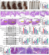
The protective effect of GDNs loaded with Cur on IR-induced intestinal injury in vivo. (a) Anatomy of the abdomen, (b) measurement of colorectal length, (c) colonic pathology characterized by H&E staining, and (d) H&E staining of small intestinal tissues in mice pretreated with GDNs, Cur, or GDN-Cur following by IR. The expression and quantification of (e–i) apoptotic proteins, and (j–m) tight junction proteins in colon tissues analyzed by WB. (n) Immunostaining of mucins in colon tissues using AB/PAS (Bellow arrow: goblet cells). *p<0.05, **p<0.01, ***p<0.001.
Figure 4.
Figure 4.
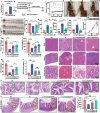
The effect of GDN-Cur on IR-induced intestinal cells injury and macrophages polarization in vitro. (a) Cell cloning analysis, and (b) quantification of clone cells of IEC-6 cells treated with GDNs, Cur, or GDN-Cur at the 4th day post-IR. (c) Cell viability assayed by CCK-8, and (d) apoptosis analysis via Annexin V/PI staining analyzed by flow cytometry of IEC-6 cells with various treatments at 24 h post-IR. ROS detected by DCFH-DA, and (e) imaged by fluorescence microscope, and (f) quantified by flow cytometry analysis in IEC-6 cells with various treatments at 6 h after IR. (g) CLSM imaging of RAW264.7 cells pretreated with Cur or equivalent GDN-Cur for 8 h. (h) Fluorescent analysis of phycoerythrin (PE)-conjugated anti-CD86 and anti-CD206 analyzed by flow cytometry, and (i–k) expression and quantification of iNOS and Arg-1 analyzed by WB of irradiated RAW 264.7 cells in the presence or absence of GDNs, Cur, or GDN-Cur (50 µg/mL) at 12 h post-IR. (l) IL-1β, (m) IL-6, (n) TNF-α, and (o) IL-10 levels in supernatant of RAW 264.7 cells with various treatments at 12 h post-IR. (p–r) Immunofluorometric imaging and quantitative assessment of colon tissues incubated with anti-CD86 and anti-CD206 in mice pretreated with or without GDNs, Cur, or GDN-Cur following by IR (5th day post-IR). *p<0.05, **p<0.01, ***p<0.001.
Figure 5.
Figure 5.
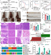
The role gut microbiota modulation in the radioprotective effects GDN-Cur. (a) Illustration of sterile verification using ABX treatment and FMT experiment. (b) The survival curve (n = 20) of mice treated with or without ABX following by IR. (c) Survival curve, (d) clinical score monitoring, (e) anatomy of the abdomen, (f) measurement of colorectal length, (g) H&E staining of tissues (blue arrow: necrosis and shedding of crypt epithelial cells, vacuolar degeneration of hepatocytes, trabeculae splenicae, or bone marrow nucleated cells; and black box: splenic white pulp, respectively) in mice received with or without FMT from GDN-Cur treated mice following by IR exposure. 16S rRNA sequencing analysis of feces in mice pretreated with or without GDN-Cur following by IR exposure, as described by (h) relative abundance, (i) graph depicts Shannon α-diversity index, (j) PCoA of composition on the genus level, (k) LefSe analysis, and (l) relative abundance of Akkermansia, Alistipes, and Parabacteroides. **p<0.01, ***p<0.001.
Figure 6.
Figure 6.
The effect of GDN-Cur related A. muciniphila on IR-induced damage. (a) RT-qPCR analysis of A. muciniphila mRNA levels of feces sample in mice pretreated with or without GDN-Cur following by IR exposure. (b) Illustration of A. muciniphila treatment and radiation experiment. (c) The survival curve, (d) anatomy of the abdomen, (e, f) measurement of colorectal length, (g–i) serum inflammatory factors levels of mice orally gavaged with A. muciniphila following by IR exposure. (j) Clinical score monitoring, (k) average dietary intake of feedstuff (10 mice per cage), (l) serum SOD levels, (m) CAT, (n) MDA levels, the pathological images of (o) liver, spleen, bone marrow, (p) colon, and (q) small intestine characterized by H&E staining, (r) the length of small intestine villi in mice pretreated with s-A. muciniphila or p-A. muciniphila following by IR exposure. *p<0.05, **p<0.01, ***p<0.001.
Figure 7.
Figure 7.
The effect of the supernatant of A. muciniphila and its metabolite 17-AAG against IRID. The expression and quantification of (a–e) apoptotic proteins analyzed by WB, (f) tight junction proteins analyzed by immunohistochemistry, (g, h) goblet cells analyzed by mucins immunostaining, and (i–k) CD86/CD206 analyzed by immunofluorescence assays in colon tissues of mice pretreated with or without s-A. muciniphila following by IR. (l) The relative content of the top 30 expressed metabolites from the supernatant of A. muciniphila. (m) The chemical structure of 17-AAG. *p<0.05, **p<0.01, ***p<0.001.
Figure 8.
Figure 8.
The effect of 17-AAG and s-A. muciniphila on the IR-induced AKT/NF-κB signaling activation. The expression and phosphorylation levels of (a, b) AKT, (c–g) IKK-α/β, IκBα, and NF-κB p65 in colonic tissues of mice pretreated with or without 17-AAG following by IR. (h–n) the expression and phosphorylation levels of AKT/NF-κB signaling pathway in colonic tissues of mice pretreated with or without s-A. muciniphila following by IR. *p<0.05, **p<0.01, ***p<0.001.

References

    1. Hauer-Jensen M, Denham JW, Andreyev HJ.. Radiation enteropathy–pathogenesis, treatment and prevention. Nat Rev Gastroenterol Hepatol. 2014;11(8):470–25. doi: 10.1038/nrgastro.2014.46. - DOI - PMC - PubMed
    1. Kim S, Shin YC, Kim TY, Kim Y, Lee YS, Lee SH, Kim M-N, O E, Kim KS, Kweon M-N, et al. Mucin degrader akkermansia muciniphila accelerates intestinal stem cell-mediated epithelial development. Gut Microbes. 2021;13(1):1–20. doi: 10.1080/19490976.2021.1892441. - DOI - PMC - PubMed
    1. Tapio S, Little MP, Kaiser JC, Impens N, Hamada N, Georgakilas AG, Simar D, Salomaa S. Ionizing radiation-induced circulatory and metabolic diseases. Environ Int. 2021;146:106235. doi: 10.1016/j.envint.2020.106235. - DOI - PMC - PubMed
    1. Yu Y, Lin X, Feng F, Wei Y, Wei S, Gong Y, Guo C, Wang Q, Shuai P, Wang T, et al. Gut microbiota and ionizing radiation-induced damage: is there a link? Environ Res. 2023;229:115947. doi: 10.1016/j.envres.2023.115947. - DOI - PubMed
    1. Yi Y, Lu W, Shen L, Wu Y, Zhang Z. The gut microbiota as a booster for radiotherapy: novel insights into radio-protection and radiation injury. Exp Hematol & Oncol. 2023;12(1):48. doi: 10.1186/s40164-023-00410-5. - DOI - PMC - PubMed

MeSH terms

Supplementary concepts

LinkOut - more resources