[Preprint]. 2025 Jul 10:rs.3.rs-6978813.
doi: 10.21203/rs.3.rs-6978813/v1.

Lysosomal Protease-Mediated APP Degradation is pH-Dependent, Mutation-Sensitive, and Facilitates Tau Proteolysis

Affiliations

Lysosomal Protease-Mediated APP Degradation is pH-Dependent, Mutation-Sensitive, and Facilitates Tau Proteolysis

Caroline Ackley et al. Res Sq. .

Abstract

Background: The accumulation and aggregation of amyloid beta (Aβ)-a peptide fragment derived from the proteolytic processing of amyloid precursor protein (APP)-is a central pathological feature of Alzheimer's disease (AD) and a current target for disease-modifying therapies. Mutations in APP can also drive early-onset AD. While the roles of α-, β-, and γ-secretases and their respective cleavage sites in APP processing are well characterized, much less is understood about the routine degradation of APP within sub-cellular compartments like the lysosome.

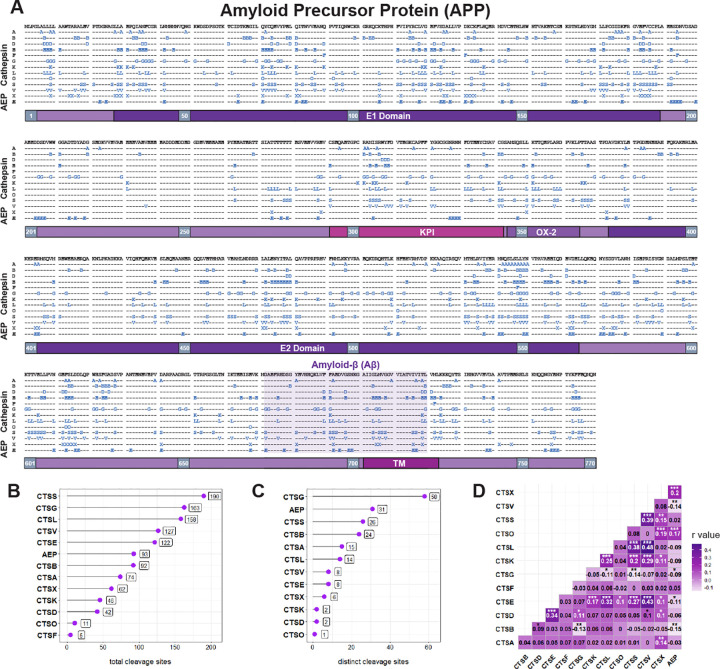
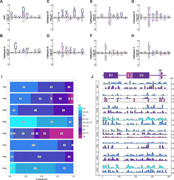
Methods: We applied Multiplexed Substrate Profiling by Mass Spectrometry (MSP-MS) to map cleavage sites within APP that may be targeted by lysosomal proteases, also known as cathepsins. We then employed cell-based and in vitro assays to examine the degradation of both wild-type and mutant APP by these enzymes.

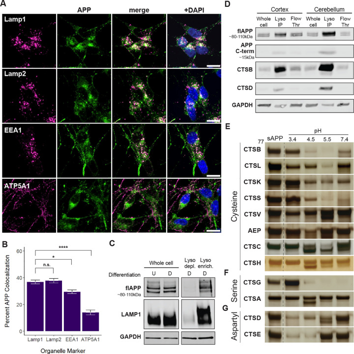
Results: Our findings confirm that APP is enriched in the endolysosomal compartment, where it is processed by many, but not all, cathepsins. Our experiments reveal that cleavages at several mapped APP sites are sensitive to both changes in pH and the presence of pathogenic variants E693G and E693Q. Additionally, we discovered that the large soluble domain of APP (sAPP) enhances tau cleavage by cathepsin G in vitro.

Conclusions: Collectively, these results underscore the importance of lysosomal processing of APP, identify a link between APP and tau, and suggest new avenues for exploring AD pathogenesis. They also point to potential therapeutic targets related to the lysosomal function of APP and its impact on neurodegenerative disease.

Keywords: APP; Alzheimer’s disease; Aβ; amyloid-beta; amyloid-precursor protein; autophagy; cathepsin; lysosome; neurodegeneration; protease; tau.


Conflict of interest statement

Competing interests A.W.K. is a Scientific Advisory Board members, and G.M.K. is an employee of Alaunus Biosciences. Authors declare that they have no conflicts of interest or competing interests.

Figures

Figure 1.
Figure 1.. APP is enriched in the endolysosomal compartment and is cleaved by lysosomal proteases in vitro.
(A) Differentiated SH-SY5Y neurons were fixed and immunostained with antibodies against organelle markers (magenta) and APP (green). Lamp1 and Lamp2, EEA1, and ATP5A1 are enriched in lysosomes, early endosomes, and mitochondria, respectively. Scale bar = 10 μm. (B) Colocalization analysis was performed as described in methods. Percent APP overlap with each organelle marker was quantified and compared using a One-Way ANOVA followed by Bonferroni post-hoc analysis. * p <0.05, ** <0.01, *** <0.001. (C) Undifferentiated (“U”) and differentiated (“D”) SH-SY5Y cells were lysed and cellular contents were separated via density gradient centrifugation. Lysosome depleted (“Lyso depl.”) and lysosome enriched (“Lyso enrich.”) fractions were compared with whole cell samples on a Western Blot after staining with antibodies against APP, Lamp1, and GAPDH. (D) Cortex and cerebellum tissue was collected from LysoTag mice and neuronal lysosomes were collected via immunoprecipitation. Purified lysosomes (“Lyso IP”) were blotted alongside the non-immunoprecipitated flow through (“Flow Thr”) and whole cell samples. (E) Silver staining of sAPP cleaved by various lysosomal proteases and at multiple pH levels. Blots of enzymes that did not cleave sAPP are shown in Figure S3.
Figure 2.
Figure 2.. MSP-MS provides a detailed cleavage map of APP by lysosomal proteases.
(A) A comprehensive cleavage map of APP generated through MSP-MS. P1 cleavage sites are indicated with a correpsonding letter (i.e. Cathepsin A = “A”), or by the symbol “Æ” for Asparagine Endopeptidase (AEP/legumain). (B) A bar graph representing the number of unique cleavage sites across the sequence of APP-770 for each enzyme. (C) Distinct cleavage sites—defined as P1 sites that were only identified for a single cathepsin—were quantified and are displayed as a bar graph. (D) A correlation plot visualizing the similarity in cleavage sites between enzymes. Darker values represent a greater degree of overlap between proteases. * p <0.05, ** <0.01, *** <0.001.
Figure 3.
Figure 3.. Lysosomal proteases exhibit unique substrate specificities that are pH-sensitive.
(A–H) IceLogos generated for select enzymes based on MSP-MS data (additional IceLogos available in Figures S6–S7). (I) Stacked barplots representing the number of cleavage sites tallied at each pH (J) Graphical overview of the APP-770 protein and a map of cleavage patterns across APP at varying pH levels. Dotted lines occur every 100 amino acids. E1, E2 = E1 and E2 domains, respectively. KPI = Kunitz Protease Inhibitor domain. TM = transmembrane. Histograms were generated at each experimental pH value and protease combination tested using MSP-MS. Bar heights indicate the relative number of cleavage sites for each condition (ie. the area under each plot = 100%).
Figure 4.
Figure 4.. Disease-associated mutations alter cathepsin cleavage of Aβ region.
(A) Expanded Aβ region of the MSP-MS cleavage map from Figure 2. Dotted lines represent known β, α, and γ cleavage sites. Protective (green) and deleterious (red) variants are plotted above the primary sequence. Aβ numbering is shown in black. Site E693 is highlighted with a grey bar. (B–G) E693 variants alter cleavage by lysosomal proteases. Wild-type, Arctic (E693G), and Dutch (E693Q) fluorogenic peptides were incubated with various cathepsins. Cleavage activity was determined by quantifying fluorescence over time. (H) Maximum reaction velocity (Vmax) of the cleavage assays shown in (B–G). RFU/min are plotted on a logarithmic scale. (I) Differentiated wild-type and E693 variant SH-SY5Y cells were collected at 0 and 5 days following doxycycline induction of APP expression. (J) Band intensity from (I) was quantified and normalized to GAPDH on Western Blots (8 replicates). * p <0.05, ** <0.01, *** <0.001.
Figure 5:
Figure 5:. APP augments Tau cleavage by Cathepsin G.
(A) Activity assay measuring cleavage of a fluorogenic APP peptide in the presence of increasing Trypsin concentrations. (B–C) Activity assays measuring the cleavage of a fluorogenic tau-peptide (B) and FITC-labelled tau (C) by Cathepsin G over time in the presence of either sAPP or BSA. (D) Recombinant human tau-441 was cleaved in vitro by Cathepsin G in the presence of either sAPP or BSA and analyzed via Western Blot. (E) Quantification of (D), measuring tau (55 kDa) as well as a prominent cleavage product (40 kDa) (3 replicates). * p <0.05, ** <0.01, *** <0.001.

Similar articles

References

    1. García-González L, Pilat D, Baranger K, Rivera S. Emerging Alternative Proteinases in APP Metabolism and Alzheimer’s Disease Pathogenesis: A Focus on MT1-MMP and MT5-MMP. Frontiers in Aging Neuroscience. 2019;11. - PMC - PubMed
    1. Kimura A, Hata S, Suzuki T. Alternative Selection of β-Site APP-Cleaving Enzyme 1 (BACE1) Cleavage Sites in Amyloid β-Protein Precursor (APP) Harboring Protective and Pathogenic Mutations within the Aβ Sequence*. Journal of Biological Chemistry. 2016. Nov;291(46):24041–24053. 10.1074/jbc.M116.744722. - DOI - PMC - PubMed
    1. Hampel H, Hardy J, Blennow K, Chen C, Perry G, Kim SH, et al. The Amyloid-β Pathway in Alzheimer’s Disease. Molecular Psychiatry. 2021. Oct;26(10):5481–5503. 10.1038/s41380-021-01249-0. - DOI - PMC - PubMed
    1. Andrew RJ, Kellett KAB, Thinakaran G, Hooper NM. A Greek Tragedy: The Growing Complexity of Alzheimer Amyloid Precursor Protein Proteolysis*. Journal of Biological Chemistry. 2016. Sep;291(37):19235–19244. 10.1074/jbc.R116.746032. - DOI - PMC - PubMed
    1. Chen AC, Kim S, Shepardson N, Patel S, Hong S, Selkoe DJ. Physical and functional interaction between the α- and γ-secretases: A new model of regulated intramembrane proteolysis. Journal of Cell Biology. 2015. Dec;211(6):1157–1176. 10.1083/jcb.201502001. - DOI - PMC - PubMed

Publication types

LinkOut - more resources