. 2025 Mar;67(1-2):65-116.
doi: 10.4002/040.067.0104. Epub 2025 Mar 25.

PIONOCONUS: A PISCIVOROUS SUBGENUS OF CONUS GASTROPODS

Affiliations

PIONOCONUS: A PISCIVOROUS SUBGENUS OF CONUS GASTROPODS

Maren Watkins et al. Malacologia. 2025 Mar.

Abstract

Alan Kohn showed that two cone-snail species-Conus striatus Linnaeus, 1758, and Conus catus Hwass, 1792-hunt fish as their primary prey. In the seven decades since then, it has been established that these two species belong to the subgenus Pionoconus Mörch, 1852, a well-defined lineage of Conus Linnaeus, 1758, and one of eight piscivorous cone-snail clades. In this review, an integrated multidisciplinary framework for the biology of Pionoconus is presented, based on the numerous research papers published since Kohn's seminal discovery. The molecular biology, phylogeny, biogeography, life history, and prey-capture strategy of Pionoconus are among the topics reviewed, along with more specialized subjects, e.g., human envenomation, culinary recipes, shell-collecting history, and biomedical applications. These illustrate the continuing impact of Kohn's scientific contributions.

Keywords: Pionoconus; clades; conotoxins; envenomation; taxonomy.


Figures

FIGS. 35–37.
FIGS. 35–37.
Cone-snail recipes: (35) Sinabawan nag pakinhason (cone-snail broth); (36) Pancit bihon (noodles with cone snail); (37) Ginataang sihi (Marinduque cone-snail coconut-milk stew).
FIG. 38.
FIG. 38.
Filipino fishermen using tangle nets to harvest mollusks. Photographs courtesy of Noel Saguil.
FIG. 1.
FIG. 1.
Phylogenetic tree of fish-hunting cone snails constructed using house-keeping genes (HKG) derived from transcriptomic analysis. Eight clades of piscivorous snails are shown. Pionoconus forms a distinct branch, and its closest relatives are snails from Textilia and Embrikena. See Chase et al., (2022) for details of the phylogenetic analysis. Species not otherwise mentioned in the text are: Conus brettinghami Coomans, Moolenbeek, & Wils, 1982; Conus bullatus Linnaeus, 1758; Conus cebuensis Wills, 1990; Conus dusaveli (H. Adams, 1872); Conus ermineus Born, 1778; Conus cf. flavus Röckel, 1985; Conus kinoshitai (Kuroda, 1956); Conus lynceus G. B. Sowerby II, 1858; Conus mucronatus Reeve, 1843; Conus neocostatus Olivera, Watkins, Puillandre, & Tenorio, 2021; Conus pergrandis (Iredale, 1937); Conus radiatus Gmelin, 1791; Conus rolani Röckel, 1986; Conus ochroleucus tmetus Tomlin, 1937.
FIGS. 2–4.
FIGS. 2–4.
Specimens representing major phylogenetic branches in Pionoconus. (2) Conus striatus Linnaeus, 1758 (left), and Conus gubernator Hwass, 1792 (right). These define the earliest diverging branch of Pionoconus. (3) The “fragmented clade.” Shells of six Pionoconus species form a monophyletic branch in the phylogenetic tree. The figured specimens are (left to right): Conus stercusmuscarum Linnaeus, 1758; Conus circumscisus Born, 1778; Conus gauguini Richard & Salvat, 1973; Conus aurisiacus Linnaeus, 1758; Conus floccatus G. B. Sowerby I, 1841; and Conus barthelemyi Bernardi, 1861. (4) The Conus magus/catus clade. The eight species that form a monophyletic branch of Pionoconus, including Conus magus Linnaeus, 1758, the type species of Pionoconus, and Conus catus Hwass, 1792, one of the smallest species. The shells of each of the eight species provisionally assigned to this clade are (left to right): Conus magus; Conus catus; Conus fulmen Reeve, 1843; Conus striolatus Kiener, 1848; Conus fischoederi Röckel & da Motta, 1983; Conus achatinus Gmelin, 1791; Conus consors G. B. Sowerby I, 1833; and Conus monachus Linnaeus, 1758. Scale bar 1 cm.
FIG. 5.
FIG. 5.
Phylogenetic tree based on three mitochondrial markers, as described by Puillandre et al. (2014). This tree shows all available Pionoconus for which molecular data have been obtained (14 of the 16 species listed by Röckel et al., 1995). This phylogenetic tree defines three major clades within Pionoconus, labeled A, B, and C, each discussed in the text. Two species, Conus fischoederi and Conus fulmen, lack the molecular data required for inclusion in the phylogenetic tree. The black square indicates the position of the last common ancestor of Pionoconus.
FIG. 6.
FIG. 6.
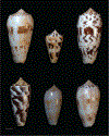
Pionoconus morphospecies related to Conus striatus. Two species recognized by Röckel et al. (1995) were expanded by Monnier et al. (2018) to include five species. An example of each of these distinctive forms (see text) is illustrated. Top row (left to right): Conus striatus juliaallaryae, Conus leehmani, Conus striatus oahuensis. Bottom row (left to right): Conus gubernator, Conus floridus, Conus subfloridus. Scale bar 1 cm.
FIGS. 7–11.
FIGS. 7–11.
Species that belong to major branch B in the Pionoconus phylogenetic tree shown in Figure 5. Examples of variation in the shell pattern are shown for each species: (7) Conus circumcisus Born, 1778; (8) Conus stercusmuscarum Linnaeus, 1758; (9) Conus barthelemyi Bernardi, 1861; (10) Conus aurisiacus Linnaeus, 1758; (11) Conus gauguini Richard & Salvat, 1973. Scale bar 1 cm.
FIG. 12.
FIG. 12.
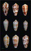
Morphological variation of Conus floccatus G. B. Sowerby I, 1841; correlation to biogeography. The nine specimens shown come from different oceanic localities. Specimens in the top row are from the southwestern edge of the oceanic distribution of Conus floccatus, from the Arafura Sea to the Solomon Islands. Specimens in the middle row are from the northeastern range of the species from Guam to the Marshall Islands. Specimens in the bottom row are from islands off Surigao Province, southeastern Philippines.
FIG. 13.
FIG. 13.
Map of the oceanic localities in which the Conus floccatus specimens illustrated in Figure 12 were collected. Map numbers are 1, West Reef, Kwajalein Atoll (ex Abbey Shells); 2, Ocean Reef, Guam, 37 mi (59 km) southwest of Guam, 120 ft (36.5 km) (dead collected; shell lip eaten away, collected February 14, 1973); 3, Jaluit Atoll, Marshall Islands, collected by Navy Commander Norman Currin, 1958 (ex Tidepool Gallery); 4, Truk Lagoon, Caroline Islands (ex Marshall collection); 5, Arafura Sea, collected by Hugh Morrison off of a dive boat; 6, Siargao Island, Surigao Province, eastern Mindanao, Philippines (ex R. Pagobo); 7, Marau Sound, Guadalcanal, Solomon Islands, diver on a sandy slope at night (ex E. Uhle collection); 8, Malaita, Solomon Islands, in dead coral (ex D. Thorne). Inset: Jaluit Atoll, Marshall Islands, specimens washed ashore after a typhoon were collected at this locality by Commander Norman Currin (image courtesy of National Aeronautics and Space Administration).
FIGS. 14–19.
FIGS. 14–19.
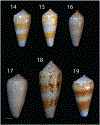
Conus consors G. B. Sowerby I, 1833, and Conus magus Linnaeus, 1758, have similar morphology. The figures show shells that were regarded by Röckel et al. (1995) as Conus consors and distinct variants; (14, 15) typical morphologies; (16, 18) distinct local variations. Some variants of Conus magus (e.g., Figs. 178 and 19) can be very similar morphologically to Conus consors. Scale bar 1 cm.
FIG. 20.
FIG. 20.
Conus magus Linnaeus, 1758. This is one of the most variable species of cone snails for which every locality seems to have its own distinctive variant. For this reason, its taxonomy is extremely confusing. A series of local variations, all from the Philippines, are shown in the top row. The bottom row shows some of the more distinctive forms, proposed by Monnier et al. (2018) to be separable at the species level. The two specimens on the lower left are Conus leobottonii Lorenz, 2006, the two in the middle are Conus epistomoides Weinkauff, 1875, and the two rightmost specimens are Conus ambaroides Shikama, 1977. The evidence for separating Conus leobottonii is robust; the two middle specimens, found in New Guinea, seem distinctive, but no molecular data are available to assess their relation to typical Conus magus from farther west. The evidence that Conus ambaroides is a separate species seems relatively weak at the present time. Scale bar 1 cm.
FIG. 21.
FIG. 21.
Conus achatinus/monachus complex. Specimens assigned to Conus monachus Linnaeus, 1758, are (left to right): Conus monachus; Conus arafurensis (Monnier, Limpalaër, & Robin, 2013); Conus koukae (Monnier, Limpalaër, & Robin, 2013); and Conus achatinus Gmelin, 1791. Scale bar 1 cm.
FIG. 22.
FIG. 22.
Conus catus Hwass, 1791. Specimens from the Red Sea to Hawaii and the Marquesas are shown. The Red Sea population (top row, left) has been separated and designated as Conus nigropunctatus G. B. Sowerby II, 1858, by some researchers. The distinctive Marquesas population is shown at the bottom left. Scale bar 1 cm.
FIG. 23.
FIG. 23.
Morphological variations of Conus striolatus Kiener, 1848. This species is sometimes confused with Conus magus Linnaeus, 1758, but adult Conus striolatus is usually smaller than Conus magus. Scale bar 1 cm.
FIG. 24.
FIG. 24.
Conus fischoederi Rockel & da Motta, 1983. Specimens similar to the second from the left and the second from the right have been separated into a separate species, Conus robini (Limpalaër & Monnier, 2012), by Monnier et al. (2018). Scale bar 1 cm.
FIG. 25.
FIG. 25.
Conus fulmen Reeve, 1843, is a highly distinctive Pionoconus species collected primarily in Japan. Scale bar 1 cm.
FIGS. 26–30.
FIGS. 26–30.
Example of piscivory by Conus consors. (26) The prey is detected by olfactory cues entering the siphon (si) and 1 min 31s after introduction of the fish into the tank, predatory behavior is initiated by extension of the proboscis (pr). (27) The snail probes the ventral fin of the prey in search of a suitable injection site. (28) A radular tooth held at the tip of the proboscis (insert, white arrowhead) is eventually fired and venom delivery causes immediate tetanic paralysis characterized by contraction of the fish fins. (29) Within < 2 min, the fish is swallowed whole inside the rostrum (ro). (30) Scanning electron micrograph of an individual radular tooth (from Rogalski et al., 2023, licensed under CC BY 4.0 <https://creativecommons.org/licenses/by/4.0/>). Note the characteristic recurved barb at the apex of the tooth that facilitates tethering fish prey. Scale bar 500 μm (30).
FIGS. 31–34.
FIGS. 31–34.
Most cone snails have a biphasic life cycle. (31). Females deposit their small, spherical eggs within semirigid capsules from which planktonic larvae emerge. (32). Note the velum extending on both sides of the larval shell. Within two weeks of planktonic development, Conus magus larvae eventually settle and metamorphose into benthic, carnivorous juveniles. (33). The transition from vermivory to piscivory in Conus magus is marked by profound morphological changes, including the shape of the radular tooth. (34) Scanning electron micrographs of juvenile (left) and adult (right) radular teeth. Note the recurved barb absent from the juvenile tooth. Scale bars 20 mm (31), 1 mm (33), 0.5 mm (32, 34 right), 10 μm (34 left). Adapted from Rogalski et al. (2023), licensed under CC BY 4.0 <https://creativecommons.org/licenses/by/4.0/>).

Similar articles

References

    1. Abalde S, Tenorio MJ, Afonso CML, & Zardoya R, 2018, Conotoxin diversity in Chelyconus ermineus (Born, 1778) and the convergent origin of piscivory in the Atlantic and Indo-Pacific cones. Genome Biology and Evolution, 10(10): 2643–2662, 10.1093/gbe/evy150. - DOI - PMC - PubMed
    1. Adams DJ, & Berecki G, 2013, Mechanisms of conotoxin inhibition of N-type (Ca(v)2.2) calcium channels. Biochimica et Biophysica Acta, 1828(7): 1619–1628, 10.1016/j.bbamem.2013.01.019. - DOI - PubMed
    1. Akondi KB, Muttenthaler M, Dutertre S, Kaas Q, Craik DJ, Lewis RJ, & Alewood PF, 2014, Discovery, synthesis, and structure-activity relationships of conotoxins. Chemical Reviews, 114(11): 5815–5847, https://pubs.acs.org/doi/10.1021/cr400401e. - DOI - PMC - PubMed
    1. Al-Amrani S, Al-Jabri Z, Al-Zaabi A, Alshekaili J, & Al-Khabori M, 2021, Proteomics: concepts and applications in human medicine. World Journal of Biological Chemistry, 12(5): 57–69, 10.4331/wjbc.v12.i5.57. - DOI - PMC - PubMed
    1. Ali JR, & Aitchison JC, 2008, Gondwana to Asia: plate tectonics, paleogeography and the biological connectivity of the Indian sub-continent from the Middle Jurassic through latest Eocene (166–35 Ma). Earth-Science Reviews, 88(3): 145–166, 10.1016/j.ear-scirev.2008.01.007. - DOI

LinkOut - more resources