. 2025 Jul 12:18:2317-2330.
doi: 10.2147/DMSO.S516173. eCollection 2025.

Metformin Activation of Sirtuin 3 Signaling Regulates Mitochondrial Function Improves Diabetes-Associated Cognitive Impairment

Affiliations

Metformin Activation of Sirtuin 3 Signaling Regulates Mitochondrial Function Improves Diabetes-Associated Cognitive Impairment

Jiang-Fei An et al. Diabetes Metab Syndr Obes. .

Abstract

Context: Diabetes-associated cognitive impairment (DACD) is a prevalent complication of diabetes mellitus, with a strong correlation to both the severity and duration of the disease. While metformin has demonstrated a significant impact on mitigating DACD, the precise mechanisms underlying its therapeutic effects remain inadequately understood.

Objective: This study aims to examine the protective effects of metformin (MET) on DACD and to elucidate the underlying mechanisms involved.

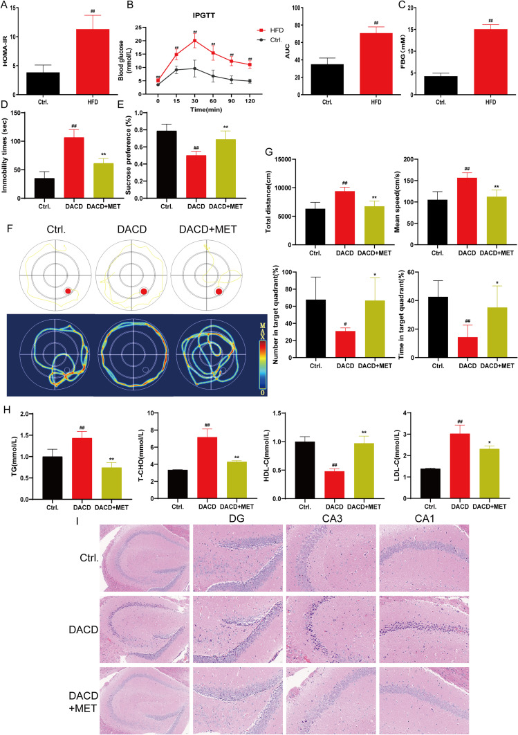
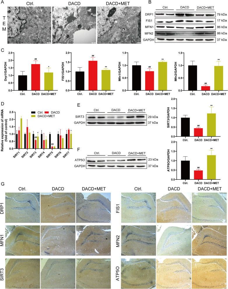
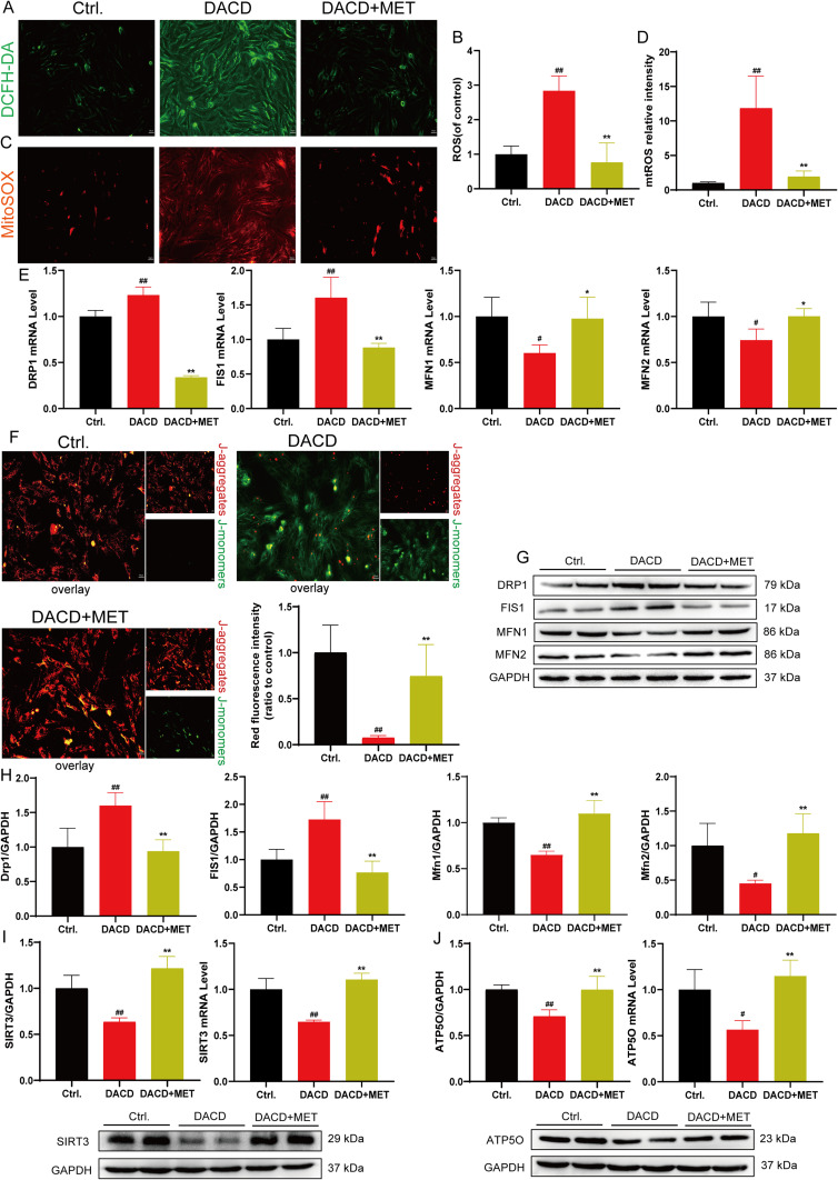
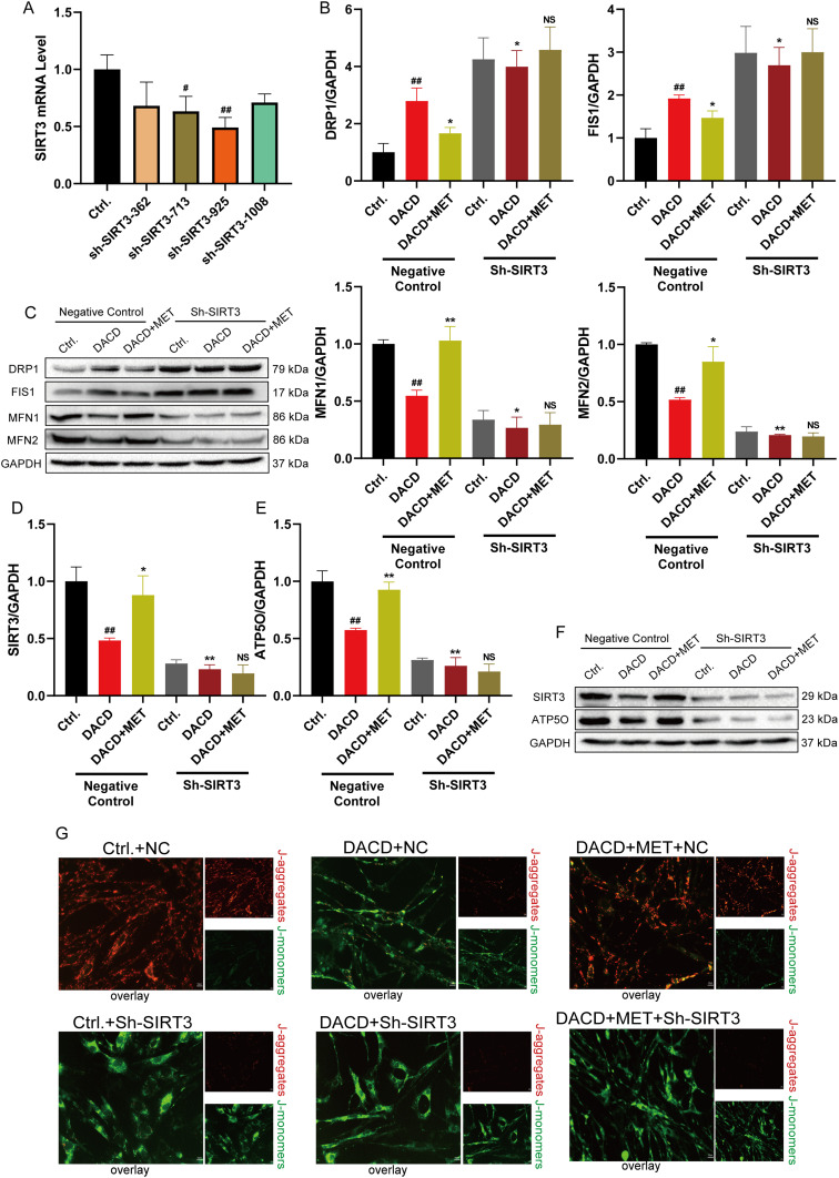
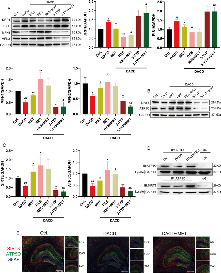
Materials and methods: C57BL/6J male mice from in vivo animal experiments established DACD by high-fat diet (HFD) for 12 weeks, combined with intraperitoneal injection of low-dose streptozotocin (STZ, 40 mg/kg). Subsequently, DACD mice were administered MET for 2 months. The expression levels of proteins related to mitochondrial function were analyzed using immunohistochemical staining, immunofluorescence double staining, qRT-PCR, and Western blot. Furthermore, the mechanism underlying the improvement of DACD by MET was validated by using the Sirtuin 3 (SIRT3) agonist resveratrol (RES), the inhibitor 3-TYP, and sh-SIRT3 on astrocytes.

Results: Our findings indicate that MET significantly ameliorated mitochondrial dysfunction in DACD mice, accompanied by an upregulation of SIRT3 expression. Furthermore, comparable results were noted with the SIRT3 agonist RES. Meanwhile, suppressing SIRT3 expression via sh-SIRT3 or SIRT3 inhibitor 3-TYP in astrocytes largely abolished MET's ability to restore mitochondrial function.

Conclusion: It has been demonstrated that MET ameliorates mitochondrial dysfunction by activating the SIRT3 signaling pathway to rescue DACD.

Keywords: astrocytes; diabetes-associated cognitive dysfunction; metformin; mitochondria; sirtuin 3.


Conflict of interest statement

The authors declare that they have no conflict of interest.

Figures

Figure 1
Figure 1
MET rescues the HFD-induced DACD in mice. (A) Insulin resistance indices in mice (n =6). (B) OGTT in different groups, AUC of blood glucose in mice (n =6). (C) Fasting blood glucose (FBG) in mice (n =6). (D) Mouse immobility time (n =6). (E) Sugar and water consumption rate (n =6). (F) Representative swimming path of the mice. (G) The statistics of the Total distance, Mean speed, Number in target quadrant and Time in target quadrant (n =6). (H) MET on the serum levels of TG, T-CHO, HDL-C and LDL-C (n =6). (I) Hippocampal tissue specimens were dyed with H&E (scale bar = 20 µm). #p < 0.05, ##p < 0.01 versus the control group; *p < 0.05, **p < 0.01 versus the model group.
Figure 2
Figure 2
MET improves mitochondrial function in DACD mice. (A) Representative Transmission electron microscopy (TEM) images of mitochondria in the hippocampal tissue (scale bar = 500 nm). (B and C) Western blotting analysis of DRP1, FIS1, MFN1 and MFN2 protein levels in mouse hippocampal tissues (n = 4). (D) Detection of tissue SIRT1-SIRT7 mRNA levels in the hippocampus (n = 3). (E and F) SIRT3 and ATP5O protein levels were analysed by Western blotting (n = 4). (G) Representative images of DRP1, FIS1, MFN1 and MFN2 immunohistochemical dyeing (scale bar = 50 µm). #p < 0.05, ##p < 0.01 versus the control group; *p < 0.05, **p < 0.01 versus the model group.
Figure 3
Figure 3
MET relies on SIRT3 to alleviate mitochondrial dysfunction in HG/PA-induced AST. (A) DCFH-DA fluorescence staining of AST (scale bar = 50 µm). (B) Quantitative analysis of the ROS (n = 5). (C) MitoSOX fluorescence staining of AST (scale bar = 50 µm). (D) Quantitative analysis of the MitoROS (n = 5). (E) Detection of DRP1, FIS1, MFN1 and MFN2 mRNA levels in the AST (n = 3). (F) Representative images of JC-1 dyeing and quantitative analysis (scale bar = 50 µm). (G and H) Western blotting analysis of DRP1, FIS1, MFN1 and MFN2 protein levels in AST (n = 4). (I and J) Analysis of SIRT3 and ATP5O protein and mRNA levels (n = 3). #p < 0.05, ##p < 0.01 versus the control group; *p < 0.05, **p < 0.01 versus the model group.
Figure 4
Figure 4
Transfection of SIRT3 shRNA to verify the regulatory mechanism of mitochondrial function by MET. (A) qRT-PCR for SIRT3 proteins in AST transfected with negative control and SIRT3 shRNA (n = 3). (B and C) Western blotting analysis of DRP1, FIS1, MFN1 and MFN2 protein levels after SIRT3 shRNA treatment in AST (n = 3). (DF) Western blotting analysis of SIRT3 and ATP5O proteins in SIRT3 shRNA-transfected AST (n = 3). (G) Representative images of JC-1 dyeing (scale bar = 50 µm). #p < 0.05, ##p < 0.01 versus the control group; *p < 0.05, **p < 0.01 versus the model group.
Figure 5
Figure 5
MET up-regulates SIRT3 to ameliorate mitochondrial dysfunction in the DACD. (A) The expression level of DRP1, FIS1, MFN1 and MFN2 proteins after RES or 3-TYP pre-treatment in NMVMs (n = 3). (B and C) Western blotting analysis of SIRT3 and ATP5O proteins (n = 3). (D) The interaction between SIRT3 and ATP5O in AST analysed by co-immunoprecipitation. (E) Immunofluorescence double staining of SIRT3 and ATP5O in the hippocampus (scale bar = 625 µm, scale bar = 200 µm). #p < 0.05, ##p < 0.01 versus the control group; *p < 0.05, **p < 0.01 versus the model group; &p < 0.05, &&p < 0.01 versus the CVB-D group.

Similar articles

References

    1. Liu J, Liu L, Han Y, et al. The molecular mechanism underlying mitophagy-mediated hippocampal neuron apoptosis in diabetes-related depression. J Cell Mol Med. 2021;25(15):7342–7353. doi: 10.1111/jcmm.16763 - DOI - PMC - PubMed
    1. Gao Y, Xiao Y, Miao R, et al. The prevalence of mild cognitive impairment with type 2 diabetes mellitus among elderly people in China: a cross-sectional study. Arch Gerontol Geriatr. 2016;62:138–142. doi: 10.1016/j.archger.2015.09.003 - DOI - PubMed
    1. Jiang Z, Liu B, Lu T, et al. SGK1 drives hippocampal demyelination and diabetes-associated cognitive dysfunction in mice. Nat Commun. 2025;16(1):1709. doi: 10.1038/s41467-025-56854-2 - DOI - PMC - PubMed
    1. Jiang T, Li Y, He S, et al. Reprogramming astrocytic NDRG2/NF-kappaB/C3 signaling restores the diabetes-associated cognitive dysfunction. EBioMedicine. 2023;93:104653. doi: 10.1016/j.ebiom.2023.104653 - DOI - PMC - PubMed
    1. Habib N, Mccabe C, Medina S, et al. Disease-associated astrocytes in Alzheimer’s disease and aging. Nat Neurosci. 2020;23(6):701–706. doi: 10.1038/s41593-020-0624-8 - DOI - PMC - PubMed

LinkOut - more resources