[Preprint]. 2025 Jul 11:2025.07.08.663680.
doi: 10.1101/2025.07.08.663680.

ENHANCED CLEAVAGE OF GENOMIC CCR5 USING CASX2Max

Affiliations

ENHANCED CLEAVAGE OF GENOMIC CCR5 USING CASX2Max

Christine A Hodge et al. bioRxiv. .

Abstract

Development of novel CRISPR/Cas systems enhances opportunities for gene editing to treat infectious diseases, cancer, and genetic disorders. We evaluated CasX2 (PlmCas12e), a class II CRISPR system derived from Planctomycetes, a non-pathogenic bacterium present in aquatic and terrestrial soils. CasX2 offers several advantages over Streptococcus pyogenes Cas9 (SpCas9) and Staphylococcus aureus Cas9 (SaCas9), including its smaller size, distinct protospacer adjacent motif (PAM) requirements, staggered cleavage cuts that promote homology-directed repair, and no known pre-existing immunity in humans. A recent study reported that a three amino acid substitution in CasX2 significantly enhanced cleavage activity (1). Therefore, we compared cleavage efficiency and double-stranded break repair characteristics between the native CasX2 and the variant, CasX2Max, for cleavage of CCR5, a gene that encodes the CCR5 receptor important for HIV-1 infection. Two CasX2 single guide RNAs (sgRNAs) were designed that flanked the 32 bases deleted in the natural CCR5 Δ32 mutation. Nanopore sequencing demonstrated that CasX2 using sgRNAs with spacers of 17 nucleotides (nt), 20 nt or 23 nt in length were ineffective at cleaving genomic CCR5. In contrast, CasX2Max using sgRNAs with 20 nt and 23 nt spacer lengths, enabled robust genomic cleavage of CCR5. Structural modeling indicated that two of the CasX2Max substitutions enhanced sgRNA-DNA duplex stability, while the third improved DNA strand alignment within the catalytic site. These structural changes likely underlie the increased activity of CasX2Max in cellular gene excision. In sum, CasX2Max consistently outperformed native CasX2 across all assays and represents a superior gene-editing platform for therapeutic applications.

Keywords: CCR5; CRISPR/Cas; CasX2; guide RNA.


Figures

Figure 1.
Figure 1.. Schematic of CasX2 and SaCas9 CCR5 gRNAs and their editing abilities.
A. Location of tested gRNAs on chromosome 3. SaCas9 gRNA spacer binding sites CCR5-A and CCR5-B are shown with green arrows, and CasX2 gRNA spacer binding sites sg5 and sg10 sites are indicated with blue arrows. B. Gel electrophoresis was used to analyze a T7 endonuclease assay assessing the activity of CasX2 gRNAs sg5 and sg10 of varying guide lengths (17 nt, 20 nt or 23 nt) in HEK293-FT cells. C. PCR to detect excision of genomic CCR5 following transfection of HEK293-FT cells with either CasX2 and sg5, CasX2 and sg10, SaCas9 and CCR5-A, SaCas9 and CCR5-B, or respective controls of Cas enzyme but no gRNA.
Figure 2.
Figure 2.. CasX2Max demonstrates increased cleavage activity of isolated DNA targets compared to CasX2.
A. RNPs were generated by incubating CasX2 or CasX2Max with purified sgRNAs. RNPs were then incubated with target CCR5 DNA at 37°C for 60 minutes, and DNA cleavage assessed by gel electrophoresis. The “*” denotes the unbound sgRNA. B. Densiometric analysis of gel images of three independent cleavage assays were processed using Image Lab, and statistics calculated using Prism.
Figure 3.
Figure 3.. Improved targeting of CCR5 by CasX2Max compared to CasX2.
A. T7 endonuclease assay of CasX2 and CasX2Max editing of a CCR5 plasmid target in HEK293-FT cells using sg5 and sg10 gRNAs with varying spacer lengths (17 nt, 20 nt, and 23 nt). B. Quantification of plasmid editing by densitometry. Mean editing is based on n=3 replicates, with error bars indicating standard deviation. C. T7 endonuclease assay of CasX2 and CasX2Max editing of genomic CCR5 in HEK293-FT cells by sg5 and sg10 gRNAs of varying spacer lengths (17 nt, 20 nt, and 23 nt). Arrows indicate expected edited bands. Asterisks “*” indicate a product resulting from T7 endonuclease targeting of the heteroduplexes resulting from the presence of the single CCR5 Δ32 allele in HEK293-FT cells.
Figure 4.
Figure 4.. Improved targeting of CCR5 with CasX2Max.
A. Editing of a transfected CCR5 target plasmid following co-transfection of HEK293-FT cells with plasmids encoding CasX2 or CasX2Max and sg5 with the indicated spacer length. Editing was assessed by Nanopore sequencing and editing efficiency was assessed using CRISPResso2. B. The most frequently detected edits are shown for CasX2 (i-iii) and CasX2Max (iv-vi), and for spacer lengths of 17 nt (i, iv), 20 nt (ii, v), and 23 nt (iii, vi). C. Editing efficiency of genomic CCR5 following transfection of HEK293-FT cells with plasmids encoding CasX2 or CasX2Max and sg10 with the indicated spacer length. Editing was assessed by Nanopore sequencing and editing efficiency was assessed using CRISPResso2. D. The most frequently detected edits are shown for CasX2 (i-iii) and CasX2Max (iv-vi), and for spacer lengths of 17 nt (i, iv), 20 nt (ii, v) and 23 nt (iii, vi). E. Editing of genomic CCR5 following transfection of HEK293-FT cells with plasmids encoding CasX2 or CasX2Max and sg5 with the indicated spacer length. Editing was assessed by Nanopore sequencing and editing efficiency by CRISPResso2. F. Editing of genomic CCR5 following transfection of HEK293-FT cells with plasmids encoding CasX2 or CasX2Max and sg10 with the indicated spacer lengths. Editing was assessed by Nanopore sequencing and editing efficiency by CRISPResso2 analysis. G. The most frequently detected edits are shown for CasX2Max plus sg5 of spacer lengths of 20 nt (v) and 23 nt (vi). H. The most frequently detected edits are shown for CasX2Max plus sg10 of spacer lengths of 20 nt (v) and 23 nt (vi). The “*” indicates summation of reads with edits in the region shown but individually comprising less than 1% of the edited reads. “Ref” indicates the reference sequence.
Figure 5.
Figure 5.. Comparable targeting of genomic CCR5 with CasX2Max and SaCas9.
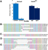
A. Editing of genomic CCR5 following transfection of HEK293-FT cells with plasmids encoding SaCas9 and sgRNAs CCR5-A or CCR5-B, or CasX2Max and sgRNAs sg5 or sg10, was assessed by Nanopore sequencing and CRISPResso2 analysis. B. The most frequently detected edits are shown for SaCas9 CCR5-A at the 20 nt spacer length. C. The most frequently detected edits are shown for SaCas9 CCR5-B at the 20 nt spacer length. D. The most frequently detected edits are shown for CasX2Max with sg5 at the 23 nt spacer length. E. The most frequently detected edits are shown for CasX2Max with sg10 at the 23 nt spacer length. The “*” indicates summation of reads with edits in the region shown but individually comprising less than 1% of the edited reads.
Figure 6.
Figure 6.. Disruption of CCR5 using CasX2Max and paired sg5 and sg10 sgRNAs.
A. PCR amplification of genomic CCR5 after transfection of HEK293-FT cells with scaffold only-plasmids (Ctrl), or with a combination of both sg5 and sg10 with either CasX2 or CasX2Max. B. Minimap2 alignments of Nanopore sequencing data obtained from high-fidelity PCR were displayed in Integrative Genomics Viewer (IGV), demonstrating excision of genomic CCR5 between the sg5 and sg10 binding sites. The target sites for sg5 and sg10 are indicated, as is the location of the CCR5 D32 allele.
Figure 7.
Figure 7.. Structural modeling reveals enhanced PAM-proximal DNA interactions mediated by T26R and K610R substitutions in CasX2Max
A. Positioning of T26 in CasX2. (B). Positioning of R26 in CasX2Max. Panels A and B show (in green) the site where double-stranded DNA (dsDNA) separates into single strands. C. Positioning of K610 in CasX2. D. Positioning of R610 in CasX2Max. Panels C and D show the location near the DNA fork adjacent to the PAM duplex. The target strand (TS, white) and non-target strand (NTS, dark grey) are shown with the NTS PAM nucleotides (TTCA) highlighted in pink. Gold nucleotides represent the spacer region of the sgRNA, complementary to the TS protospacer. Individual nucleotide positions are labeled according to their strand designation (e.g., TS12, NTS20).
Figure 8.
Figure 8.. Structural context of K808R in CasX2 and CasX2Max across catalytic states I and II.
A. In CasX2 state I, K808 is positioned near the RuvC catalytic triad and the zinc ribbon motif. B. K808 forms a single 3.8 Å hydrogen bond with the NTS phosphate backbone at NTS20. It resides within a polar pocket composed of nearby basic residues that help stabilize the displaced NTS for catalysis. C. In CasX2Max state I, the K808R substitution enables dual hydrogen bonds (2.0 Å and 2.2 Å) with the NTS phosphate at NTS20 increasing backbone contact compared to native CasX2. D. The K808R substitution also contributes a larger, more highly charged guanidinium group, subtly reshaping the local electrostatic environment within the same structural pocket. E. In CasX2 state II, K808 reorients toward the TS and forms a single 2.9 Å hydrogen bond with the phosphate backbone at TS12. F. The K808 residue remains within the same polar environment seen in state I, now positioned to support TS engagement during the cleavage step. G. In CasX2Max state II, R808 engages the phosphate backbone at TS12 through dual hydrogen bonds (3.1 Å and 3.2 Å), increasing contact with the substrate strand. H. The additional positive charge and extended reach of the arginine side chain further enriches the local electrostatic surface, potentially enhancing retention of the TS near the RuvC active site. Residues and strands are colored as follows: K808 or R808 side chains (purple); basic polar residues (red); zinc-coordinating cysteines of the zinc ribbon motif (yellow); RuvC catalytic residues D659, E756, and D922 (cyan); DNA target strand (TS) (white); and non-target strand (NTS) (dark gray), with non-bridging oxygens (red) and phosphorous atoms (orange) to indicate the phosphodiester backbone.
Figure 9.
Figure 9.. Zinc ribbon collapse in state III and associated structural shifts.
A. Root mean square deviation (RMSD) values (Å) for α-carbon atoms across the CasX2 sequence, comparing state I and state III. Structural divergence is highest in the bridge helix (BH), RuvC, and TSL domains. Labeled positions indicate CasX2Max substitutions (T26R, K610R, K808R) and selected catalytic and zinc-coordinating residues. Values for the Helical-II domain were excluded due to the absence of resolvable structure in this region in State III. B. In CasX2 state I, residues C810, C813, C912, and C915 coordinate a Zn2+ ion in a tetrahedral configuration, forming the basis of a stable zinc ribbon that anchors adjacent loops and helices. C. In state III, oxidation of C810 and C912 leads to disulfide bond formation, displacing the zinc ion and collapsing the zinc ribbon motif. This rearrangement disrupts the local scaffold, altering the surrounding loop geometry and destabilizing the structural support that normally anchors the adjacent RuvC and TSL domains. D. Structural features of CasX2 in state III shown as a ribbon-and-tube backbone model (blue), with bound sgRNA (black ribbon), non-target strand (NTS) (gray), and target strand (TS) (white). The four cysteines comprising the zinc ribbon motif (C810, C813, C912, C915) are shown (yellow). These cysteines form a disulfide bond between C810 and C912 in state III. Residues undergoing >2 Å displacement relative to state I are shown in orange, revealing that the majority of structural rearrangements are concentrated around the oxidized zinc ribbon cysteines. The only major exception is the bridge helix, that connects to the unresolved Helical-II domain. To provide spatial context, the position of the Helical-II domain from state I is shown (transparent tan surface).

Similar articles

References

    1. Chen Y, Hu Y, Wang X, Luo S, Yang N, Chen Y, et al. Synergistic engineering of CRISPR-Cas nucleases enables robust mammalian genome editing. Innovation (Camb). 2022;3(4):100264. - PMC - PubMed
    1. Pannunzio NR, Watanabe G, Lieber MR. Nonhomologous DNA end-joining for repair of DNA double-strand breaks. J Biol Chem. 2018;293(27):10512–23. - PMC - PubMed
    1. Liu JJ, Orlova N, Oakes BL, Ma E, Spinner HB, Baney KLM, et al. CRISPR-CasX is an RNA-dominated enzyme active for human genome editing. Nature. 2019;566(7743):218–23. - PMC - PubMed
    1. Burstein D, Harrington LB, Strutt SC, Probst AJ, Anantharaman K, Thomas BC, et al. New CRISPR-Cas systems from uncultivated microbes. Nature. 2017;542(7640):237–41. - PMC - PubMed
    1. Armstrong DA, Hudson TR, Hodge CA, Hampton TH, Howell AL, Hayden MS. PlmCas12e (CasX2) cleavage of CCR5: impact of guide RNA spacer length and PAM sequence on cleavage activity. RNA Biol. 2023;20(1):296–305. - PMC - PubMed

Publication types