

This is a preprint.
Development of enterovirus trans-encapsidation assays as tools to understand viral entry
- PMID: 40672215
- PMCID: PMC12265522
- DOI: 10.1101/2025.07.11.664324
Development of enterovirus trans-encapsidation assays as tools to understand viral entry
Abstract
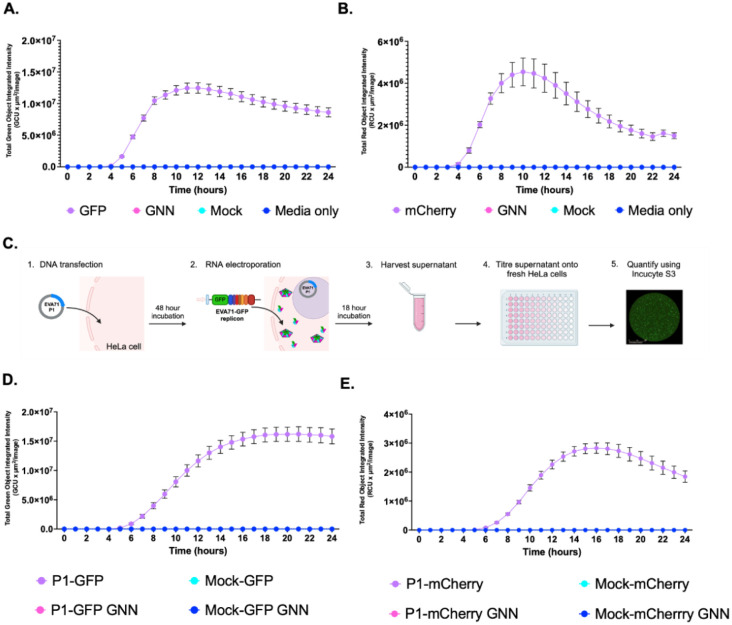
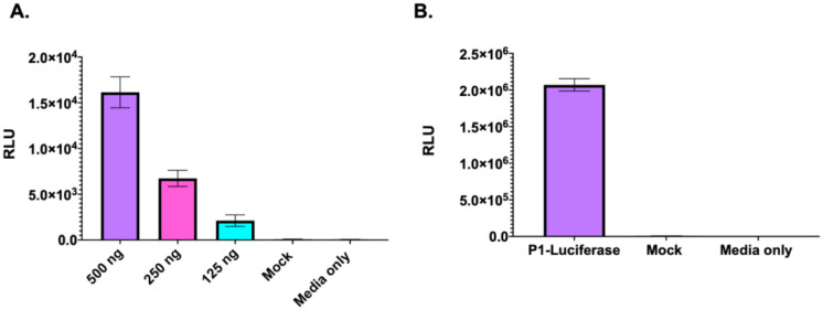
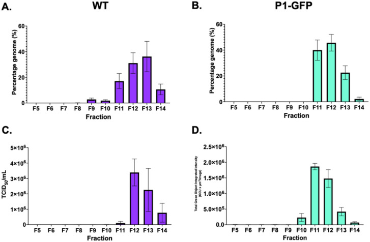
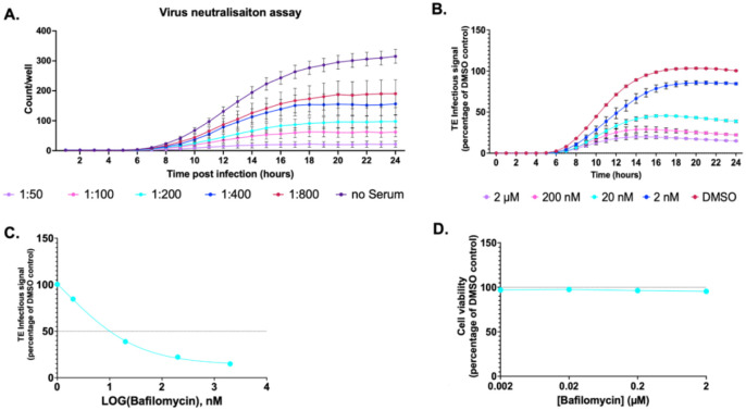
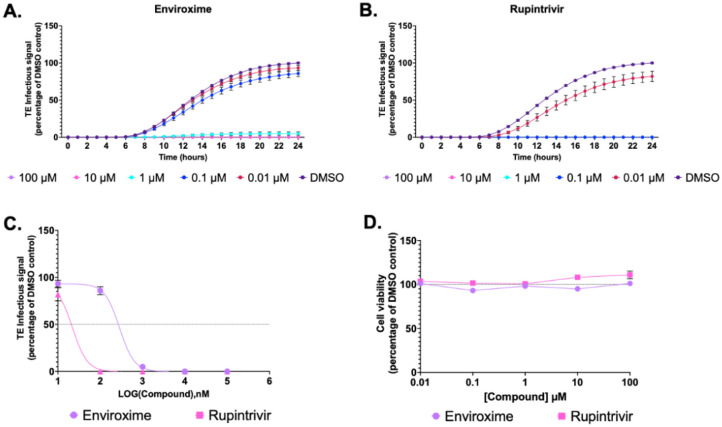
Enteroviruses (EVs) are globally important human and animal pathogens which cause a diverse spectrum of disease, ranging from febrile illness to paralysis. Despite decades of research, parts of the EV lifecycle remain poorly understood. Replicons, in which reporter genes replace the structural protein coding region, have proved useful for the study of EV biology. However, it is not possible to study the molecular mechanism(s) of entry, capsid uncoating and genome release without the production of virus particles. To utilise the benefits provided by replicons for the study of viral cell entry, it would be necessary to supply the structural proteins in trans. Here, we present an EV trans-encapsidation (TE) system in which reporter replicons are transfected into cells modified to express the viral structural proteins. The nascent replicons are packaged in trans to form virus particles containing fluorescent or luminescent replicon genomes. This enables the real-time assessment of EV entry and replication through quantification of fluorescence using live-cell imaging. We demonstrate that these TE particles are biologically accurate proxies to EVA71 virions and show utility for the study of EV entry, uncoating and replication. Additionally, we demonstrate the use of TE particles as platforms for drug discovery and immunological screening, applicable to the development of antiviral therapeutics and assessment of immunisation outcomes.
Conflict of interest statement
Competing interests: The authors declare no competing interests.
Figures
Similar articles
-
The use of sialic acids as attachment factors is a common feature of Enterovirus-D species.J Virol. 2025 Jun 17;99(6):e0042925. doi: 10.1128/jvi.00429-25. Epub 2025 May 13. J Virol. 2025. PMID: 40358210 Free PMC article.
-
Short-Term Memory Impairment.2024 Jun 8. In: StatPearls [Internet]. Treasure Island (FL): StatPearls Publishing; 2025 Jan–. 2024 Jun 8. In: StatPearls [Internet]. Treasure Island (FL): StatPearls Publishing; 2025 Jan–. PMID: 31424720 Free Books & Documents.
-
A rapid and systematic review of the clinical effectiveness and cost-effectiveness of paclitaxel, docetaxel, gemcitabine and vinorelbine in non-small-cell lung cancer.Health Technol Assess. 2001;5(32):1-195. doi: 10.3310/hta5320. Health Technol Assess. 2001. PMID: 12065068
-
The Black Book of Psychotropic Dosing and Monitoring.Psychopharmacol Bull. 2024 Jul 8;54(3):8-59. Psychopharmacol Bull. 2024. PMID: 38993656 Free PMC article. Review.
-
Comparison of Two Modern Survival Prediction Tools, SORG-MLA and METSSS, in Patients With Symptomatic Long-bone Metastases Who Underwent Local Treatment With Surgery Followed by Radiotherapy and With Radiotherapy Alone.Clin Orthop Relat Res. 2024 Dec 1;482(12):2193-2208. doi: 10.1097/CORR.0000000000003185. Epub 2024 Jul 23. Clin Orthop Relat Res. 2024. PMID: 39051924
References
-
- Kreuter JD, Barnes A, McCarthy JE, Schwartzman JD, Oberste MS, Rhodes CH, et al. A fatal central nervous system enterovirus 68 infection. Arch Pathol Lab Med. 2011;135(6):793–6. - PubMed
Publication types
Grants and funding
LinkOut - more resources
Full Text Sources