[Preprint]. 2025 Jul 8:2025.04.14.648540.
doi: 10.1101/2025.04.14.648540.

Nuclear and cytosolic J-domain proteins provide synergistic control of Hsf1 at distinct phases of the heat shock response

Affiliations

Nuclear and cytosolic J-domain proteins provide synergistic control of Hsf1 at distinct phases of the heat shock response

Carmen Ruger-Herreros et al. bioRxiv. .

Abstract

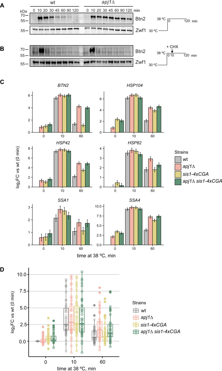
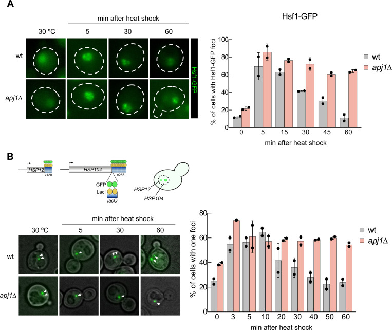
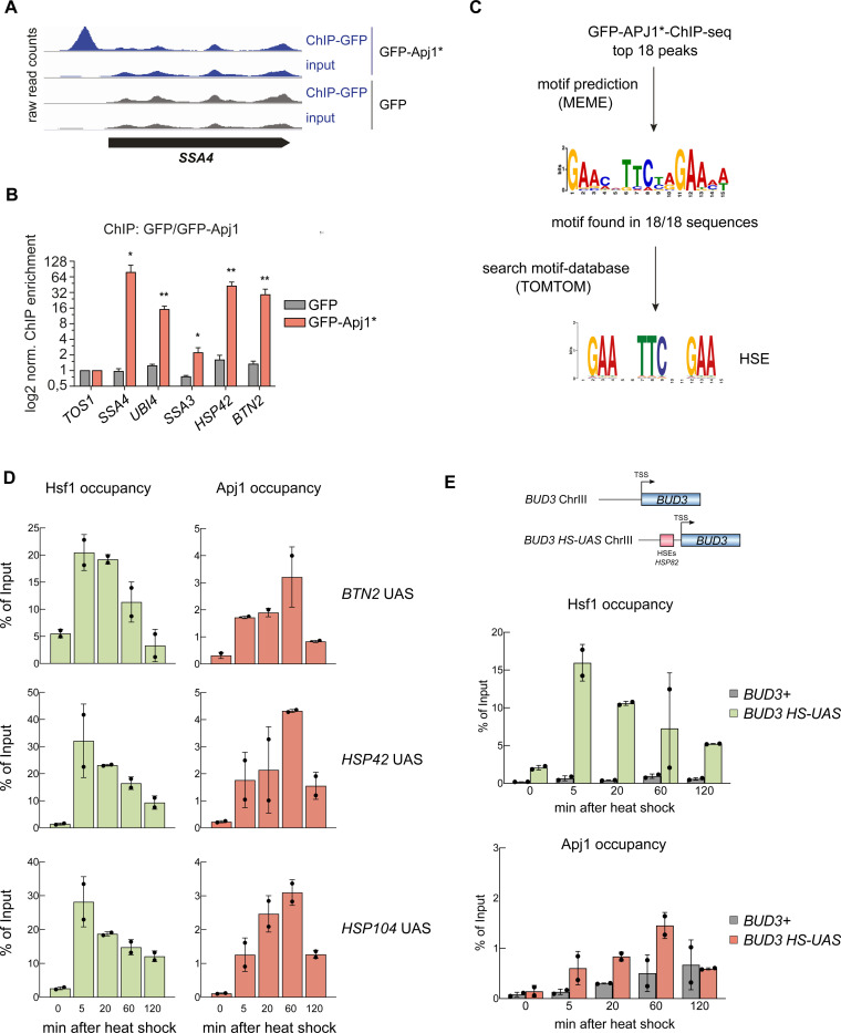
The heat shock response (HSR) is the major defense mechanism against proteotoxic stress in the cytosol and nucleus of eukaryotic cells. Initiation and attenuation of the response are mediated by stress-dependent regulation of heat shock transcription factors (HSFs). Saccharomyces cerevisiae encodes a single HSF (Hsf1), facilitating the analysis of HSR regulation. Hsf1 is repressed by Hsp70 chaperones under non-stress conditions, and becomes activated under proteotoxic stress, directly linking protein damage and its repair to the HSR. J-domain proteins (JDPs) are essential for targeting of Hsp70s to their substrates, yet the specific JDP(s) regulating Hsf1 and connecting protein damage to HSR activation remain unclear. Here we show that the yeast nuclear JDP Apj1 primarily controls the attenuation phase of the HSR by promoting Hsf1's displacement from heat shock elements in target DNA. In apj1Δ cells, HSR attenuation is significantly impaired. Additionally, yeast cells lacking both Apj1 and the major JDP Ydj1 exhibit increased HSR activation even in non-stress conditions, indicating their distinct regulatory roles. Apj1's role in both nuclear protein quality control and Hsf1 regulation underscores its role in directly linking nuclear proteostasis to HSR regulation. Together these findings establish the nucleus as key stress-sensing signaling hub.


Figures

Figure 1
Figure 1
Attenuation of the Hsf1-mediated heat shock response is defective in S. cerevisiae apj1Δ cells. (A-B) S. cerevisiae wt and apj1Δ cells were grown at 30°C till logarithmic growth phase and shifted to 38°C. After 10 min Cycloheximide (CHX) was added to inhibit protein synthesis (B). Total cell extracts were prepared and levels of the heat shock protein Btn2 were determined by western blot analysis at the indicated timepoints after heat shock. Zwf1 levels were determined as loading control. (C) Changes in expression levels of selected Hsf1-target genes were determined by ribosome profiling in indicated yeast strains grown at 30°C and subjected to heat shock at 38°C for 0, 10 or 60 min. Levels of translated mRNAs were normalized to respective levels determined in wild-type cells prior to heat shock and shown in a log2-fold change-scale (log2FC) with the standard error (n=2). (D) Changes in expression levels of all 46 Hsf1 targets were determined and normalized to respective levels determined in wild-type cells prior heat shock. Significance was determined by Wilcox test (**, P<0.01; ***, P <0.001).
Figure 2
Figure 2
Persistent activation of Hsf1 in apj1Δ cells upon heat shock. (A) S. cerevisiae wt and apj1Δ cells expressing Hsf1-GFP were grown at 30°C and heat shocked to 38°C. Cellular localizations of Hsf1-GFP were determined at indicated time points and the proportions of cells showing two or more nuclear Hsf1-GFP foci were determined (n>221 for wt, n>133 for apj1Δ). (B) S. cerevisiae wt and apj1Δ cells expressing LacI-GFP and harboring HSP12 and HSP104 gene loci linked to 128 and 256 repeats of the lacO operator sequence, respectively, were grown at 30°C and heat shocked to 38°C. The percentage cells showing one or two LacI-GFP foci, reporting on coalescence of HSP104 and HSP12 gene loci, were determined at indicated timepoints (n>82 for wt, n>35 for apj1Δ).
Figure 3
Figure 3
Chromatin occupancy of Hsf1 and Apj1 at Heat Shock Response genes is anti-correlated. (A) ChIP experiments using S. cerevisiae cells grown at 30°C and expressing GFP or GFP-Apj1* (GFP-Apj1-34AAA37). Cells were shifted to 42°C for 30 min, crosslinked and processed for ChIP. Raw read counts of inputs and IPs for the heat shock gene SSA4 (HSP70) are depicted. (B) ChIP enrichment (vs TOS1 as control) of GFP-Apj1* and GFP for the indicated Hsf1 targets and the control TOS1 are shown (n=3). Significance was determined by unpaired two-tailed t-test (*, P<0.05; **, P <0.01). (C) Top 18 binding peaks of GFP-Apj1* ChIP experiments were subjected to sequence analysis using MEME and searched for binding sites of transcription factors (using TOMTOM), revealing the Hsf1 target HSE (heat shock element). (D) Hsf1 and Apj1 occupancies at UAS regions of Hsf1-dependent heat shock gene loci. Occupancies were determined at the indicated time points after heat shock (30°C to 39°C) by a ChIP assay. The percentage of input was calculated, and the mean values were plotted with SD (n=2). Statistical significance was determined relative to T=0 min sample by an unpaired two-tailed t-test. ns, P >0.05; *, P<0.05; **, P <0.01; ***, P <0.001. (E) Hsf1 and Apj1 occupancies of UAS regions of the native BUD3 gene and of BUD3 that was placed under Hsf1 control (BUD3-HSE). The latter was accomplished by fusion of the HSP82 promoter (containing three HSEs) with the minimal promoter region of BUD3 (Chowdhary et al, 2019). Occupancies were determined at the indicated time points after heat shock (30°C to 39°C) as above. The percentage of input was calculated, and the mean values were plotted with SD (n=2).
Figure 4
Figure 4
Hsf1 binding to UAS regions of heat shock genes is prolonged in apj1Δ cells. Hsf1 occupancies were determined S. cerevisiae wt and apj1Δ cells at the indicated time points after heat shock (30°C to 38°C) by a ChIP assay. The percentage of input was calculated, and the mean values were plotted with SD (n=4). Statistical significance (relative to T=0 min sample) was determined by One-way ANOVA test: ns, P >0.05; *, P<0.05; **, P <0.01; ***, P <0.001.
Figure 5
Figure 5
Loss of Apj1 and Ydj1 triggers almost full Hsf1 activation. (A) Changes in expression levels of selected Hsf1-target genes were determined by ribosome profiling in indicated yeast strains grown at 25°C. Levels of translated mRNAs in mutant strains were normalized to respective levels determined in wild-type cells and are shown in a log2-scale with the standard error (n=2–3). (B) Indicated yeast strains were grown at 25°C and then heat shocked at 35°C for 30 min. Total cell extracts were prepared and levels of the Hsf1 targets Btn2, Hsp42 and Apj1 were determined by western blot analysis. Levels of histone H3 are provided as loading control. (C) S. cerevisiae wt and apj1Δ ydj1Δ cells expressing Hsf1-GFP were grown at 30°C and heat shocked to 38°C. Cellular localizations of Hsf1-GFP were determined at indicated time points and the proportions of cells showing nuclear Hsf1-GFP foci were determined (n>221 for wt, n>203 for apj1Δ). (D) S. cerevisiae wt and apj1Δ ydj1Δ cells expressing LacI-GFP and harboring HSP12 and HSP104 gene loci linked to 128 and 256 repeats of the lacO operator sequence, respectively, were grown at 30°C and heat shocked to 38°C. The percentage of cells showing one or two LacI-GFP foci, reporting on coalescence of HSP104 and HSP12 gene loci, were determined at indicated timepoints (n>82 for wt, n>169 for apj1Δydj1Δ).
Figure 6
Figure 6
High Hsf1 activity rescues growth of apj1 ydj1 mutant cells. (A/B) Serial dilutions of indicated yeast strains were spotted on YPD plates and incubated at indicated temperatures for 3 d. (C) S. cerevisiae wt, apj1Δ, ydj1Δ and apj1Δ ydj1Δ cells overexpressing SIS1 (TDH3:Sis1) from plasmid or harboring an empty vector (EV) were grown at 25°C and levels of the Hsf1 targets Btn2 and Hsp42 were determined by western blot analysis. Levels of histone H3 were determined as loading control. (D) Serial dilutions of indicated yeast strains overexpressing Sis1 (TDH3::SIS1) from plasmid or harboring an empty vector (EV) were spotted on SC-Leu plates and incubated at indicated temperatures for 3 days.
Figure 7
Figure 7
Regulation of Hsf1 activity via diverse J-domain proteins (JDPs). Hsp70 is targeted to Hsf1 in non-stressed cells by diverse J-domain proteins, including Ydj1, Sis1 and Apj1, to repress heat shock gene expression. Stress conditions trigger protein misfolding and aggregation. Binding of JDPs/Hsp70 to misfolded and aggregated proteins liberates Hsf1 to bind to heat shock elements (HSE) located upstream of heat shock genes, triggering their expression. Apj1 re-targets Hsp70 to HSE-bound Hsf1 during the attenuation phase, triggering Hsf1 dissociation and reducing heat shock gene expression. The HSR ultimately returns to the repressed state.

Similar articles

References

    1. Albert B, Kos-Braun IC, Henras AK, Dez C, Rueda MP, Zhang X, Gadal O, Kos M, Shore D (2019) A ribosome assembly stress response regulates transcription to maintain proteome homeostasis. eLife 8 - PMC - PubMed
    1. Ali A, Garde R, Schaffer OC, Bard JAM, Husain K, Kik SK, Davis KA, Luengo-Woods S, Igarashi MG, Drummond DA et al. (2023) Adaptive preservation of orphan ribosomal proteins in chaperone-dispersed condensates. Nat Cell Biol 25: 1691–1703 - PMC - PubMed
    1. Anders S, Pyl PT, Huber W (2015) HTSeq--a Python framework to work with high-throughput sequencing data. Bioinformatics 31: 166–169 - PMC - PubMed
    1. Aparicio O, Geisberg JV, Sekinger E, Yang A, Moqtaderi Z, Struhl K (2005) Chromatin immunoprecipitation for determining the association of proteins with specific genomic sequences in vivo. Curr Protoc Mol Biol Chapter 21: Unit 21 23 - PubMed
    1. Atencio DP, Yaffe MP (1992) MAS5, a yeast homolog of DnaJ involved in mitochondrial protein import. Mol Cell Biol 12: 283–291 - PMC - PubMed

Publication types

LinkOut - more resources