[Preprint]. 2025 Jul 11:2025.07.10.660754.
doi: 10.1101/2025.07.10.660754.

Preventing vision loss in a mouse model of Leber Congenital Amaurosis by engineered tRNA

Affiliations

Preventing vision loss in a mouse model of Leber Congenital Amaurosis by engineered tRNA

Enes Akyuz et al. bioRxiv. .

Abstract

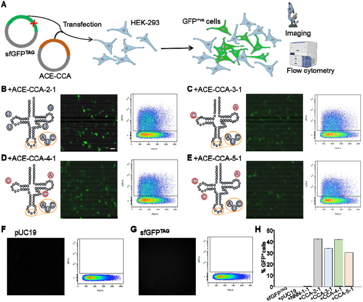
Premature termination codons (PTCs) are associated with rare genetic disorders. Inducing targeted read-through of these 'nonsense mutations' presents a potential therapeutic strategy for modifying disease outcomes. We previously reported that one such PTC, W53X, in the KCNJ13 gene causes blindness and Leber congenital amaurosis type-16 (LCA-16) due to loss of function of the inwardly rectifying potassium channel 7.1 (Kir7.1). Here, we present the proof of concept of a therapeutic approach based on anticodon-engineered transfer RNA (ACE-tRNA). The ACE-tRNA encodes the amino acid tryptophan (Trp) and suppresses the W53X PTC, restoring full-length protein expression. We used helper-dependent adenovirus (HDAd) to deliver the ACE-tRNATrp.UAG (tRNATrp.UAG) and rescue Kir7.1 function and physiology in patient-specific human induced pluripotent stem cell-derived retinal pigment epithelium (hiPSC-RPE) cells. Furthermore, in a W53X mouse model of LCA16, HDAd delivery of tRNATrp.UAG resulted in durable restoration of vision as measured by retinography. This study provides the first example of the therapeutic application of ACE-tRNA for treating an inherited form of blindness.

Keywords: Anticodon engineered tRNA; Kir7.1; Leber Congenital Amaurosis; RPE; electrophysiology; gene therapy; iPSC.


Figures

Fig. 1:
Fig. 1:. Selection of tRNATrp.UAG-tRNA isodecoders using sfGFPTAG readthrough.
A, Overview of the experiments in HEK293 cells transfected with sfGFPTAG (green plasmid) and various codon-edited ACE-CCA-tRNA (red plasmid) to assay GFP read-through efficiency by fluorescence imaging and flow cytometry. Co-transfection of sfGFPTAG along with the codon-edited tRNAs B, ACE-CCA-2-1, C, ACE-CCA-3-1/3-2/3-3, D, ACE-CCA-4-1, E, and ACE-CCA-5-1 show representative structures and varied amounts of GFP fluorescence expression in HEK293T cells along with flow cytometry plots. Cells transfected with F, pUC19, or G sfGFPTAG alone showed no GFP expression. H, Percentage comparison of the different isodecoders for tryptophan tRNA (ACE-CCA) to read through the GFPTAG by flow cytometry assay. ACE-CCA stands for engineered tRNA isodecoder type tRNA-Trp-CcA (2-1 through 5-1). Scale bar=50 μm.
Fig. 2:
Fig. 2:. Suppression of ectopic KCNJ13 disease mutation W53X by ACE-tRNATrp. UAG.
A. Schematic representation of W53X and ACE-tRNATrp. UAG (tRNATrp.UAG) plasmids were co-transfected into HEK293 cells, which resulted in the expression of any combination of tRNATrp.UAG, truncated 53 amino acid polypeptide, and full-length Kir7.1 protein channel. Cells showing positive tdTomato reporter expression represent successful tRNATrp.UAG gene expression, as shown in Supplemental Fig. 2. B, Confocal images show the expression of GFP fluorescence at the cell membrane due to proper localization of GFP-fused WT Kir7.1 in the cytoplasm due to truncated GFP-W53X polypeptide and a mix of membrane and cytoplasmic locations due to tRNATrp.UAG readthrough GFP-Kir7.1 protein. Both the middle and lower panels are immunocytochemistry images of cells (same as shown in the upper panel), probed with a C-terminal anti-Kir7.1 antibody (Gray) that detects only full-length protein that colocalizes with Na-K-ATPase (magenta). Scale bars in B, 20 μm. C, Membrane colocalization of Kir7.1 and Na-K-ATPase was measured by calculating Pearson's coefficient for WT (black quadrangles), W53X (orange circles), and W53X + ACE-tRNATrp. UAG (purple triangles). An identical selection area was used on the cell membrane for the ROI to ensure consistency across measurements. Data points in C are individual ROIs that were compared for statistical significance using two-tailed Student's t-test; n > 3 biological replicates. Statistical significance was determined as ***P<0.001. D, Representative whole-cell current traces for cells measured in the presence of physiological K+ or high concentrations of Rb+ in the extracellular solution. Current responses were generated by applying 50 mV voltage steps (−150 to +50 mV, see inset) for 500 ms from a holding potential of 0 mV for WT, W53X alone, or W53X co-transfected with tRNATrp.UAG cells. The vertical scale bars indicate 150 pA for the K+ current and 500 pA for the Rb+ currents, whereas the horizontal scale bar is 100 ms. E, Average I-V plot (I, current density) for the Kir current measured in physiological K+ in WT (black quadrangles), W53X mutant (orange circles), and W53X + tRNATrp.UAG (purple triangles) cells. The blue rectangle represents the inward current density in F, and the green arrow shows the resting membrane potential in I. F, Comparison of K+ current densities for all recorded cells, as in E, measured at −150 mV. G, Current density plot for Rb+ ions recorded in cells as in E. Orange rectangle was included for the measured current increase in H. H, Rb+ enhances the inward current at −150 mV, which is represented as a fold-increase. I, Plot of resting membrane potentials for individual cells as in E. Data are individual recordings from n>3 biological repeats. Significance was determined as *P<0.05, **P<0.01, and ***P<0.001 using one way ANOVA. WT, GFP-Kir7.1; W53X, GFP-Kir7.1W53X; +tRNATrp.UAG, GFP-Kir7.1W53X + tRNATrp.UAG.
Fig. 3:
Fig. 3:. ACE-tRNATrp.UAG mediated readthrough of endogenous Kir7.1 W53X in LCA16 hiPSC-RPE cells.
A, Schematic of LCA16 hiPSC-RPE grown to a mature tight monolayer of pigmented cells transduced with HDAd virus-packaged ACE-tRNATrp. UAG (HD tRNATrp.UAG). B, Confocal images of WT hiPSC-RPE, LCA16 hiPSC-RPE, and LCA16 hiPSC-RPE transduced with HD tRNATrp.UAG shows the immunofluorescence localization of Kir7.1 (gray) and Na-K-ATPase (magenta). Scale bar, 20 μm. C, Quantitative fluorescence colocalization of Kir7.1 and Na-K-ATPase proteins by Pearson’s correlation. The co-localization of membrane and membrane proteins was analyzed by ROI selection using confocal microscopy. As shown in Fig. 3C, an identical area on the membrane was selected for the ROI, ensuring consistency across the measurements (N = 36 cells). D, Representative current traces in the presence of physiological K+ or high Rb+ extracellular solutions generated in response to 50 mV voltage steps from −150 to 50 mV from a holding potential of 0 mV (inset) for WT hiPSC-RPE WT, LCA16 hiPSC-RPE, and LCA16 hiPSC-RPE cells treated with HD tRNATrp.UAG. The vertical scale bars indicate 100 pA for K+ currents and 250 pA for Rb+ currents, whereas the horizontal scale bar indicates 50 ms. E, K+ current densities were measured at −150 mV in individual WT hiPSC-RPE (black quadrangles), LCA16 hiPSC-RPE (orange circles), and LCA16 hiPSC-RPE cells treated with HD tRNATrp.UAG (purple triangles) groups. F, Inward current fold increase by extracellular Rb+ measured at −150 mV, and G, resting membrane potentials for individual cells, as in E. The data corresponds to individual cells in more than three experimental repeats, and significance was measured as *P<0.05, **P<0.01, and ***P<0.001 when LCA16 hiPSC-RPE was compared with the WT hiPSC-RPE or HD tRNATrp.UAG treated cells using one-way ANOVA.
Fig. 4:
Fig. 4:. Readthrough of nonsense mutation in Kcnj13W53X/ΔR mouse by HDAd mediated tRNATrp.UAG delivery.
A, Schematic of HDAd virus-mediated delivery of sfGFP, sfGFPTAG, or sfGFPTAG with 4X tRNATrp.UAG to the mouse eye via the subretinal route and RPE floret imaging. B, Representative RPE floret images showing fluorescence expression in mice injected with sfGFP, sfGFPTAG, and sfGFPTAG plus 4X tRNATrp.UAG. The upper panels show magnified views of the indicated areas, the middle panel images show whole florets, and the lower panel shows the percentage of fluorescence area coverage due to transduction. Scale bars=50 μm for 20X image (top) and 500 μm for the floret images (middle). C, Time course of the in vivo experiment: The WT allele of Kcnj13W53X/+ was disrupted after the baseline ERG (t1), followed by the delivery of the HDAd virus carrying therapeutic 4X tRNATrp.UAG (t2). Follow-up ERG was performed on mice at different intervals until week 14 (t3). D, In vivo experimental demonstration of RPE genotype-phenotype correlations in the Kcnj13W53X/AR mouse model used for 4X tRNATrp.UAG-mediated nonsense readthrough. E, The ERG c-wave comparison in five individual eyes at baseline (Kcnj13W53X/+; t1; gray), after disruption of WT allele (Kcnj13W53X/ΔR; t2; orange) and the injection of the HD 4X tRNATrp.UAG to Kcnj13W53X/ΔR (t3; purple). F, A comparison of the average a-wave and b-wave amplitude of Kcnj13W53X/ΔR mice after the HD 4X tRNATrp.UAG at 8 wks and 14 wks. G, Comparison of the average c-wave amplitude measured during the experimental time course shows progressive recovery for the mice injected with HD 4X tRNATrp.UAG at 4, 6, 8, and 14 weeks after treatment.

References

    1. Cartegni L., Chew S. L. & Krainer A. R. Listening to silence and understanding nonsense: exonic mutations that affect splicing. Nat Rev Genet 3, 285–298 (2002). 10.1038/nrg775 - DOI - PubMed
    1. Mort M., Ivanov D., Cooper D. N. & Chuzhanova N. A. A meta-analysis of nonsense mutations causing human genetic disease. Hum Mutat 29, 1037–1047 (2008). 10.1002/humu.20763 - DOI - PubMed
    1. Finkel R. S. Read-through strategies for suppression of nonsense mutations in Duchenne/ Becker muscular dystrophy: aminoglycosides and ataluren (PTC124). J Child Neurol 25, 1158–1164 (2010). 10.1177/0883073810371129 - DOI - PMC - PubMed
    1. Keeling K. M., Xue X., Gunn G. & Bedwell D. M. Therapeutics based on stop codon readthrough. Annu Rev Genomics Hum Genet 15, 371–394 (2014). 10.1146/annurev-genom-091212-153527 - DOI - PMC - PubMed
    1. Spelier S., van Doorn E. P. M., van der Ent C. K., Beekman J. M. & Koppens M. A. J. Readthrough compounds for nonsense mutations: bridging the translational gap. Trends Mol Med 29, 297–314 (2023). 10.1016/j.molmed.2023.01.004 - DOI - PubMed

Publication types