[Preprint]. 2025 Jul 12:2025.07.10.663928.
doi: 10.1101/2025.07.10.663928.

PARP1 promotes replication-independent DNA double-strand break formation after acute DNA-methylation damage

Affiliations

PARP1 promotes replication-independent DNA double-strand break formation after acute DNA-methylation damage

Anne Marie McMahon et al. bioRxiv. .

Abstract

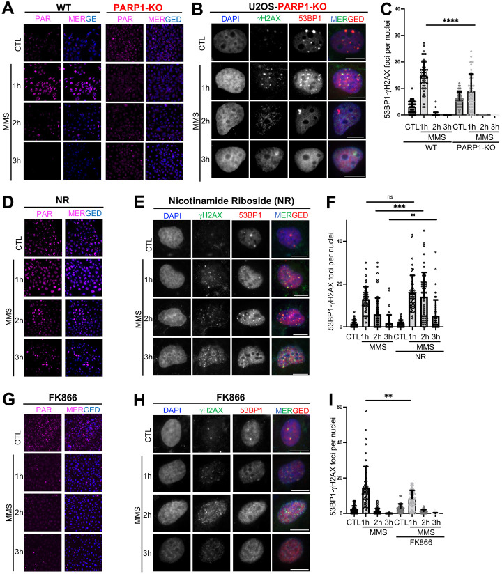
Poly-ADP-Ribose Polymerase 1 (PARP1) is a potent regulator of DNA damage response signaling through the recruitment of DNA damage repair proteins to damage sites, and its catalytic function of converting Nicotinamide adenine dinucleotide (NAD+) into poly-ADP-ribose (PAR) which covalently modifies hundreds of protein substrates in a process known as PARylation. However, PARP1's role in the recognition, processing, and intracellular signaling downstream of DNA damage in cells remains incompletely understood, especially in a replication-independent context. Here, we show that cells exposed to high doses of the methylating agent Methyl Methanesulfonate (MMS) generate DNA double-strand breaks (DSBs) in a base excision repair (BER)-dependent and DNA replication-independent manner. The capacity of cells to generate DSBs after MMS exposure relies heavily on intracellular NAD+ availability and PARP1's catalytic production of PAR. In our experimental system, we show that acute MMS exposure causes NAD+ exhaustion in a PARP1-dependent manner, which results in a temporal-dependent loss of downstream PARP1 activity. This functional loss of PARP1 signaling in later timepoints leads to the loss of BER-dependent single-strand break (SSB)-to-DSB conversion, as well as silencing of ATR-Chk1 signaling in both cycling and non-cycling cells, demonstrating a novel PARP1-dependent regulatory mechanism for both ATR-Chk1 signaling and BER-associated processes following methylation challenge. Additionally, we provide experimental evidence supporting the role of PARP1 and NAD+ in promoting the exonuclease-mediated SSB-to-DSB conversion. These findings support a previously uncharacterized mechanism of PARP1-mediated replication-independent DSB generation and provide insight into checkpoint signaling by integrating DDR with PARP1's consumption of NAD+ and production of PAR.


Conflict of interest statement

DECLARATION OF INTERESTS S.Y. and A.M.M. report a pending patent application related to this manuscript titled "Modulation of DNA double-strand break formation and repair by Poly-ADP-Ribose Polymerase 1, ADP-Ribosylation, and/or NAD+ availability to treat cancer" (PCT#63/783,282). Other authors declare that they have no competing interests.

Figures

Figure 1.
Figure 1.. MMS generates DSB in a BER-dependent manner.
A, U2OS cells were used for immunofluorescent microscopy using DAPI (nuclei staining), γH2AX (AF488), and 53BP1 (AF594) after exposing cells to one, two, and three hours of 3mM MMS respectively. B, Overlapping/colocalizing γH2AX-53BP1 foci were quantified on a per nuclei basis using n=50 cells. C, γH2AX foci per cell were quantified for n=50 cells after MMS exposure. D, Comet assay under neutral condition was performed after indicated 3mM MMS exposure. E, Tail moment was quantified using Comet Assay IV Lite software for n=50 cells from D. F, Comet assay under alkaline condition was performed after indicated 3mM MMS exposure. G, Tail moment was quantified using Comet Assay IV Lite software for n-=50 cells from F. H, U2OS MPG-KO cells were used for immunofluorescent microscopy using DAPI (nuclei staining), γH2AX (AF488), and 53BP1 (AF594) after exposing cells to one, two, and three hours of 3mM MMS respectively. I, Overlapping/colocalizing γH2AX-53BP1 foci were quantified on a per nuclei basis using n=50 cells from H. J, γH2AX foci per nuclei were quantified for n=50 cells from H. K, U2OS APE1-KO cells were used for immunofluorescent microscopy using DAPI (nuclei staining), γH2AX (AF488), and 53BP1 (AF594) after exposing cells to one, two, and three hours of 3mM MMS respectively. L Overlapping/colocalizing γH2AX-53BP1 foci were quantified on a per nuclei basis using n=50 cells from K. M, γH2AX foci per nuclei were quantified for n=50 cells from K. N, Immunoblot confirmation of APE1-KO cells was performed in parallel with WT U2OS cells. For all experiments, the Kolmogorov-Smirnov test was used to determine statistical significance and is demonstrated as follows: ****, p<0.0001; ns, no significance. Scale bar = 10μm.
Figure 2.
Figure 2.. NAD+ availability and PARP1-dependent PAR production promote MMS-induced DSB formation.
A, PAR (magenta) was imaged using immunofluorescence (AF488) and DAPI (nuclei) staining after indicated exposure to 3mM MMS in both WT and PARP1-KO U2OS cells. B, U2OS PARP1-KO cells were treated with MMS in parallel with WT U2OS cells and stained for DAPI, 53BP1 (AF594) and γH2AX (AF488). C, Overlapping/colocalizing γH2AX-53BP1 foci were quantified on a per nuclei basis using n=50 cells from B. D, PAR was imaged after supplementation with Nicotinamide Riboside Chloride (NR) (200μM for 24 hours pre-treatment) and subsequent MMS challenge. E, NR treatment was performed before immunofluorescent microscopy using DAPI (nuclei staining), γH2AX (AF488), and 53BP1 (AF594) after exposing cells to one, two, and three hours of 3mM MMS respectively in parallel with untreated controls. F, Overlapping/colocalizing γH2AX-53BP1 foci were quantified on a per nuclei basis using n=50 cells from E. G, PAR was imaged after treatment with FK866 (50μM for 24 hours pre-treatment) and MMS challenge as indicated. H FK866 treatment was performed before immunofluorescent microscopy using DAPI (nuclei staining), γH2AX (AF488), and 53BP1 (AF594) after exposing cells to one, two, and three hours of 3mM MMS, respectively, in parallel with untreated controls. I Overlapping/colocalizing γH2AX-53BP1 foci were quantified on a per nuclei basis using n=50 cells from H. For all experiments, Kolmogorov-Smirnov test was used to determine statistical significance, and statistical significance is demonstrated as follows: *, p<0.05; **, p<0.01; ***, p<0.001; ****, p<0.0001; ns, no significance. Scale bar = 10μm.
Figure 3.
Figure 3.. NAD+ availability and PAR retention maintains ATR-Chk1 signaling.
A, Immunoblotting was performed on total cell lysates after 15-minute increment samples were collected after 3mM MMS exposure, for a total of three hour of continuous MMS exposure. B, Percent change of Chk1-P-S345 and DNA-PKcs-P-S2056 was quantified from three independent biological replicates using ImageJ after normalization to respective total Chk1 OR DNA-PKcs signals. C, Three-hour treatment schema for media change experiment. D, Immunoblotting was performed after 3mM MMS exposure with media change every hour. E, NR pre-treatment was performed (200μM for 24 hours) before 3mM MMS exposure for the indicated times, followed by subsequent immunoblotting. F, Quantification of Chk1-P-S345 normalized to total Chk1 comparing untreated and NR-treated samples. G, FK866 pre-treatment was performed (50μM for 24 hours) before 3mM MMS exposure for the indicated times and subsequent immunoblotting. H, Quantification of Chk1-P-S345 normalized to total Chk1 comparing untreated and FK866-treated samples. I, PARG inhibitor pre-treatment was performed before 3mM MMS exposure for the indicated times and subsequent immunoblotting. J, Quantification of Chk1-P-S345 normalized to total Chk1 comparing untreated and PARGi-treated samples.
Figure 4.
Figure 4.. PARP1, APE1, and APE2 promote the exonuclease activity of nuclear extracts following MMS challenge.
A, Nuclear extract was prepared from cells after MMS exposure for indicated times. 20μg of nuclear extract was incubated with the DNA construct for indicated times at 37°C, followed by denaturing EMSA assay. B, FAM-labeled AP-site mimic construct and SSB-to-DSB model. C, Nuclear extracts were prepared from WT and PARP1-KO U2OS cells after indicated MMS exposures. 20 μg of nuclear extract was incubated with the DNA construct for the indicated times at 37°C, followed by denaturing EMSA assay. D, Nuclear extracts were prepared from untreated and NR-treated WT U2OS cells (200μM for 24 hours pre-treatment) after indicated MMS exposures. 20 μg of nuclear extract was incubated with the DNA construct for the indicated times at 37°C, followed by a denaturing EMSA assay. E, Nuclear extracts were prepared from untreated and FK866-treated WT U2OS cells (50μM for 24 hours pre-treatment) after indicated MMS exposures. 20 μg of nuclear extract was incubated with the DNA construct at 37°C for the indicated times, followed by denaturing EMSA assay. F, Nuclear extracts were prepared from WT U2OS cells. 20 μg of nuclear extract was incubated with indicated concentrations of AR03 for 30 minutes prior to the addition of the DNA construct for indicated times at 37°C, followed by denaturing EMSA assay. G, Nuclear extracts were prepared from WT U2OS cells. 20 μg of nuclear extract was incubated with indicated concentrations of Celastrol for 30 minutes prior to the addition of the DNA construct for indicated times at 37°C, followed by denaturing EMSA assay.
Figure 5.
Figure 5.. PARP1 promotes DSB formation in a DNA replication-independent manner.
A, U2OS WT cells were synchronized in G1 phase for immunofluorescent microscopy using DAPI (nuclei staining), γH2AX (AF488), and 53BP1 (AF594) after exposing cells to one, two, and three hours of 3mM MMS respectively. B, Overlapping/colocalizing γH2AX-53BP1 foci were quantified on a per nuclei basis using n=50 cells from A. C, γH2AX foci per nuclei were quantified for n=50 cells from A. D, U2OS WT cells were synchronized to G1 phase before neutral Comet assay was performed after indicated 3mM MMS exposure. E, Tail moment was quantified using Comet Assay IV Lite software for n=50 cells from D. F, EdU labelling was performed on WT and PARP1-KO U2OS cells for 2 hours prior to one hour of MMS exposure as indicated. DAPI, EdU (AF488), and 53BP1 (AF594) were labeled and imaged under appropriate channels. G, WT U2OS quantification for 53BP1 foci per cell, stratified based on cell cycle stage (n=25 cells were quantified from 3 biological replicates for a total n=75 cells, bars represent the mean of these three replicates and error bars represent standard deviation between replicates). H, PARP1-KO U2OS quantification for 53BP1 foci per cell, stratified based on cell cycle stage (n=25 cells were quantified from 3 biological replicates for a total n=75 cells, bars represent the mean of these three replicates and error bars represent standard deviation between replicates). For all experiments, the Kolmogorov-Smirnov test was used to determine statistical significance and is demonstrated as follows: *, p<0.05; **, p<0.01; ****, p<0.0001; ns, no significance. Scale bar = 10μm.

Similar articles

References

    1. Beranek D.T. (1990). Distribution of methyl and ethyl adducts following alkylation with monofunctional alkylating agents. Mutat Res 231, 11–30. 10.1016/0027-5107(90)90173-2. - DOI - PubMed
    1. Beranek D.T., Weis C.C., and Swenson D.H. (1980). A comprehensive quantitative analysis of methylated and ethylated DNA using high pressure liquid chromatography. Carcinogenesis 1, 595–606. 10.1093/carcin/1.7.595. - DOI - PubMed
    1. Brink A., Schulz B., Stopper H., and Lutz W.K. (2007). Biological significance of DNA adducts investigated by simultaneous analysis of different endpoints of genotoxicity in L5178Y mouse lymphoma cells treated with methyl methanesulfonate. Mutat Res 625, 94–101. 10.1016/j.mrfmmm.2007.05.007. - DOI - PubMed
    1. Nikolova T., Ensminger M., Lobrich M., and Kaina B. (2010). Homologous recombination protects mammalian cells from replication-associated DNA double-strand breaks arising in response to methyl methanesulfonate. DNA Repair (Amst) 9, 1050–1063. 10.1016/j.dnarep.2010.07.005. - DOI - PubMed
    1. Gates K.S., Nooner T., and Dutta S. (2004). Biologically relevant chemical reactions of N7-alkylguanine residues in DNA. Chem Res Toxicol 17, 839–856. 10.1021/tx049965c. - DOI - PubMed

Publication types

LinkOut - more resources