. 2025 Jul 15;6(8):e70301.
doi: 10.1002/mco2.70301. eCollection 2025 Aug.

Combining Intramuscular and Intranasal Immunization With the MF59-Adjuvanted Respiratory Syncytial Virus Pre-Fusion Protein Subunit Vaccine Induces Potent Humoral and Cellular Immune Responses in Mice

Affiliations

Combining Intramuscular and Intranasal Immunization With the MF59-Adjuvanted Respiratory Syncytial Virus Pre-Fusion Protein Subunit Vaccine Induces Potent Humoral and Cellular Immune Responses in Mice

Jie Shi et al. MedComm (2020). .

Abstract

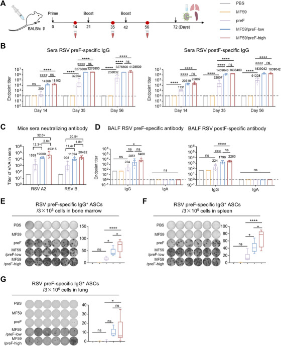
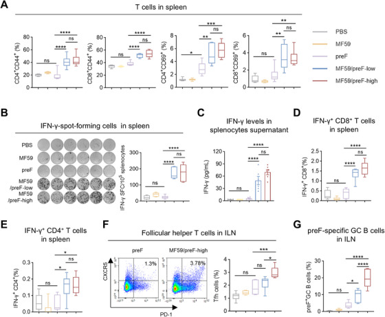
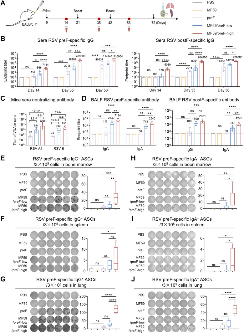
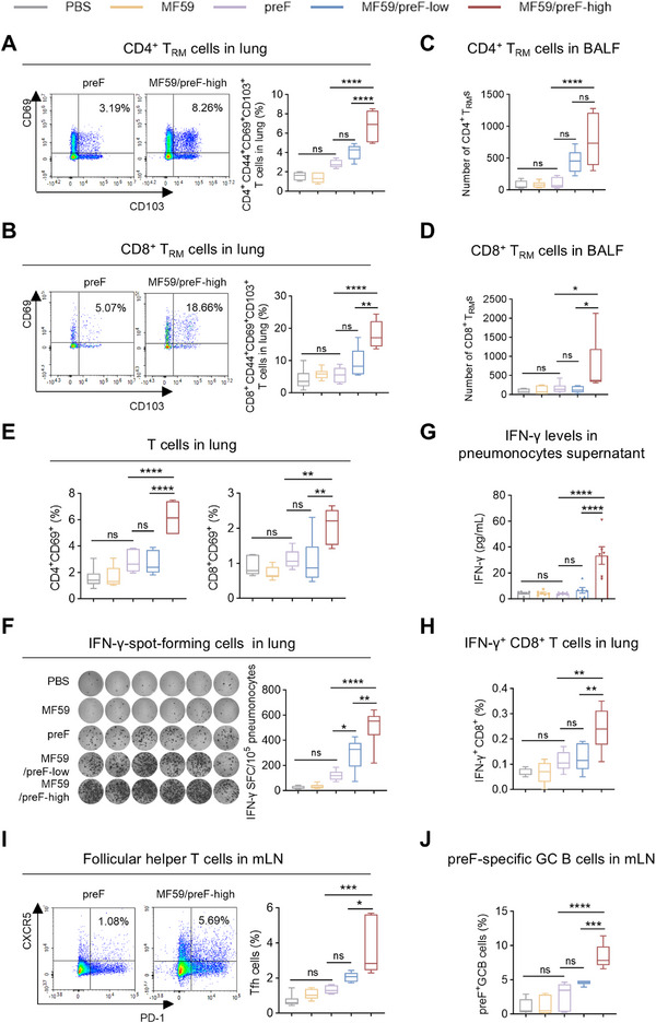
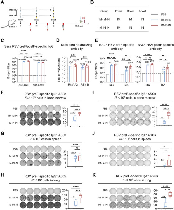
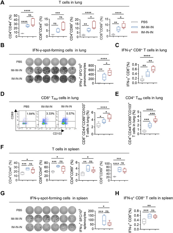
Respiratory syncytial virus (RSV) ranks as the second leading cause of infant death globally and a significant contributor to morbidity and mortality among adults over 60 years old. The development of effective RSV vaccines and immunoprophylaxis remains a key focus. In our research, we formulated a protein-based vaccine known as MF59/preF, which combines the RSV pre-fusion (preF) antigen with an MF59-like oil-in-water adjuvant. Intramuscular (IM) or intranasal (IN) immunization of the MF59-adjuvanted preF protein vaccine elicited robust immune responses and neutralizing antibodies against both RSV A2 and RSV B strains, with the IM showing a particularly pronounced effect. Notably, IN immunization with MF59/preF demonstrated superior mucosal immunity, characterized by elevated levels of IgA antibodies and an increased frequency of tissue-resident memory T (TRM) cells locally. More importantly, the combined IM and IN delivery of the MF59/preF vaccine synergistically enhanced antigen-specific humoral and cellular immune responses at both systemic and mucosal sites. Our study highlights the crucial impact of the route of administration and adjuvanted-protein subunit vaccines on triggering strong humoral and cellular immunity in mice.

Keywords: MF59 adjuvant; RSV pre‐fusion (preF) protein; intranasal immunization; mucosal immunity; respiratory syncytial virus (RSV).


Conflict of interest statement

The authors declare no conflicts of interest.

Figures

FIGURE 1
FIGURE 1
IM immunization with MF59/preF vaccine induced systemic humoral and B‐cell immune responses. (A) The schematic representation of the mouse immunization and sample collection protocol. Mice were immunized intramuscularly with PBS, MF59, preF, MF59/preF‐low, or MF59/preF‐high on Days 0, 21, and 42. Sera were collected on Days 14, 35, and 56, and BALF, spleen, lung, and ILN were harvested on day 72. (B) Endpoint titers of anti‐preF/postF IgG in sera from mice intramuscularly immunized with adjuvanted preF on Days 14, 35, and 56. (C) Titers of virus neutralizing antibody (VNA) against RSV A2 and RSV B in sera collected on Day 56. (D) Endpoint titers of anti‐preF/postF IgG and IgA in BALF collected on Day 72. (E–G) The representative images and quantitative analysis of preF‐specific IgG+ ASCs in bone marrow (E), spleen (F), and lung (G). Data are presented as geometric mean values ± SD in B–D. The middle line indicates the median while the whisker shows the data range in E–G. n = 6 mice per group. p values were conducted by One‐way ANOVA analysis followed by Tukey's multiple comparisons test in B, and D–G. ****p < 0.0001; ***p < 0.001; **p < 0.01; *p < 0.05; ns, not significant.
FIGURE 2
FIGURE 2
IM immunization with MF59/preF vaccine induced systemic T‐cell immune responses. (A) The percentages of antigen‐experienced (CD44+) and activated (CD69+) CD4+ and CD8+ T cells in the spleen. (B) IFN‐γ‐spot‐forming cells in the spleen. Images (left) and quantification (right) of IFN‐γ‐spot‐forming cells were displayed. (C) The levels of IFN‐γ in the supernatants of splenic lymphocytes. (D–E) The percentages of antigen‐specific IFN‐γ producing memory CD8+ (D) and CD4+ (E) T cells in the spleen. (F–G) The percentages of Tfh cells (CD4+CXCR5+PD‐1+) (F) and preF‐specific GC B cells (B220+GL‐7+CD95+) (G) in ILN. Data are presented as mean values ± SEM in C. The middle line indicates the median while the whisker shows the data range in A–B and D–G. n = 6 mice per group. p values were conducted by One‐way ANOVA analysis followed by Tukey's multiple comparisons test in A–G. ****p < 0.0001; ***p < 0.001; **p < 0.01; *p < 0.05; ns, not significant.
FIGURE 3
FIGURE 3
IN immunization with MF59/preF vaccine induced local humoral and B‐cell immune responses. (A) The schematic representation of the mouse immunization and sample collection protocol. Mice were immunized intranasally with PBS, MF59, preF, MF59/preF‐low, or MF59/preF‐high on Days 0, 21, and 42. Sera were collected on Days 14, 35, and 56, and BALF, spleen, lung, and mLN were harvested on Day 72. (B) Endpoint titers of anti‐preF/postF IgG in sera from mice intranasally immunized with adjuvanted preF on Days 14, 35, and 56. (C) Titers of virus neutralizing antibody (VNA) against RSV A2 and RSV B in sera collected on Day 56. (D) Endpoint titers of anti‐preF/postF IgG and IgA in BALF collected on Day 72. (E–J) The representative images and quantitative analysis of preF‐specific IgG+ (left) and IgA+ (right) ASCs in bone marrow (E, H), spleen (F, I), and lung (G, J). Data are presented as geometric mean values ± SD in B–D. The middle line indicates the median while the whisker shows the data range in E–J. n = 6 mice per group. p values were conducted by One‐way ANOVA analysis followed by Tukey's multiple comparisons test in B, and D–J. ****p < 0.0001; ***p < 0.001; **p < 0.01; *p < 0.05; ns, not significant.
FIGURE 4
FIGURE 4
IN immunization with MF59/preF vaccine induced local T‐cell immune responses. (A–D) The proportion of CD4+ (A) and CD8+ (B) TRM cells in the lung and the absolute number of CD4+ (C) and CD8+ (D) TRM cells in BALF. TRM cells were gated on CD4+CD44+CD69+CD103+ or CD8+CD44+CD69+CD103+. (E) The percentages of activated (CD69+) CD4+ and CD8+ T cells in the lung. (F) IFN‐γ‐spot‐forming cells in the lung. Images (left) and quantification (right) of IFN‐γ‐spot‐forming cells were displayed. (G) The levels of IFN‐γ in the supernatants of lung single‐cell suspensions. (H) The percentages of antigen‐specific IFN‐γ‐producing memory CD8+ T cells in the lung. (I‐J) The percentages of Tfh cells (CD4+CXCR5+PD‐1+) (I) and preF‐specific GC B cells (B220+GL‐7+CD95+) (J) in mLN. Data are presented as mean values ± SEM in G. The middle line indicates the median while the whisker shows the data range in A– F and H–J. n = 6 mice per group. p values were conducted by One‐way ANOVA analysis followed by Tukey's multiple comparisons test in A–J. ****p < 0.0001; ***p < 0.001; **p < 0.01; *p < 0.05; ns, not significant.
FIGURE 5
FIGURE 5
Combination of IM and IN immunization using MF59/preF vaccine elicited both local and systemic humoral and B‐cell immune responses. (A–B) The schematic representation of the mouse immunization and sample collection protocol. BALB/c mice received two intramuscular doses of the MF59/preF vaccine followed by a single intranasal dose (IM‐IM‐IN), or one intramuscular dose followed by two intranasal doses (IM‐IN‐IN). Sera were collected on Days 14, 35, and 56, and BALF, spleen, lung, and mLN were harvested on Day 72. (C) Endpoint titers of anti‐preF/postF IgG in sera on Day 56. (D) Titers of virus neutralizing antibody (VNA) against RSV A2 and RSV B in sera collected on Day 56. (E) Endpoint titers of anti‐preF/postF IgG and IgA in BALF collected on Day 72. (F‐K) The representative images and quantitative analysis of preF‐specific IgG+ (left) and IgA+ (right) ASCs in bone marrow (F, I), spleen (G, J), and lung (H, K). Data are presented as geometric mean values ± SD in C–E. The middle line indicates the median while the whisker shows the data range in F–K. n = 6 mice per group. p values were conducted by One‐way ANOVA analysis followed by Tukey's multiple comparisons test in C–K. ****p < 0.0001; ***p < 0.001; **p < 0.01; *p < 0.05; ns, not significant.
FIGURE 6
FIGURE 6
Combination of IM and IN immunization using MF59/preF vaccine elicited both local and systemic T‐cell immune responses. (A) The percentages of antigen‐experienced (CD44+) and activated (CD69+) CD4+ and CD8+ T cells in the lung. (B) IFN‐γ‐spot‐forming cells in the lung. Images (left) and quantification (right) of IFN‐γ‐spot‐forming cells were displayed. (C) The percentages of antigen‐specific IFN‐γ‐producing memory CD8+ T cells in the lung. (D‐E) The proportion of CD8+ (D) and CD4+ (E) TRM cells in the lung. TRM cells were gated on CD4+CD44+CD69+CD103+ or CD8+CD44+CD69+CD103+. (F) The percentages of antigen‐experienced (CD44+) and activated (CD69+) CD4+ and CD8+T cells in the spleen. (G) IFN‐γ‐spot‐forming cells in the spleen. Images (left) and quantification (right) of IFN‐γ‐spot‐forming cells were displayed. (H) The percentages of antigen‐specific IFN‐γ‐producing memory CD8+ T cells in the spleen. The middle line indicates the median while the whisker shows the data range in A–H. n = 6 mice per group. p values were conducted by One‐way ANOVA analysis followed by Tukey's multiple comparisons test in A–H. ****p < 0.0001; ***p < 0.001; **p < 0.01; *p < 0.05; ns, not significant.

Similar articles

References

    1. Wang X., Li Y., Shi T., et al., “Global Disease Burden of and Risk Factors for Acute Lower Respiratory Infections Caused by Respiratory Syncytial Virus in Preterm Infants and Young Children in 2019: A Systematic Review and Meta‐Analysis of Aggregated and Individual Participant Data,” Lancet 403, no. 10433 (2024): 1241–1253. - PubMed
    1. Rago A. R. P., D'Arrigo S. F., Osmani M., Espinosa C. M., Torres C. M., “Respiratory Syncytial Virus: Epidemiology, Burden of Disease, and Clinical Update,” Advances in Pediatrics 71, no. 1 (2024): 107–118. - PubMed
    1. Ruckwardt T. J., Morabito K. M., Graham B. S., “Immunological Lessons From Respiratory Syncytial Virus Vaccine Development,” Immunity 51, no. 3 (2019): 429–442. - PubMed
    1. Dalziel S. R., Haskell L., O'Brien S., et al., “Bronchiolitis,” Lancet 400, no. 10349 (2022): 392–406. - PubMed
    1. Li Y., Wang X., Blau D. M., et al., “Global, Regional, and National Disease Burden Estimates of Acute Lower Respiratory Infections due to Respiratory Syncytial Virus in Children Younger Than 5 Years in 2019: A Systematic Analysis,” Lancet 399, no. 10340 (2022): 2047–2064. - PMC - PubMed

LinkOut - more resources