Review
. 2025 Jul 2:18:1602343.
doi: 10.3389/fnmol.2025.1602343. eCollection 2025.

The neurological pathology of peroxisomal ACBD5 deficiency - lessons from patients and mouse models

Affiliations
Review

The neurological pathology of peroxisomal ACBD5 deficiency - lessons from patients and mouse models

Michael L Dawes et al. Front Mol Neurosci. .

Abstract

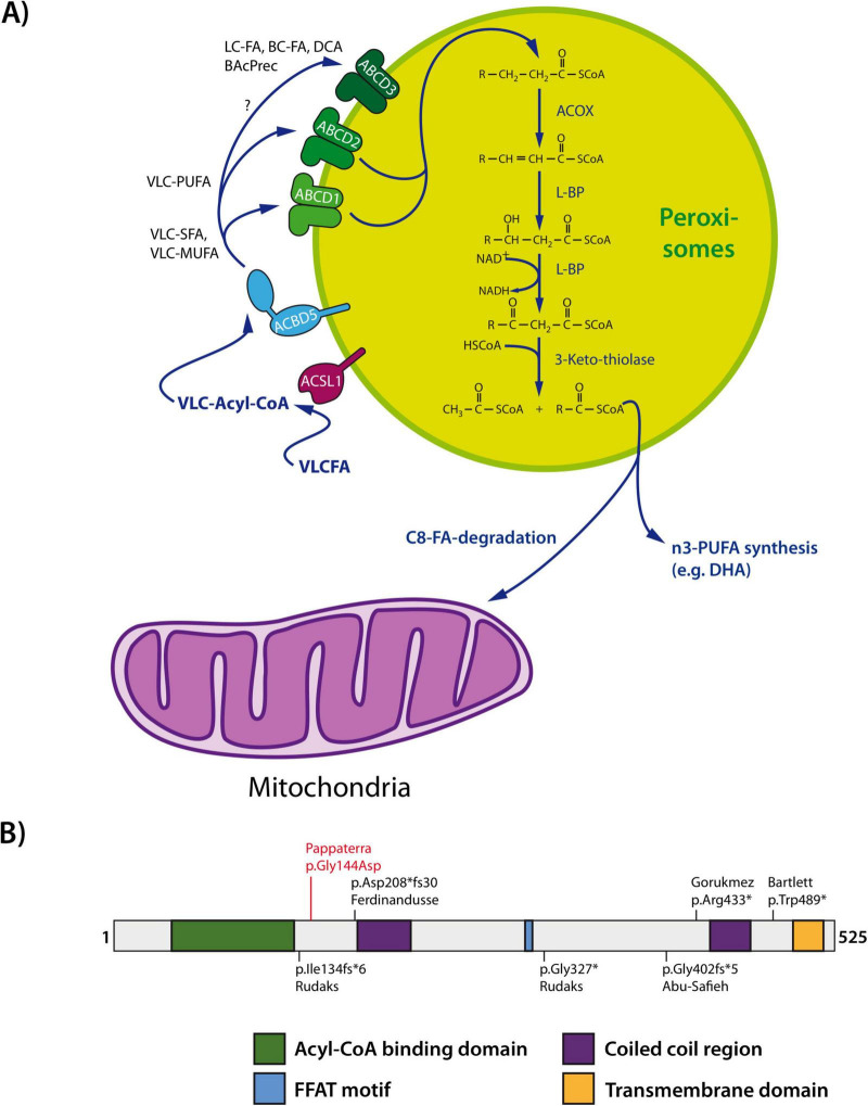
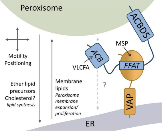
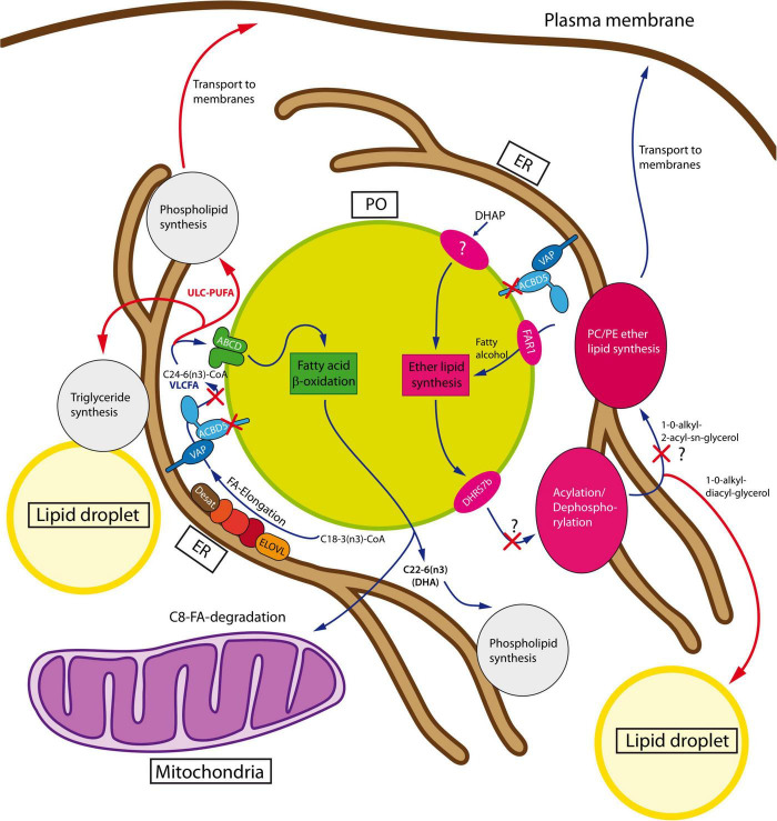
The absence or dysfunction of the peroxisomal membrane protein Acyl-CoA Binding Domain-Containing Protein 5 (ACBD5) is the cause of the most recently discovered peroxisomal disorder "Retinal Dystrophy with Leukodystrophy" (RDLKD). ACBD5 is a tail-anchored protein, anchored by its C-terminus into the peroxisomal membrane; hence, the bulk of its amino acid sequence faces the cytosol. With respect to ACBD5's molecular functions, RDLKD is unique since it is not only an accessory protein for the import of very-long-chain fatty acids (VLCFAs) into peroxisomes but also the first identified peroxisomal tethering protein facilitating membrane contacts with the endoplasmic reticulum (ER). Consequently, RDLKD is neither a peroxisomal biogenesis disorder nor single enzyme deficiency, since a deficiency in ACBD5 likely affects several aspects of peroxisomal function including VLCFA degradation, ether lipid synthesis, docosahexaenoic acid synthesis but also the transfer of membrane lipids from the ER to peroxisomes. Hence, RDLKD appears to be a multifactorial disorder leading to a mosaic pathology, combining symptoms caused by the disruption of several pathways. In this review, we will highlight recent findings obtained from case reports of RDLKD patients as well as insights from ACBD5-deficient mouse models to better understand its complex retinal and brain pathology. Moreover, we will discuss the possible contribution of the different dysregulated metabolites in the neurological pathogenesis of this latest peroxisomal disorder.

Keywords: ACBD5; RDLKD; VAP; fatty acid metabolism; membrane contact sites; peroxisomes.


Conflict of interest statement

The authors declare that the research was conducted in the absence of any commercial or financial relationships that could be construed as a potential conflict of interest.

Figures

FIGURE 1
FIGURE 1
(A) Overview of peroxisomal very-long-chain fatty acid (VLCFA) β-oxidation. Long chain fatty acid- CoA ligase (ACSL1) activates VLCFAs generating VLC-acyl-CoA. ACBD5 can capture VLC-acyl-CoA via its acyl-CoA binding domain and hands it over to the peroxisomal ABC transporters for VLCFAs (e.g., ABCD1 and ABCD2). VLCFAs are then imported into the peroxisomal matrix and degraded by the peroxisomal β- oxidation pathway in four consecutive steps: (i) dehydrogenation catalyzed by acyl-CoA oxidase (ACOX), (ii) hydration and (iii) second dehydrogenation mediated by L-bifunctional protein (L-BP) with enoyl-CoA hydratase and 3-hydroxyacyl-CoA dehydrogenase activity, and (iv) thiolytic cleavage by 3-ketothiolase. The chain-shortened acyl-CoAs can be routed to mitochondria for complete degradation via the mitochondrial β-oxidation pathway or used for the synthesis of omega-3 polyunsaturated fatty acids (PUFAs) such as docosahexaenoic acid (DHA). (B) ACBD5 domain structure. Known mutations are indicated. FFAT, two phenylalanines (FF) in an acidic tract (adapted from Schrader et al., 2020). BAcPrec, bile acid precursors; BC, branched-chain; DCA, dicarboxylic acid; FA, fatty acid; LC, long-chain, MUFA, monounsaturated fatty acid; SFA, saturated fatty acid; VLC, very-long-chain.
FIGURE 2
FIGURE 2
Overview of ACBD5 cellular functions. The FFAT motif of peroxisomal ACBD5 interacts with the major sperm protein (MSP) domain of ER-resident VAP to mediate peroxisome (PO)-ER membrane contacts. The PO-ER membrane contacts have been implicated in peroxisome motility and positioning, the transfer of ether lipid/plasmalogen precursors for further synthesis in the ER, and the transfer of membrane lipids for peroxisome membrane expansion and proliferation. ACBD5 possesses an acyl-CoA binding (ACB) domain which can capture very-long-chain acyl CoA (VLCFA) for peroxisomal β-oxidation (adapted from Kors et al., 2022b).
FIGURE 3
FIGURE 3
Peroxisomal metabolic interaction with the ER. The ether lipid synthesis pathway (shown in magenta) is initiated in peroxisomes by acylation of dihydroxyacetone phosphate (DHAP). In/at peroxisomes, 1-acyl-DHAP is converted stepwise to 1-alkyl-DHAP and 1-alkyl-2-hydroxy-glycerophosphate (GPA), which must be transferred to the ER for completion of the pathway. At the ER, GPA is supplied with an acyl-chain at the sn2-position and a phosphatidylethanolamine (PE) or phosphatidylcholine (PC) head group to yield ether phospholipids. Alternatively, the ether lipids can receive a second acyl group at sn3 to be stored in lipid droplets. Consequently, a disruption of the pathway might lead to GPA accumulation, which has not yet been experimentally analyzed. Peroxisomal β-oxidation is required for the synthesis of n3-polyunsaturated fatty acids like DHA (pathway in green). At the ER, α-linolenic acid is elongated and desaturated to tetracosahexaenoic acid (C24:6n-3), which must be delivered to peroxisomes to be shortened to DHA (C22:6n3) in one single round of β-oxidation. Loss of the ACBD5-mediated tethering complex could presumably lead to local enrichment of C24:6n3 at the ER to be further elongated and desaturated by the ER fatty acid elongation system. This could lead to the accumulation of VLC-PUFA to be finally incorporated into phospholipids or triglycerides (adapted from Darwisch et al., 2020). For details see text. FA, fatty acid; PUFA, polyunsaturated fatty acid; UCL, ultra-long-chain.
FIGURE 4
FIGURE 4
Graph depicting variability of the earliest onset of symptoms in patients. Note that cerebellar and visual dysfunction commonly occur as first signs for an ACBD5-deficiency. “Extrapyramidal” excludes motor dysfunctions, which can be related to a cerebellar degeneration.

Similar articles

References

    1. Abu-Safieh L., Alrashed M., Anazi S., Alkuraya H., Khan A. O., Al-Owain M., et al. (2013). Autozygome-guided exome sequencing in retinal dystrophy patients reveals pathogenetic mutations and novel candidate disease genes. Genome Res. 23 236–247. 10.1101/gr.144105.112 - DOI - PMC - PubMed
    1. Al Shamsi B., Ganesh A., Harikrishna B., Al Zuhaibi S., Markovic I., Mansy A., et al. (2025). Retinal dystrophy and leukodystrophy caused by ACBD5 deficiency in five Omani patients: A case series. Oman. Med. J. 39. 10.5001/omj.2025.34 - DOI
    1. Ambesh P., Paliwal V. K., Shetty V., Kamholz S. (2017). The babinski sign: A comprehensive review. J. Neurol. Sci. 372 477–481. 10.1016/j.jns.2016.10.041 - DOI - PubMed
    1. Bartlett M., Nasiri N., Pressman R., Bademci G., Forghani I. (2021). First reported adult patient with retinal dystrophy and leukodystrophy caused by a novel ACBD5 variant: A case report and review of literature. Am. J. Med. Genet. A 185 1236–1241. 10.1002/ajmg.a.62073 - DOI - PubMed
    1. Berger J., Dorninger F., Forss-Petter S., Kunze M. (2016). Peroxisomes in brain development and function. Biochim. Biophys. Acta 1863 934–955. 10.1016/j.bbamcr.2015.12.005 - DOI - PMC - PubMed

LinkOut - more resources