

4D-CTA image and geometry dataset for kinematic analysis of abdominal aortic aneurysms
- PMID: 40673181
- PMCID: PMC12266568
- DOI: 10.1016/j.dib.2025.111797
4D-CTA image and geometry dataset for kinematic analysis of abdominal aortic aneurysms
Abstract
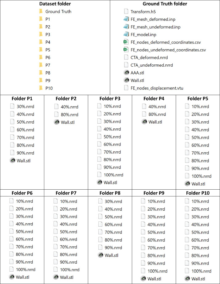
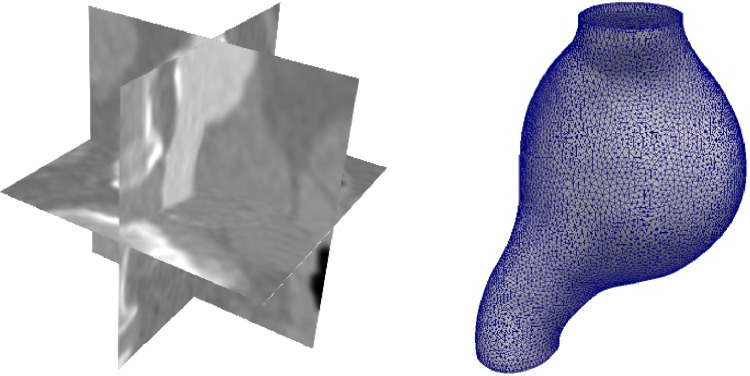
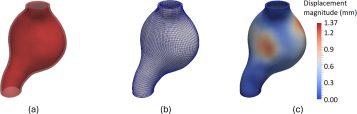
This article presents a dataset used in the article "Kinematics of Abdominal Aortic Aneurysms" [1], published in the Journal of Biomechanics. The dataset is publicly available for download from the Zenodo data repository (10.5281/zenodo.15477710). The dataset includes time-resolved 3D computed tomography angiography (4D-CTA) images of abdominal aortic aneurysm (AAA) captured throughout the cardiac cycle from ten patients diagnosed with AAA, along with ten patient-specific AAA geometries extracted from these images. Typically, the 4D-CTA dataset for each patient contains ten electrocardiogram (ECG)-gated 3D-CTA image frames acquired over a cardiac cycle, capturing both the systolic and diastolic phases of the AAA configuration. For method verification, the dataset also includes synthetic ground truth data generated from Patient 1's 3D-CTA AAA image in the diastolic phase. The ground truth data includes the patient-specific finite element (FE) biomechanical model and a synthetic systolic 3D-CTA image. The synthetic systolic image was generated by warping Patient 1's diastolic 3D-CTA image using the realistic displacement field obtained from the AAA biomechanical FE model. The images were acquired at Fiona Stanley Hospital in Western Australia and provided to the researchers at the Intelligent Systems for Medicine Laboratory at The University of Western Australia (ISML-UWA), where image-based AAA kinematic analysis was performed using a newly created algorithm, as described in [1]. The AAA geometries were extracted using an automated image processing pipeline comprising AI-based segmentation with PRAEVAorta software by NUREA (https://www.nurea-soft.com/), automated post-processing with the ISML-UWA in-house code (https://arxiv.org/abs/2403.07238), and surface model extraction using the freely available BioPARR (Biomechanics-based Prediction of Aneurysm Rupture Risk) (https://bioparr.mech.uwa.edu.au/) and 3D Slicer (https://www.slicer.org/) software packages [2,3]. Our dataset enabled the analysis of AAA wall displacement and strain throughout the cardiac cycle using a non-invasive, in vivo, image registration-based approach [1]. The use of widely adopted, open-source file formats-NRRD for images and STL for geometries-facilitates broad applicability and reusability in AAA biomechanics studies that require patient-specific geometry and information about AAA kinematics during cardiac cycle.
Keywords: Abdominal aortic aneurysm; Biomechanics; Computed tomography angiography; Image registration; Non-invasive method; Patient-specific analysis; Wall displacement; Wall strain.
© 2025 The Author(s).
Figures
Similar articles
-
Laparoscopic surgery for elective abdominal aortic aneurysm repair.Cochrane Database Syst Rev. 2017 May 4;5(5):CD012302. doi: 10.1002/14651858.CD012302.pub2. Cochrane Database Syst Rev. 2017. PMID: 28471523 Free PMC article.
-
An open-source deep learning framework for respiratory motion monitoring and volumetric imaging during radiation therapy.Med Phys. 2025 Jul;52(7):e18015. doi: 10.1002/mp.18015. Med Phys. 2025. PMID: 40665474 Free PMC article.
-
Endovascular treatment for ruptured abdominal aortic aneurysm.Cochrane Database Syst Rev. 2017 May 26;5(5):CD005261. doi: 10.1002/14651858.CD005261.pub4. Cochrane Database Syst Rev. 2017. PMID: 28548204 Free PMC article.
-
Duplex ultrasound for diagnosing symptomatic carotid stenosis in the extracranial segments.Cochrane Database Syst Rev. 2022 Jul 11;7(7):CD013172. doi: 10.1002/14651858.CD013172.pub2. Cochrane Database Syst Rev. 2022. PMID: 35815652 Free PMC article.
-
Systematic review and meta-analysis of the growth and rupture rates of small abdominal aortic aneurysms: implications for surveillance intervals and their cost-effectiveness.Health Technol Assess. 2013 Sep;17(41):1-118. doi: 10.3310/hta17410. Health Technol Assess. 2013. PMID: 24067626 Free PMC article.
References
-
- Fedorov A., Beichel R., Kalpathy-Cramer J., Finet J., Fillion-Robin J.-C., Pujol S., Bauer C., Jennings D., Fennessy F., Sonka M., Buatti J., Aylward S., Miller J.V., Pieper S., Kikinis R. 3D slicer as an image computing platform for the quantitative imaging network. Magn. Reson. Imaging. 2012;30(9):1323–1341. doi: 10.1016/j.mri.2012.05.001. - DOI - PMC - PubMed
-
- Joldes G.R., Wittek A., Warfield S.K., Miller K. Performing brain image warping using the deformation field predicted by a biomechanical model. Comput. Biomech. Med.: Deform. Flow. 2012:89–96. doi: 10.1007/978-1-4614-3172-5_10. - DOI
-
- Wanhainen A., Verzini F., Herzeele I.Van, Allaire E., Bown M., Cohnert T., Dick F., Herwaarden J.van, Karkos C., Koelemay M., Kölbel T., Loftus I., Mani K., Melissano G., Powell J., Szeberin Z., Committee E.G., Borst G.J.de, Chakfe N., Debus S., Hinchliffe R., Kakkos S., Koncar I., Kolh P., Lindholt J.S., Vega M.de, Vermassen F., Document R., Björck M., Cheng S., Dalman R., Davidovic L., Donas K., Earnshaw J., Eckstein H.-H., Golledge J., Haulon S., Mastracci T., Naylor R., Ricco J.-B., Verhagen H. Editor's choice – European society for vascular surgery (ESVS) 2019 clinical practice guidelines on the management of abdominal aorto-iliac artery aneurysms. Eur. J. Vasc. Endovasc. Surg. 2019;57(1):8–93. doi: 10.1016/j.ejvs.2018.09.020. - DOI - PubMed
LinkOut - more resources
Full Text Sources
