An anterior mediastinal cystic lesion pathologically confirmed as a mediastinal pancreatic pseudocyst after thoracoscopic resection: a rare case report and literature review
- PMID: 40673205
- PMCID: PMC12263553
- DOI: 10.3389/fped.2025.1613764
An anterior mediastinal cystic lesion pathologically confirmed as a mediastinal pancreatic pseudocyst after thoracoscopic resection: a rare case report and literature review
Abstract
Background: Mediastinal lesions have diverse etiologies, with thymoma, cystic teratoma, and lymphoma being relatively prevalent. In contrast, a pancreatic pseudocyst within the mediastinum is exceedingly rare and can often be mistaken for a thymic cyst or teratoma.
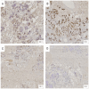
Case presentation: A 17-year-old female presented with a cough and sputum production. Chest CT revealed an anterior mediastinal mass, initially raising the suspicion of a thymic cyst. Thoracoscopic exploration and resection revealed a cystic lesion with a thick wall and brownish fluid. Both frozen section and final histopathological analysis confirmed a mediastinal cyst. Immunohistochemical markers (SYN positive, CK7 positive) led to a diagnosis of mediastinal pancreatic pseudocyst. The patient experienced significant recovery post-surgery, with a marked improvement in symptoms.
Conclusion: This case highlights the importance of including mediastinal pancreatic pseudocyst in the differential diagnosis of anterior mediastinal cystic lesions. A thorough clinical and radiological assessment, along with surgical pathology and immunohistochemical profiling, is essential for accurate diagnosis and appropriate management.
Keywords: CT; anterior mediastinal cystic mass; mediastinal pancreatic lesion; pancreatic pseudocyst; x-ray.
© 2025 Zhai, Miao, Xue, Yuan, Jia, Chen and Zha.
Conflict of interest statement
The authors declare that the research was conducted in the absence of any commercial or financial relationships that could be construed as a potential conflict of interest.
Figures


Similar articles
-
Mature Cystic Teratoma of Anterior Mediastinum in a Child: A Case Report and Literature Review.J Investig Med High Impact Case Rep. 2024 Jan-Dec;12:23247096241274510. doi: 10.1177/23247096241274510. J Investig Med High Impact Case Rep. 2024. PMID: 39230157 Free PMC article. Review.
-
Coexistence of mediastinal teratoma and intrapulmonary bronchogenic cyst: a case report.J Med Case Rep. 2025 Jul 4;19(1):320. doi: 10.1186/s13256-025-05391-z. J Med Case Rep. 2025. PMID: 40616167 Free PMC article.
-
Renal hydatid cyst mimicking malignancy: a case report.Int J Surg Case Rep. 2025 Jul;132:111506. doi: 10.1016/j.ijscr.2025.111506. Epub 2025 Jun 13. Int J Surg Case Rep. 2025. PMID: 40517679 Free PMC article.
-
Thymoma with Intravascular Tumor Thrombus in the Left Brachiocephalic Vein: A Case Report.Surg Case Rep. 2025;11(1):25-0118. doi: 10.70352/scrj.cr.25-0118. Epub 2025 Jul 1. Surg Case Rep. 2025. PMID: 40620846 Free PMC article.
-
Intraoperative frozen section analysis for the diagnosis of early stage ovarian cancer in suspicious pelvic masses.Cochrane Database Syst Rev. 2016 Mar 1;3(3):CD010360. doi: 10.1002/14651858.CD010360.pub2. Cochrane Database Syst Rev. 2016. PMID: 26930463 Free PMC article.
References
-
- Karamouzos V, Karavias D, Siagris D, Kalogeropoulou C, Kosmopoulou F, Gogos C, et al. Pancreatic mediastinal pseudocyst presenting as a posterior mediastinal mass with recurrent pleural effusions: a case report and review of the literature. J Med Case Rep. (2015) 9:110. 10.1186/s13256-015-0582-z - DOI - PMC - PubMed
Publication types
LinkOut - more resources
Full Text Sources
Research Materials
Miscellaneous

