. 2025 Jul 20;45(7):1451-1459.
doi: 10.12122/j.issn.1673-4254.2025.07.11.

[S1PR5 activation or overexpression enhances barrier function of mouse brain microvascular endothelial cells against OGD/R injury by modulating oxidative stress]

[Article in Chinese]
Affiliations

[S1PR5 activation or overexpression enhances barrier function of mouse brain microvascular endothelial cells against OGD/R injury by modulating oxidative stress]

[Article in Chinese]
Jingxian Wang et al. Nan Fang Yi Ke Da Xue Xue Bao. .

Abstract

Objectives: To investigate the role of sphingosine-1-phosphate receptor 5 (S1PR5) in modulating barrier function of mouse brain microvascular endothelial cells with oxygen-glucose deprivation and reoxygenation (OGD/R).

Methods: Mouse brain microvascular endothelial cells (bEnd.3) were exposed to OGD/R to induce barrier dysfunction following treatment with S1PR5-specific agonist A971432 or lentivirus-mediated transfection with a S1PR5-specific siRNA, a S1PR5-overexpressing plasmid, or their respective negative control sequences. The changes in viability and endothelial barrier permeability of the treated cells were evaluated with CCK-8 assay and FITC-dextran permeability assay; the levels of intracellular reactive oxygen species (ROS) and localization and expression levels of the proteins related with barrier function and oxidative stress were detected using immunofluorescence staining, DCFH-DA probe and Western blotting.

Results: S1PR5 activation obviously enhanced viability of bEnd.3 cells exposed to OGD/R (P<0.0001). Both activation and overexpression of S1PR5 reduced FITC-dextran leakage, while S1PR5 knockdown significantly increased FITC-dextran leakage in the exposed bEnd.3 cells. Activation and overexpression of S1PR5 both increased the cellular expressions of the barrier proteins ZO-1 and occludin, while S1PR5 knockdown produced the opposite effect. In cells exposed to OGD/R, ROS production was significantly reduced by S1PR5 activation and overexpression but increased following S1PR5 knockdown. Overexpression of S1PR5 obviously increased the expressions of the antioxidant proteins Nrf2, HO-1 and SOD2 in the exposed cells.

Conclusions: S1PR5 activation and overexpression significantly improve cell viability and reduce permeability of a mouse brain microvascular endothelial cell model of OGD/R, the mechanism of which may involve the reduction in ROS production and upregulation of the antioxidant proteins.

目的: 探讨鞘氨醇1-磷酸受体5(S1PR5)对氧糖剥夺及复氧复糖(OGD/R)诱导的小鼠脑微血管内皮细胞屏障功能的影响及相关机制。方法: 体外培养小鼠脑微血管内皮细胞bEnd.3,使用OGD/R诱导屏障功能障碍,分别使用S1PR5特异性激动剂A971432、敲低S1PR5的siRNA及过表达S1PR5的慢病毒进行干预。设置对照组:bEnd.3正常培养;OGD/R组:bEnd.3进行OGD/R;A971432组:OGD/R组+A971432;siNC组:转染siNC+OGD/R;siS1PR5组:转染siS1PR5+OGD/R;OE NC组:感染慢病毒LV5-NC+OGD/R;OE S1PR5组:感染慢病毒LV5-S1PR5+OGD/R。RT-qPCR法分别检测敲低或过表达S1PR5的效率;采用CCK-8检测在不同培养条件下bEnd.3细胞的活力;采用FITC-dextran渗透法检测内皮屏障渗透性;采用细胞免疫荧光法检测蛋白的定位及表达;DCFH-DA探针法检测细胞内活性氧水平;Western blotting法检测蛋白的表达水平。结果: CCK-8结果显示激动S1PR5可以增加OGD/R下调的细胞活力(P<0.0001),FITC-dextran渗透法结果显示激动和过表达S1PR5可减少FITC-dextran的渗漏(P<0.001),而敲低S1PR5增加FITC-dextran的渗漏(P<0.001)。Western blotting及免疫荧光结果显示,与OGD/R组相比,激动和过表达S1PR5可以增加屏障蛋白ZO-1(P<0.05)、Occludin(P<0.05)的表达,而敲低S1PR5会下调ZO-1和Occludin(P<0.05)的表达。ROS检测结果显示,激动和过表达S1PR5能减少ROS的产生,而敲低S1PR5会增加ROS产生。Western blotting检测发现过表达S1PR5可以增加抗氧化蛋白Nrf2(P<0.0001)、HO-1(P<0.0001)、SOD2(P<0.01)的表达。结论: S1PR5受体的激动剂干预及基因过表达可显著改善OGD/R模型诱导的活力减少及通透性增加,而基因敲低S1PR5则加剧OGD/R诱导的功能损伤,其机制可能与减少ROS,上调抗氧化蛋白表达相关。.

Keywords: blood-brain barrier dysfunction; oxidative stress; oxygen-glucose deprivation and reoxygenation; sphingosine 1-phosphate receptor 5.


Conflict of interest statement

The authors declare no competing interests.

Figures

图1
图1
OGD处理降低bEnd.3细胞活性并下调细胞间紧密连接蛋白的表达 Fig.1 Oxygen-glucose deprivation (OGD) decrease viability of bEnd.3 cells and down-regulate expressions of intercellular tight junction proteins. A: Representative images of bEnd.3 cells after OGD treatment. B: Viability of b.End3 cells assessed with CCK-8 assay (n=6). C: Western blotting of tight junction proteins (ZO-1 and occludin) and VE-cadherin after OGD. D-F: Quantification of ZO-1, VE-cadherin, and occludin protein expression levels (n=3). *P<0.05, **P<0.01, ***P<0.001, ****P<0.0001 vs Con.
图2
图2
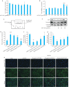
激动S1PR5有助于恢复OGD下调的细胞活性并上调细胞间紧密连接蛋白的表达 Fig.2 Activation of S1PR5 restores viability of OGD-damaged cells and up-regulates expressions of intercellular tight junction proteins. A: CCK8 assay for assessing viability of cultured bEnd.3 cells treated with different concentrations of A971432. B: CCK8 assay for assessing viability of bEnd.3 cells exposed to OGD after treatment with different concentrations of A971432 (****P<0.0001 vs 0 μmol/L A971432). C: Schematic diagram for detecting endothelial permeability of bEnd.3 cells using FITC-Dextran. D: Detection of endothelial permeability using FITC-Dextran. E: Western blotting of ZO-1 and occluding expressions in bEnd.3 cells with OGD/R after S1PR5 activation. F, G: Quantitative analysis of the expression levels of ZO-1 and occludin. H: Changes in the fluorescence intensity of tight junction proteins caused by activation of S1PR5 in bEnd.3 cells with OGD/R (scale bar= 50 μm). *P<0.05, **P<0.01, ****P<0.0001 vs Con; # P<0.05, ## P<0.01, #### P<0.0001 vs OGD/R.
图3
图3
敲低S1PR5加剧OGD诱导的细胞活力下降及紧密连接蛋白的减少 Fig.3 S1PR5 knockdown exacerbates the reduction of cell viability and tight junction protein expressions in OGD-induced bEnd.3 cells. A: Efficiency of S1PR5 knockdown assessed by RT-qPCR. B: Endothelial permeability of the transfected cells detected by FITC-Dextran. C: Alterations in the fluorescence intensity of the tight junction proteins in bEnd.3 cells with S1PR5 knockdown following OGD/R. D-F: Western blotting for detecting the expression levels of ZO-1 and occluding in bEnd.3 cells with OGD/R after S1PR5 knockdown. *P<0.05, **P<0.01 vs Con; # P<0.05, ### P<0.001 vs OGD/R.
图4
图4
过表达S1PR5抑制OGD诱导的细胞活力下降及紧密连接蛋白的减少 Fig.4 Overexpression of S1PR5 enhances viability and tight junction protein expressions in OGD-induced bEnd.3 cells. A: RT-qPCR for assessing efficiency of lentivirus-mediated S1PR5 overexpression (**P<0.01 vs OENC). B: Endothelial permeability of the cells detected by FITC-Dextran. C: Changes in the fluorescence intensity of tight junction proteins in OGD-induced bEnd.3 cells with S1PR5 overexpression. D-F: Western blotting for detecting expression levels of ZO-1 and occludin in OGD-induced bEnd.3 cells with S1PR5 overexpression. *P<0.05, **P<0.01, ****P<0.0001 vs Con; # P<0.05, ## P<0.01, ### P<0.001 vs OGD/R.
图5
图5
S1PR5介导Nrf-2/HO-1通路发挥抗氧化作用 Fig.5 S1PR5 overexpression produces antioxidant effect in OGD-induced bEnd.3 cells via the Nrf-2/HO-1 pathway. A: Effects of S1PR5 knockdown and overexpression on intracellular reactive oxygen species in OGD-induced bEnd.3 cells (scale bar=50 μm). B-E: Western blotting for detecting expression levels of Nrf2, HO-1 and SOD2 in OGD-induced bEnd.3 cells with S1PR5 knockdown or overexpression. *P<0.05 vs Con, ## P<0.01, #### P<0.0001 vs OGD/R.

References

    1. Zhao Z, Nelson AR, Betsholtz C, et al. Establishment and dysfunction of the blood-brain barrier[J]. Cell, 2015, 163(5): 1064-78. doi: 10.1016/j.cell.2015.10.067 - DOI - PMC - PubMed
    1. Ding QQ, Liu SW, Yao YD, et al. Global, regional, and national burden of ischemic stroke, 1990-2019[J]. Neurology, 2022, 98(3): e279-90. doi: 10.1212/wnl.0000000000013115 - DOI - PubMed
    1. Munji RN, Soung AL, Weiner GA, et al. Profiling the mouse brain endothelial transcriptome in health and disease models reveals a core blood-brain barrier dysfunction module[J]. Nat Neurosci, 2019, 22(11): 1892-902. doi: 10.1038/s41593-019-0497-x - DOI - PMC - PubMed
    1. Cartier A, Hla T. Sphingosine 1-phosphate: Lipid signaling in pathology and therapy[J]. Science, 2019, 366(6463): eaar5551. doi: 10.1126/science.aar5551 - DOI - PMC - PubMed
    1. Obinata H, Hla T. Sphingosine 1-phosphate and inflammation[J]. Int Immunol, 2019, 31(9): 617-25. doi: 10.1093/intimm/dxz037 - DOI - PMC - PubMed

Publication types

LinkOut - more resources