Age and sex are associated with Alzheimer's disease neuropathology in Down syndrome
- PMID: 40673442
- PMCID: PMC12268376
- DOI: 10.1002/alz.70408
Age and sex are associated with Alzheimer's disease neuropathology in Down syndrome
Abstract
Introduction: This study investigates the association of age and biological sex with Alzheimer's disease (AD) neuropathology in Down syndrome (DS).
Methods: We examined the frontal/occipital cortex in people with DS (n = 14/13, 1-39 years), DS with AD (DSAD) neuropathology (n = 18/19, 42-61 years), late-onset AD (n = 15/16, 72-96 years), and age-matched controls (n = 50/47)(n = 156). The area occupied by AT8 and 6E10 immunolabeling, representing tangle and plaque loads, respectively, was used for segmented linear regression analyses.
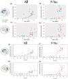
Results: There was elevated neuropathology after age 35 in DSAD, with inflection points at ∼31 years (amyloid-β [Aβ]) and ∼28 (phosphorylated tau [p-tau]) in the frontal cortex and ∼36 years (both Aβ and p-tau) in the occipital cortex. Occipital p-tau was higher in women relative to men with DS. Aβ and p-tau pathology were correlated in women with DS but not in men with DS in the occipital cortex.
Discussion: Women with DS may show a more advanced stage of tau pathology relative to men with DS.
Highlights: Amyloid-β (Aβ) and phosphorylated tau (p-tau) Alzheimer's disease (AD) pathology emerge after 30 years of age in the frontal cortex, followed 7 years later by pathology in the occipital cortex in Down syndrome (DS). Women with DS show a more rapid progression of AD neuropathology seen by trends in higher p-tau relative to men, despite similar levels of Aβ. Women with DS show a stronger association between Aβ and tau in the occipital but not frontal cortex relative to men with DS, independent of age.
Keywords: Alzheimer disease progression; beta‐amyloid; neuropathology; tau; trisomy 21.
© 2025 The Author(s). Alzheimer's & Dementia published by Wiley Periodicals LLC on behalf of Alzheimer's Association.
Conflict of interest statement
The authors declare no conflicts of interest. Author disclosures are available in the Supporting Information.
Figures


Similar articles
-
Amyloid-β peptide signature associated with cerebral amyloid angiopathy in familial Alzheimer's disease with APPdup and Down syndrome.Acta Neuropathol. 2024 Jul 18;148(1):8. doi: 10.1007/s00401-024-02756-4. Acta Neuropathol. 2024. PMID: 39026031 Free PMC article.
-
Joint spatial associations of amyloid beta and tau pathology in Down syndrome and preclinical Alzheimer's disease: Cross-sectional associations with early cognitive impairments.Alzheimers Dement. 2025 Jul;21(7):e70424. doi: 10.1002/alz.70424. Alzheimers Dement. 2025. PMID: 40588723 Free PMC article.
-
Timeline to symptomatic Alzheimer's disease in people with Down syndrome as assessed by amyloid-PET and tau-PET: a longitudinal cohort study.Lancet Neurol. 2024 Dec;23(12):1214-1224. doi: 10.1016/S1474-4422(24)00426-5. Lancet Neurol. 2024. PMID: 39577922
-
CSF tau and the CSF tau/ABeta ratio for the diagnosis of Alzheimer's disease dementia and other dementias in people with mild cognitive impairment (MCI).Cochrane Database Syst Rev. 2017 Mar 22;3(3):CD010803. doi: 10.1002/14651858.CD010803.pub2. Cochrane Database Syst Rev. 2017. PMID: 28328043 Free PMC article.
-
Current advances and unmet needs in Alzheimer's disease trials for individuals with Down syndrome: Navigating new therapeutic frontiers.Alzheimers Dement. 2025 Jun;21(6):e70258. doi: 10.1002/alz.70258. Alzheimers Dement. 2025. PMID: 40528298 Free PMC article. Review.
References
-
- Prasher VP, Farrer MJ, Kessling AM, et al. Molecular mapping of Alzheimer‐type dementia in Down's syndrome. Ann Neurol. 1998;43(3):380‐383. - PubMed
-
- Margallo‐Lana ML, Moore PB, Kay DWK, et al. Fifteen‐year follow‐up of 92 hospitalized adults with Down's syndrome: incidence of cognitive decline, its relationship to age and neuropathology. J Intellect Disabil Res. 2007;51(Pt. 6):463‐477. - PubMed
MeSH terms
Substances
Grants and funding
- RF1AG079519/NIH/NIA
- AG/NIA NIH HHS/United States
- U54 HD087011/HD/NICHD NIH HHS/United States
- U24 AG21886/National Centralized Repository for Alzheimer Disease and Related Dementias
- NIHR Cambridge Biomedical Research Centre
- CPFT
- P50 AG005681/AG/NIA NIH HHS/United States
- UL1 TR001414/TR/NCATS NIH HHS/United States
- P50 AG008702/AG/NIA NIH HHS/United States
- Fulbourn Hospital, Cambridge
- U01 AG051412/AG/NIA NIH HHS/United States
- EH/PJL/AMB
- P50 AG005133/AG/NIA NIH HHS/United States
- Eunice Kennedy Shriver National Institute of Child Health and Human Development
- NH/NIH HHS/United States
- DS-Connect
- UL1 TR002345/TR/NCATS NIH HHS/United States
- Windsor Research Unit
- UL1 TR001857/TR/NCATS NIH HHS/United States
- P30 AG062421/AG/NIA NIH HHS/United States
- U19 AG068054/AG/NIA NIH HHS/United States
- P30 AG062715/AG/NIA NIH HHS/United States
- T32AG000096/NIH/NIA
- P50 HD105353/HD/NICHD NIH HHS/United States
- U54 HD090256/HD/NICHD NIH HHS/United States
- P30 AG066519/AG/NIA NIH HHS/United States
- UL1 TR002373/TR/NCATS NIH HHS/United States
- UL1 TR001873/TR/NCATS NIH HHS/United States
- P50 AG16537/Alzheimer's Disease Research Centers Program
- U01 AG051406/AG/NIA NIH HHS/United States
LinkOut - more resources
Full Text Sources
Medical

