. 2025 Aug 5;13(8):e0336824.
doi: 10.1128/spectrum.03368-24. Epub 2025 Jul 17.

Amino acid insertion in the Meq protein of Marek's disease virus, an avian oncogenic herpesvirus, accelerates tumorigenesis

Affiliations

Amino acid insertion in the Meq protein of Marek's disease virus, an avian oncogenic herpesvirus, accelerates tumorigenesis

Jumpei Sato et al. Microbiol Spectr. .

Abstract

Marek's disease virus (MDV) causes lymphomas (Marek's disease) in chickens. Despite vaccination, MDV field strains exhibit increased virulence, and sporadic outbreaks are still reported. An insertion/deletion in Meq has been identified in several MDV strains, and our previous study using recombinant MDV (rMDV) demonstrated that an insertion in Meq enhanced MDV virulence, whereas a deletion reduced its virulence. However, the mechanisms by which these indels in Meq alter MDV virulence remain elusive. We aimed to clarify the impact of the insertion in Meq on pathogenesis. We compared the effects of Meq and Meq with the insertion, termed L-Meq, on transcriptional regulation, the dynamics of transformed T cells and relevant T-cell subsets, and the patterns of gene expression in tumor lesions of infected chickens. Reporter assays revealed that insertion increased the transactivation activity on the infected-cell protein 4 promoter in the MDV genome and bcl-2/cd30 promoters in the host genome. rMDV encoding L-Meq (vRB-1B_L-Meq) exhibited higher mortality and tumor incidence than rMDV encoding Meq (vRB-1B_Meq), and vRB-1B_L-Meq infection increased the proportion of CD4+ T cells, which are targets for transformation by MDV, in the early post-infection phase, compared with vRB-1B_Meq. RNA-seq analysis revealed that similar genes were modulated in tumor lesions of chickens infected with vRB-1B_Meq or vRB-1B_L-Meq. These findings suggest that although the insertion in Meq does not alter the target genes for transcriptional regulation, it accelerates the process of tumorigenesis by enhancing the transactivation activity.IMPORTANCEMarek's disease, an avian lymphoproliferative disease, is caused by Marek's disease virus (MDV). Meq, an MDV oncoprotein, regulates the expression of viral and host genes. Meq with an insertion, termed L-Meq, has been identified as a factor that enhances MDV virulence. However, the mechanisms by which the insertion in Meq alters MDV virulence remain unknown. Our study clarified that the insertion enhances the transactivation activity of Meq on the host promoters related to tumorigenesis. Notably, the transcriptomes of tumor lesions in chickens infected with recombinant MDV (rMDV) with L-Meq and those infected with rMDV encoding Meq without the insertion were similar; however, chickens infected with rMDV harboring L-Meq exhibited higher proportions of CD4+ T cells and regulatory T cells, which are targets for transformation by MDV, in the early post-infection phase, suggesting accelerated tumorigenesis. This study contributes to the current understanding of the mechanisms underlying MDV virulence.

Keywords: L-Meq; Marek's disease; Marek's disease virus; Meq; insertion; transactivation activity; tumorigenesis; virulence.


Conflict of interest statement

The authors declare no competing interests.

Figures

Fig 1
Fig 1
Analysis of transcriptional regulation by the Meq and L-Meq proteins. (A) Schematic representation of the Meq isoforms. The structures of Meq from RB-1B, long-Meq isoform (L-Meq) from CVI988, and L-Meq (RB-1B) whose sequences were matched with those of RB-1B-Meq, except for the insertion in the transactivation domain. Meq comprises a proline/glutamine (Pro/Gln)-rich domain followed by a basic region (BR) and leucine zipper (ZIP) at the N-terminal region and a transactivation domain at the C-terminal region. The Meq isoforms include amino acid polymorphisms in the BR and transactivation domain. L-Meq is characterized by a 60-amino-acid insertion in the transactivation domain. (B and C) Transrepression effects of the Meq isoforms. The transrepression effects of RB-1B-Meq, wild-type L-Meq (CVI988), and L-Meq (RB-1B) on (B) the pp38 promoter and (C) the pp14 promoter-driven luciferase activities were compared. DF-1 cells in each well were transfected with 300 ng of expression plasmids for each Meq isoform, 500 ng of reporter plasmid, and 5 ng of control pRL-TK plasmid. Luciferase activities were analyzed 24 h post-transfection. Firefly luciferase activity is expressed relative to the mean basal activity in the presence of pCI-neo after normalization to Renilla luciferase activity. (D–G) Transactivation activities of the Meq isoforms. The transactivation activities of RB-1B-Meq, wild-type L-Meq (CVI988), and L-Meq (RB-1B) on (D) the icp4 promoter, (E) the gb promoter, (F) the bcl-2 promoter, and (G) the cd30 promoter-driven luciferase activities were compared. DF-1 cells in each well were transfected with 300 ng of expression plasmids for each Meq isoform, 200 ng of c-Jun expression plasmid, 500 ng of reporter plasmid, and 5 ng of control pRL-TK plasmid. Error bars indicate standard deviations. Three independent experiments were performed in triplicate. **P < 0.01, Tukey’s multiple comparison test. (H) Stability of Meq isoforms. The stability of RB-1B-Meq, wild-type L-Meq (CVI988), and L-Meq (RB-1B) was compared. DF-1 cells in each well were transfected with 150 ng of expression plasmids for each Meq isoform. The cells in each well were added 100 µg/mL of cycloheximide 24 h after transfection. The transfected cells were electrophoretically separated in 10% SDS-PAGE and then transferred onto a nitrocellulose membrane. The membrane was incubated with the rabbit anti-Meq antibody (20  µg/mL) and mouse anti-chicken actin antibody (20  µg/mL) for 1  h, and then incubated with anti-rabbit IgG secondary antibody conjugated with HRP or anti-mouse IgG1 secondary antibody conjugated with HRP for 30  min, respectively. Three independent experiments were performed in triplicate.
Fig 2
Fig 2
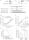
Reconstitution of recombinant Marek’s disease viruses (rMDVs) and their characterization in vivo. (A) Schematic diagrams of the constructs cloned using the RB-1B genome in this study. In the RB-1B genome cloned as the bacterial artificial chromosome (BAC) plasmid (pRB-1B), most of the internal repeat long (IRL) regions were deleted in this plasmid, designated as pRB-1B_ΔIRL, and used for mutagenesis. Meq in the terminal repeat long (TRL) was replaced with RB-1B-Meq or L-Meq (RB-1B) (encoding the long-Meq containing the insertion) by two-step red-mediated mutagenesis. (B) CEFs were infected with 50 pfu of rMDVs. The infected cells were collected daily for 6 days. The viral loads in the infected cells were analyzed by quantitative PCR (qPCR) (7–35 dpi: control group: n = 4, vRB-1B_Meq-infected group: n = 4, vRB-1B_L-Meq-infected group: n = 4). Three independent experiments were performed in triplicate. Error bars indicate standard deviations. (C) The viral loads in the whole blood of chickens infected with vRB-1B_Meq and vRB-1B_L-Meq were determined by quantitative polymerase chain reaction (qPCR). Error bars indicate standard deviations. **P < 0.01, Mann–Whitney U test. (D) Survival rate in chickens infected with rMDVs in the experimental infection. The survival rate among the groups was analyzed using the Log-rank test. (E) Tumor incidence in chickens infected with rMDVs during the study period. *P < 0.05, Fisher’s exact test.
Fig 3
Fig 3
Gating strategy for the analysis of the dynamics of major T-cell subsets in chickens infected with rMDVs. A representative gating strategy is shown for analyzing the dynamics of γδ T cells, CD8αβ+ T cells, CD4+ T cells, and mTGF-β+ CD4+ T cells. Dead cells were excluded using Fixable Viability Dye eFluor780.
Fig 4
Fig 4
Dynamics of major T cell subsets in chickens infected with rMDVs. The dynamics of CD4+ cells, mTGF-β+ CD4+ cells, CD8αβ+ cells, and γδ T cells in T cells from PBMCs and spleens of chickens infected with vRB-1B-Meq or vRB-1B-L-Meq at each time point during the experimental period (7–49 dpi: control group: n = 4, vRB-1B_Meq-infected group: n = 4, vRB-1B_L-Meq-infected group: n = 4, 56 dpi: control group: n = 4, vRB-1B_Meq-infected group: n = 7). The percentages of CD4+ cells in the T cell population from (A) PBMCs and (B) spleens, mTGF-β cells in the CD4+ T cell population from (C) PBMCs and (D) spleens, CD8αβ+ cells in the T cell population from (E) PBMCs and (F) spleens, and γδTCR+ cells in the T cell population from (G) PBMCs and (H) spleens were analyzed. Error bars indicate standard deviations. *P < 0.05, Kruskal–Wallis test (7–35 dpi), Mann–Whitney U test (49–56 dpi).
Fig 5
Fig 5
Gating strategy for the analysis of the dynamics of T-cell subsets involved in immune response in chickens infected with rMDVs. A representative gating strategy is shown for analyzing the dynamics of CD8α+ γδ T cells, CD4+ γδ T cells, and CD8αα+ T cells. Dead cells were excluded using Fixable Viability Dye eFluor780.
Fig 6
Fig 6
Dynamics of T-cell subsets involved in immune response in chickens infected with rMDVs. The dynamics of CD8α+ γδ T cells and CD8αα+ T cells from spleens of chickens infected with vRB-1B-Meq or vRB-1B-L-Meq at each time point during the experimental period (7–49 dpi: control group: n = 4, vRB-1B_Meq-infected group: n = 4, vRB-1B_L-Meq-infected group: n = 4, 56 dpi: control group: n = 4, vRB-1B_Meq-infected group: n = 7). (A) The percentages of CD8α+ T cells in the γδ T cell population from spleens, (B) the percentage of CD8αα+ cells in CD8α+ T cell population from spleens, and (C) the percentage of CD4+CD8α- T cells in CD4+ T cell population from spleens were analyzed. Error bars indicate standard deviations. *P < 0.05, Kruskal–Wallis test (7–35 dpi), Mann–Whitney U test (49–56 dpi).
Fig 7
Fig 7
Composition of T cell subsets and transcriptome analysis of tumor lesions of rMDV-infected chickens. (A–D) The composition of T cell subsets in tumor lesions from chickens that developed tumors in the kidneys or gonads during the experimental period was analyzed by flow cytometry (vRB-1B_Meq-infected group: n = 5, vRB-1B_L-Meq-infected group: n = 5). (A) The percentages of CD4+ cells in the T cell population, (B) mTGF-β+ cells in the CD4+ T cell population, (C) CD8αβ+ cells in the T cell population, and (D) γδTCR+ cells in the T cell population were analyzed. (E) The scatter plots show the Spearman rank correlation between each tumor lesion from chickens infected with vRB-1B-Meq and vRB-1B-L-Meq. r: Spearman’s rank correlation coefficient. The expression levels of (F) cox1 and (G) cox2 in tumor lesions from chickens infected with vRB-1B-Meq or vRB-1B-L-Meq at 7 dpi were determined by qPCR.

Similar articles

References

    1. Davison F, Nair V. 2005. Use of Marek’s disease vaccines: could they be driving the virus to increasing virulence? Expert Rev Vaccines 4:77–88. doi: 10.1586/14760584.4.1.77 - DOI - PubMed
    1. Schat KA. 2016. History of the first-generation Marek’s disease vaccines: the science and little-known facts. Avian Dis 60:715–724. doi: 10.1637/11429-050216-Hist - DOI - PubMed
    1. Othman I, Aklilu E. 2019. Marek’s disease herpesvirus serotype 1 in broiler breeder and layer chickens in Malaysia. Vet World 12:472–476. doi: 10.14202/vetworld.2019.472-476 - DOI - PMC - PubMed
    1. Raja A, Dhinakar Raj G, Bhuvaneswari P, Balachandran C, Kumanan K. 2009. Detection of virulent Marek’s disease virus in poultry in India. Acta Virol 53:255–260. doi: 10.4149/av_2009_04_255 - DOI - PubMed
    1. Shi MY, Li M, Wang WW, Deng QM, Li QH, Gao YL, Wang PK, Huang T, Wei P. 2020. The emergence of a vv + MDV can break through the protections provided by the current vaccines. Viruses 12:1048. doi: 10.3390/v12091048 - DOI - PMC - PubMed

MeSH terms

Substances

LinkOut - more resources