. 2025 Aug 12;97(31):17132-17140.
doi: 10.1021/acs.analchem.5c03104. Epub 2025 Jul 17.

Insights into Adeno-Associated Virus Capsid Charge Heterogeneity

Affiliations

Insights into Adeno-Associated Virus Capsid Charge Heterogeneity

Jiaqi Wu et al. Anal Chem. .

Abstract

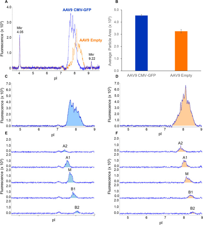
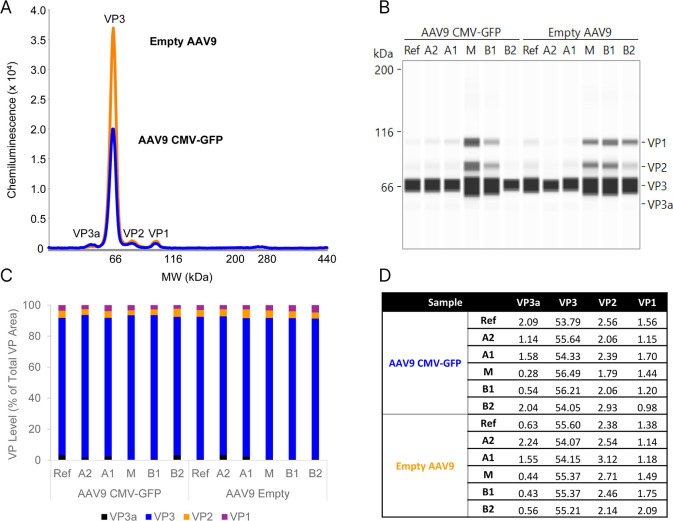
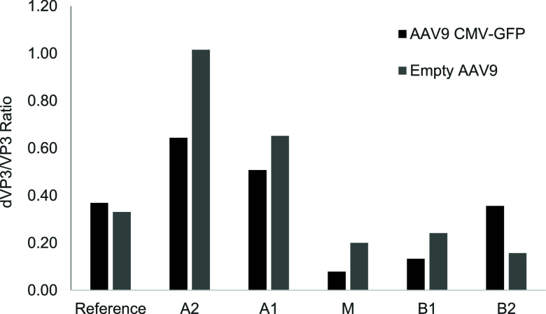
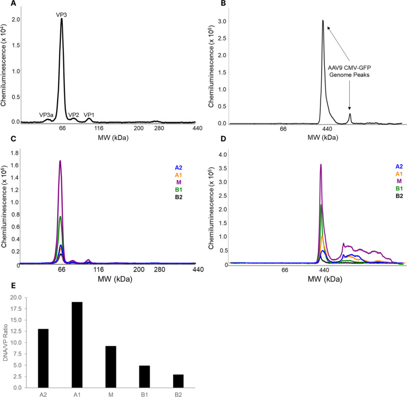
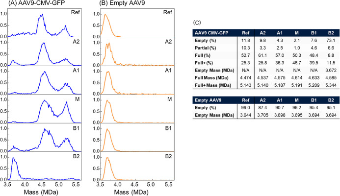
A comprehensive workflow is described to examine three contributing factors to the charge heterogeneity of Adeno-associated viruses (AAVs) from a single sample. Intact AAV9 capsids were fractionated using imaged capillary isoelectric focusing (icIEF)-based fractionation, allowing for collection of capsids with different isoelectric points (pIs). Capsid integrity of the fractions was confirmed with analytical icIEF and charge detection mass spectrometry (CD-MS). Using capillary electrophoresis (CE) immunoassays, the capsid protein ratios and capsid protein deamidation were characterized. Additionally, to analyze ssDNA content packaged in each fraction, CE-immunoassay and high-resolution CD-MS were used. This study enhances our understanding of AAVs, by examining the contributions of its attributes to capsid charge heterogeneity.


Figures

1
1
Charge heterogeneity analysis and fractionation of intact AAV9 capsids. (A) Triplicate injections of AAV9 CMV-GFP (blue) and empty AAV9 capsids (orange) are shown in overlay, detected using native fluorescence (excitation = 280 nm, emission = 325 nm, using an 80s exposure). A clear pI difference is seen between full and empty AAV9 capsids. (B) Analysis of area under the curve for triplicate injections of AAV9 CMV-GFP and empty capsids shows reproducible icIEF analysis of AAV capsids. Unfractionated AAV9-CMV-GFP (C) and empty AAV9 (D) were compared to the respective fractionation series. Each AAV9 reference sample was fractionated into five fractions for (E) AAV9-CMV-GFP for (F) empty AAV9 capsids. Fractions are labeled based on their relative pI position to the center of the unfractionated capsids, where A2 is the most acidic peak, M is the middle peak, and B2 is the most basic peak.
2
2
Fractionated AAV9 capsid protein analysis by Jess CE instrument, as described in Experimental SectionCapsid Protein Stoichiometry Analysis. (A) Comparison of reference samples (diluted 1:800) using an anti-VP1/2/3 antibody for detection. While similar levels of VP1, VP2, and VP3a are seen in the two samples, more VP3 is observed in the empty AAV9 capsids. (B) Comparison of the fractions (diluted 1:20) to reference material shows fractions have similar capsid proteins levels compared to the unfractionated samples. The molecular weight (M W) of the samples is calibrated by the M W ladder run in ref channel. (C) Quantitation of the data, represented as a stacked bar graph of the % peak areas of the three capsid proteins and the VP3a variant. (D) Capsid protein analysis of the reference samples and fractions, represented as total proteins present in a 60-mer.
3
3
VP3 deamidation profiling. Fractions of AAV9 CMV-GFP and empty AAV9 capsids were analyzed for VP3 by denatured icIEF-immunoassay. The y-axis is the ratio of deamidated VP3 peak area to amidated VP3 peak area, normalized by the capsid protein levels present in each fraction.
4
4
Capsid Content CE-Immunoassay. (A) AAV9 capsid protein analysis and (B) DNA analysis using CE-immunoassay detection of the AAV9 CMV-GFP reference sample. (C) Capsid protein analysis of the fractions and (D) DNA analysis of the fractions. As described in Experimental Section and the same as in Figure , the M W of the samples are calibrated by the M W standard ladder run in the first channel in the method. (E) Normalized DNA levels of the capsid fractions. The DNA signal for each sample was divided by the sum peak area of the capsid proteins for the same sample.
5
5
CD-MS mass histograms of reference materials (top) for (A) AAV9-CMV-GFP and (B) AAV9-Empty all normalized to 1 for ions detected within the capsid mass range. Beneath each CD-MS mass histogram of the reference are the spectra for each fractionation series, shown from acidic to basic fractions. (C) Statistics from CD-MS mass histograms of reference material and fractions. Full refers to the CMV-GFP genome, while Full+ refers to packaging of the CMV-GFP genome plus other genome content (scDNA of CMV-GFP copy). Although all fractions were buffer exchanged, they might contain trace amount of SimpleSol solvent since it was added in the fractionation sample as described in Experimental Section. The impact of the SimpleSol to the CD-MS results is minimum as shown in Figure S2.

Similar articles

References

    1. Wang J.-H., Zhan W., Gallagher T. L., Gao G.. Recombinant Adeno-Associated Virus as a Delivery Platform for Ocular Gene Therapy: A Comprehensive Review. Mol. Ther. 2024;32(12):4185–4207. doi: 10.1016/j.ymthe.2024.10.017. - DOI - PMC - PubMed
    1. Naso M. F., Tomkowicz B., Perry W. L., Strohl W. R.. Adeno-Associated Virus (AAV) as a Vector for Gene Therapy. BioDrugs. 2017;31:317–334. doi: 10.1007/s40259-017-0234-5. - DOI - PMC - PubMed
    1. Wang D., Tai P. W. L., Gao G.. Adeno-Associated Virus Vector as a Platform for Gene Therapy Delivery. Nat. Rev. Drug Discovery. 2019;18:358–378. doi: 10.1038/s41573-019-0012-9. - DOI - PMC - PubMed
    1. Wang J.-H., Gessler D. J., Zhan W., Gallagher T. L., Gao G.. Adeno-Associated Virus as a Delivery Vector for Gene Therapy of Human Diseases. Signal Transduction Targeted Ther. 2024;9(1):78. doi: 10.1038/s41392-024-01780-w. - DOI - PMC - PubMed
    1. Wright J. F.. Product-Related Impurities in Clinical-Grade Recombinant AAV Vectors: Characterization and Risk Assessment. Biomedicines. 2014;2:80–97. doi: 10.3390/biomedicines2010080. - DOI - PMC - PubMed

LinkOut - more resources