

124I-labelled BMSC-Derived Extracellular Vesicles Deliver CRISPR/Cas9 Ribonucleoproteins With a GFP-Reporter System to Inhibit Osteosarcoma Proliferation and Metastasis
- PMID: 40673793
- PMCID: PMC12269339
- DOI: 10.1002/jev2.70130
124I-labelled BMSC-Derived Extracellular Vesicles Deliver CRISPR/Cas9 Ribonucleoproteins With a GFP-Reporter System to Inhibit Osteosarcoma Proliferation and Metastasis
Abstract
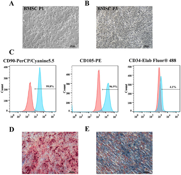
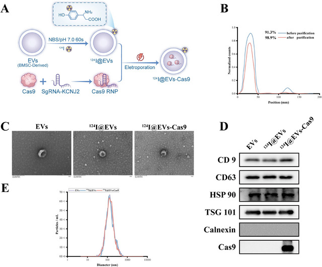
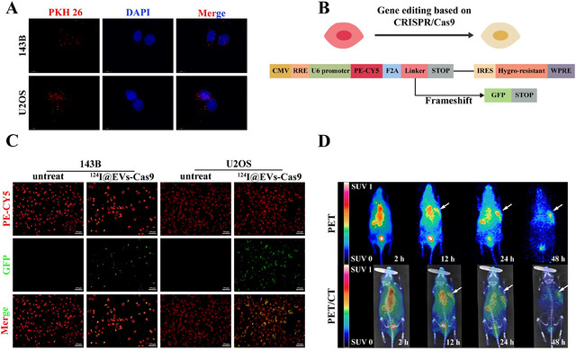
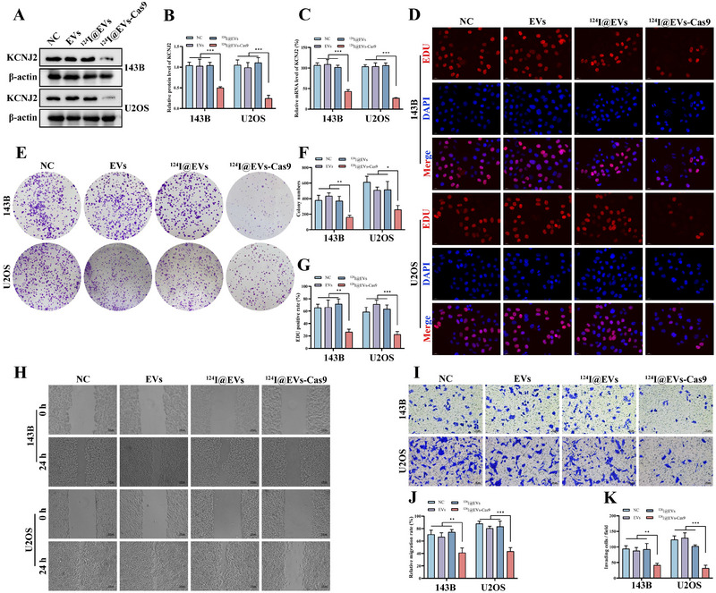
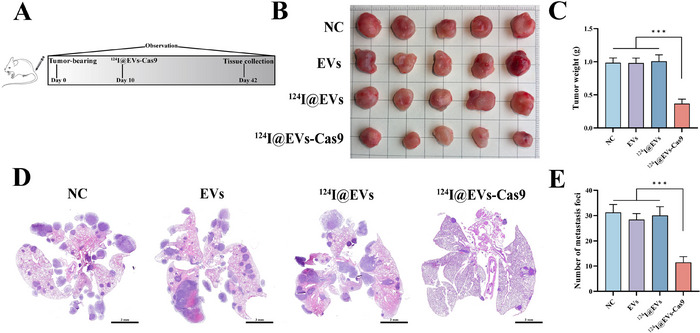
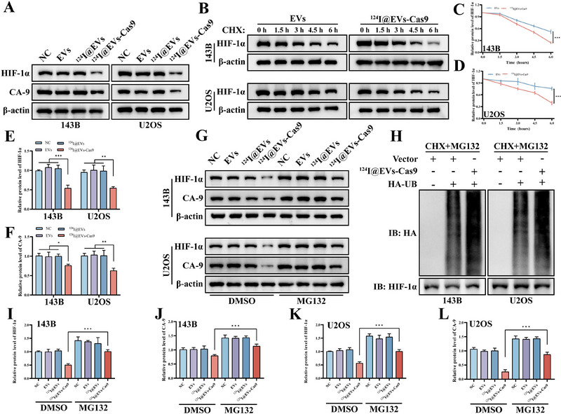
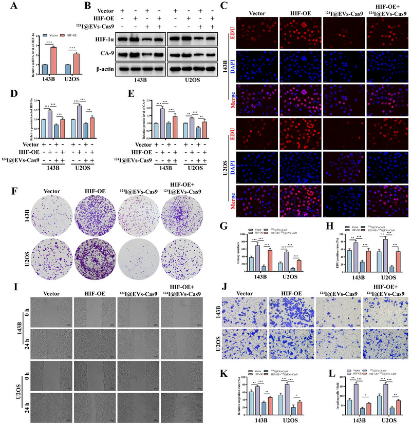
Metastasis constitutes the principal factor leading to the unfavourable prognosis of osteosarcoma patients. Hypoxia, as the inherent microenvironment of osteosarcoma, can upregulate HIF-1α via multiple pathways, thereby facilitating osteosarcoma proliferation and metastasis. Our previous research indicated that the inwardly rectifying potassium channel subfamily J member 2 (KCNJ2) inhibits the degradation of HIF-1α in osteosarcoma. Concurrently, HIF-1α upregulates the expression of KCNJ2 through a positive feedback regulatory mechanism. This positive regulatory mechanism significantly promotes the proliferation and metastasis of osteosarcoma. Therefore, the development of a KCNJ2-targeted therapeutic strategy capable of disrupting this reciprocal regulatory loop represents a crucial intervention for impeding osteosarcoma progression. The CRISPR/Cas9 targeted gene editing technology has garnered extensive attention in the field of tumour treatment due to its high efficiency and low off-target rate. Nevertheless, the relative lag of the delivery systems has restricted its application. The extracellular vesicles (EVs) secreted by bone marrow mesenchymal stem cells (BMSCs) have a natural targeting specificity for osteosarcoma and possess superior biocompatibility, making them ideal carriers for in vivo delivery. However, it is essential to confirm whether the CRISPR/Cas9 system mediated by EVs can accurately function intracellularly. Hence, we developed a fluorescence-based Cas9 editing efficiency reporter system. When CRISPR/Cas9 system induces double-strand breaks at specific target sites and results in frameshift mutations, osteosarcoma cells will stably express GFP. This system enables the transformation of gene editing events into quantifiable fluorescence signals. Furthermore, we engineered radiolabelled EVs derived from BMSCs to deliver the CRISPR/Cas9 system targeting KCNJ2. Using this reporter system, we confirmed their efficient gene-editing capabilities in vitro. Additionally, leveraging their radiolabelling properties, we validated their targeted distribution in vivo. Subsequent investigations revealed that our constructed 124I@EVs-Cas9 effectively suppresses the proliferation and metastasis of osteosarcoma by targeting the inhibition of KCNJ2 expression and promoting HIF-1α ubiquitin-dependent degradation (as depicted in Graphical Abstract).
Keywords: CRISPR‐Cas9; bone marrow mesenchymal stem cells; extracellular vesicles; osteosarcoma.
© 2025 The Author(s). Journal of Extracellular Vesicles published by Wiley Periodicals, LLC on behalf of the International Society for Extracellular Vesicles.
Conflict of interest statement
The authors declare no conflicts of interest.
Figures

Similar articles
-
Modulating binding affinity of aptamer-based loading constructs enhances extracellular vesicle-mediated CRISPR/Cas9 delivery.J Control Release. 2025 Aug 10;384:113853. doi: 10.1016/j.jconrel.2025.113853. Epub 2025 May 18. J Control Release. 2025. PMID: 40393529
-
Focused ultrasound and microbubble-mediated delivery of CRISPR-Cas9 ribonucleoprotein to human induced pluripotent stem cells.Mol Ther. 2025 Mar 5;33(3):986-996. doi: 10.1016/j.ymthe.2025.01.013. Epub 2025 Jan 10. Mol Ther. 2025. PMID: 39797397
-
Harnessing an anti-CRISPR protein for powering CRISPR/Cas9-mediated genome editing in undomesticated Bacillus strains.Microb Cell Fact. 2025 Jun 23;24(1):143. doi: 10.1186/s12934-025-02776-z. Microb Cell Fact. 2025. PMID: 40551141 Free PMC article.
-
Trojan Horse-Like Vehicles for CRISPR-Cas Delivery: Engineering Extracellular Vesicles and Virus-Like Particles for Precision Gene Editing in Cystic Fibrosis.Hum Gene Ther. 2025 Aug;36(15-16):1021-1052. doi: 10.1089/hum.2024.258. Epub 2025 Apr 28. Hum Gene Ther. 2025. PMID: 40295092 Review.
-
Management of urinary stones by experts in stone disease (ESD 2025).Arch Ital Urol Androl. 2025 Jun 30;97(2):14085. doi: 10.4081/aiua.2025.14085. Epub 2025 Jun 30. Arch Ital Urol Androl. 2025. PMID: 40583613 Review.
References
-
- Boudna, M. , Campos A. D., Vychytilova‐Faltejskova P., Machackova T., Slaby O., and Souckova K.. 2024. “Strategies for Labelling of Exogenous and Endogenous Extracellular Vesicles and Their Application for In Vitro and In Vivo Functional Studies.” Cell Communication and Signaling 22, no. 1: 171. - PMC - PubMed
-
- Chen, C. , Xie L., Ren T., Huang Y., Xu J., and Guo W.. 2021. “Immunotherapy for Osteosarcoma: Fundamental Mechanism, Rationale, and Recent Breakthroughs.” Cancer Letters 500: 1–10. - PubMed
MeSH terms
Substances
Grants and funding
- 82060491/the National Natural Science Foundation of China
- 82160568/the National Natural Science Foundation of China
- [2024]168/the Guizhou Provincial Science and Technology Projects
- [2021]395/the Guizhou Provincial Science and Technology Projects
- [2024]481/the Guizhou Provincial Science and Technology Projects
LinkOut - more resources
Full Text Sources
Medical
