. 2025 Jul;14(7):e70134.
doi: 10.1002/jev2.70134.

Extracellular Vesicles Derived From Streptococcus anginosus Aggravate Lupus Nephritis by Triggering TLR2-MyD88-NF-κB Signalling in NK Cells

Affiliations

Extracellular Vesicles Derived From Streptococcus anginosus Aggravate Lupus Nephritis by Triggering TLR2-MyD88-NF-κB Signalling in NK Cells

Ying Gong et al. J Extracell Vesicles. 2025 Jul.

Abstract

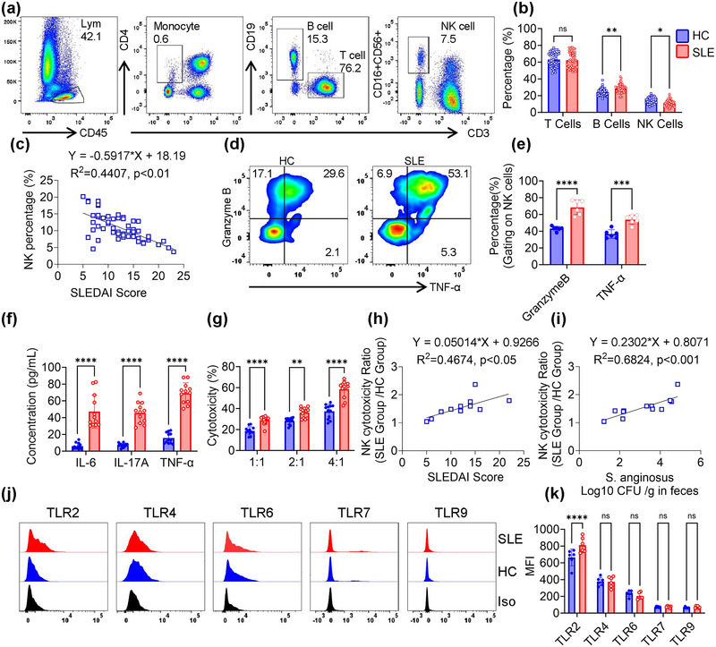
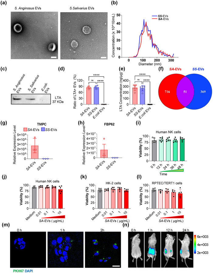
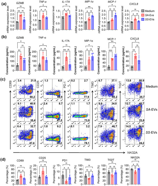
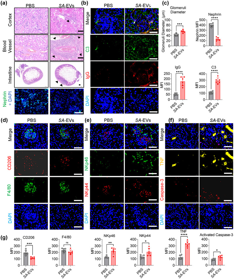
Systemic lupus erythematosus (SLE) has been linked to gut microbiome dysbiosis, notably an overabundance of Streptococcus anginosus; however, the impact of this microbial imbalance on disease pathogenesis remains unclear. Here, we investigated the contribution of S. anginosus-derived extracellular vesicles (SA-EVs) to SLE progression, with an emphasis on lupus nephritis (LN). Fifty-four SLE patients and 43 healthy controls (HC) were recruited. The faecal, blood and serum samples from participants were collected. SLE disease activity (SLEDA) was evaluated by the SLEDA Index (SLEDAI). Stool S. anginosus abundance was quantified by quantitative PCR, NK cell activation by flow cytometry and serum proinflammatory cytokines profile by ELISA. Lupus-prone MRL/lpr mice were orally administered SA-EVs to evaluate in vivo inflammatory responses, renal NK cell activation and renal histopathological changes. S. anginosus levels were significantly elevated in SLE patients relative to HC, positively correlated with SLEDAI scores and NK cell cytotoxicity. In vitro, SA-EVs stimulation of patient NK cells significantly heightened proinflammatory mediator production (granzyme B, TNF-α), increased cytotoxicity and downregulated inhibitory receptors (TIM-3, NKG2A, TIGIT) compared to control EVs from S. Salivarius (SS-EVs). Mechanistically, lipoteichoic acid (LTA) within SA-EVs engaged Toll-like receptor 2 (TLR2) on NK cells, activating MyD88/NF-κB signalling pathway. In MRL/lpr mice, SA-EVs treatment increased renal immune complex deposition, upregulated renal NK cell activation markers (NKp44, NKp46), and exacerbated LN pathology with greater immune cell infiltration and inflammatory cytokine levels. Furthermore, NK cell depletion with anti-NK1.1 antibodies significantly prolonged survival in SA-EVs administered mice. Thus, SA-EVs exacerbate SLE by hyperactivating NK cells via the TLR2-MyD88-NF-κB pathway, leading to amplified systemic inflammation and aggravated LN. These findings underscore the potential of targeting SA-EVs for therapeutic intervention in SLE.

Keywords: TLR2‐ MyD88‐NF‐κB signalling; bacterial extracellular vesicle; lupus nephritis (LN); natural killer cells (NK); systemic lupus erythematosus (SLE).


Conflict of interest statement

The authors declare no conflicts of interest.

Figures

FIGURE 1
FIGURE 1
Reduced frequency yet heightened cytotoxicity of peripheral blood NK cells in SLE patients. (a) Representative flow cytometry plots depicting gating strategy for TBNK cell populations. (b) Quantitative analysis of TBNK subpopulations in SLE patients (n = 54) versus healthy controls (HCs; n = 43). (c) Correlation of peripheral blood NK cell percentage and SLEDAI score in SLE patients. (d) Intracellular expression of Granzyme B and TNF‐α in primary human NK cells following stimulation with PMA (10 ng/mL), ionomycin (1 µg/mL) and brefeldin A (BFA; 1 µg/mL) for 12 h, analysed by flow cytometry. (e) Quantification of granzyme B and TNF‐α expression. (f) Serum concentration of IL‐6, IL‐17A and TNF‐α in the serum of HC and SLE (n = 12). (g) The cytotoxicity of NK against K‐562 cells at varying effector: target cell ratios. NK cells were purified from PBMC. NK cells and target cells were co‐cultured with tumour cells for 4 h. (h) Correlation between NK cell percentage and SLEDAI score in SLE patients (n = 12). (i) Correlation of NK cell cytotoxicity with Streptococcus anginosus abundance in SLE patients (n = 12). (j) Surface expression of TLR2, TLR4, TLR6, TLR7 and TLR9 on human NK cells. (k) Quantitative analysis of TLR receptor mean fluorescence intensity (MFI) on NK cells (n = 6). Data are shown as mean ± SD, with individual data points representing biological replicates (average of technical duplicates). Statistical comparisons were performed using one‐way ANOVA with Tukey post‐tests. *p < 0.05; **p < 0.01; ***p < 0.001; ****p < 0.0001. ns indicates not significant.
FIGURE 2
FIGURE 2
Characterisation of extracellular vesicles (EVs) derived from Streptococcus anginosus and their effects on human cells. (a) Transmission electron microscope (TEM) images of SA‐EVs. Scale bar: 100 nm. (b) Size distribution analysis of SA‐EVs by nanoparticle tracking analysis (NTA). (c) Western blot detection of lipoteichoic acid (LTA) in S. anginosus, SA‐EVs and E.coli EVs. (d) Quantitative densitometric analysis of LTA bands in SA‐EVs and E. coli EVs (ImageJ). (e) LTA concentration in SA‐EVs measured by ELISA. (f) Venn diagram illustrating peptide expression overlap between SA‐EVs and S. salivarius (SS‐EVs) as determined by DIA‐MS. (g, h) Relative expression of virulence factors TMPC and FBP62 in SA‐EVs versus SS‐EVs.(i) Time course analysis of the viability of NK cells following exposure to 1 µg/mL SA‐EVs. (j–l) Viability of NK cells, HK‐2 cells and RPTEC/TERT1 renal epithelial cells treated with varying concentrations of SA‐EVs. (m) Uptake of PKH67‐labelled SA‐EVs RPTEC/TERT1 cells. Scale bars, 50 µm. (n) Biodistribution of deep red‐labelled SA‐EVs in MRL/lpr mice following oral gavage, assessed by in vivo imaging. Data are presented as mean ± SD, with individual data points representing biological replicates (average of technical duplicates). Statistical comparisons were performed using one‐way ANOVA with Tukey post‐tests. *p < 0.05, **p < 0.01, ***p < 0.001, ****p < 0.0001. ns indicates not significant.
FIGURE 3
FIGURE 3
SA‐EVs activate human primary NK cells to produce proinflammatory cytokines. (a) RT‐ qPCR analysis of granzyme B, TNF‐α, IL‐17, MIP‐1α, MCP‐1 and CXCL8 mRNA expression in human primary NK cells after 24 h exposure to SA‐EVs (1 µg/mL) (n = 4). (b) ELISA quantification of granzyme B, TNF‐α, IL‐17, MIP‐1α, MCP‐1 and CXCL8 cytokines secreted by human primary NK cells following 48‐h exposure to SA‐EVs (1 µg/mL) (n = 4). (c) Representative flow cytometry plots depicting expression of activation (CD69, CD25) and inhibitory (NKG2A, TIM‐3, TIGIT) markers on NK cells after 48‐h incubation with SA‐EVs(1 µg/mL). (d) Quantitative analysis of activation and inhibitory marker expression in human primary NK cells treated with 1 µg/mL SA‐EVs for 48 h (n = 4). Data are presented as mean ± SD, with individual data points representing biological replicates (average of technical duplicates). Statistical differences between groups were determined using one‐way ANOVA with Tukey post‐tests.*p < 0.05, **p < 0.01, ***p < 0.001, ****p < 0.0001. ns indicates not significant.
FIGURE 4
FIGURE 4
TLR2‐MyD88‐NF‐κB pathway mediates SA‐EVs‐induced activation of NK cells. (a) Immunoprecipitation (IP) analysis demonstrating interaction between LTA and TLR2 (n = 3). (b) Quantification of IP assay results. (c) Western Blot assay of NK cells following coculture with SA‐EVs(1 µg/mL), phosphorylation of P65, IκBα and MyD88 was measured (N = 3). (d and g) The cytotoxicity of NK cells against RPTEC/TERT1 (d) and HK‐2 (g) cells with or without 1 µg/mL SA‐EVs coculture in different effector: target (E:T) ratios. Target cells were labelled with Dil membrane dye, and dead cells were identified by DAPI staining. All the cytotoxicity assay data were acquired by flow cytometry. NK cells were preincubated with 1 µg/mL SA‐EVs coculture for 24 h (N = 3). (e and h) NK cell cytotoxicity against RPTEC/TERT1 (e) and HK‐2 (h) cells in the presence of the TLR2 inhibitor C29, across varying E:T ratios (n = 3). (f, i) NK cell cytotoxicity against RPTEC/TERT1 (f) and HK‐2 (i) cells in the presence of the MyD88 inhibitor MyD88‐IN‐1, across varying E:T ratios (n = 3). Data are presented as mean ± SD, with individual data points representing biological replicates (average of technical duplicates). Statistical analysis was performed using one‐way ANOVA with Tukey post‐tests. *p < 0.05, **p < 0.01, ***p < 0.001, ****p < 0.0001. ns indicates not significant.
FIGURE 5
FIGURE 5
SA‐EVs exacerbate SLE disease severity via activation of nephritic NK cells. (a) Experimental schematic of SA‐EV oral gavage in MRL/lpr mice. Mice were received 100 µL PBS, 20 µg SS‐EVs or 20 µg SA‐EVs in 100 µL, administered every 3 days for 4 weeks. (b) Kaplan‐Meier survival curves of MRL/lpr mice following different EV treatments. (c) Time‐course analysis of urinary protein level in MRL/lpr mice subjected to different treatments. (d) Serum levels of creatinine (CRE), blood urea nitrogen (BUN), uric acid (UA), urea (UREA), anti‐dsDNA antibody (Anti‐dsDNA) and anti‐ANA antibodies (Anti‐ANA) after 4 weeks of treatment. (e) Flow cytometry analysis of nephritic infiltration NK cells; representative plots of NK cell markers NKp44 and NKp46. (f) Quantification of NK cells percentage, NKp44+ and NKp46+ cells among nephritic infiltration lymphocytes (n = 6). (g) Serum concentrations of proinflammatory cytokines TNF, IL‐6, IL‐17A, IFN‐1β and MIP‐1α (n = 4). (h) Kaplan‐Meier survival curves of MRL/lpr mice following specific immune cell depletions. Data is shown as mean ± SD, with dots representing individual donors (average of technical duplicates). Statistical differences between groups were determined using one‐way ANOVA with Tukey post‐tests.*p < 0.05, **p < 0.01, ***p < 0.001, ****p < 0.0001. ns indicates not significant.
FIGURE 6
FIGURE 6
SA‐EVs exacerbate lupus nephritis histopathological changes in an experimental SLE mouse model. (a) Representative histopathological images of kidney cortex, blood vessels, intestinal lymph nodes and glomerular nephrin expression in MRL/lpr mice. Haematoxylin and eosin (H&E) staining; scale bar = 50 µm. Black triangles indicate infiltrating inflammatory immune cells. (b) immunofluorescence images showing glomerular deposition of IgG and complement C3 in kidneys of MRL/lpr mice; scale bars = 50 µm. (c) Quantitative analysis of the glomerular size, the expression of nephrin and the mean fluorescence intensity of IgG and C3. (d) Expression of CD206 and F4/80 markers in the glomeruli of the kidneys from MRL/lpr lupus mice. Scale bars = 50 µm. (e) Expression of NK cell activation markers NKp44 and NKp46 in the kidneys from MRL/lpr lupus mice. Scale bars = 50 µm. (f) Expression of TNF and activated caspase‐3 in the kidneys from MRL/lpr lupus mice. Scale bars = 50 µm. (g) Quantitative analysis of the expression of CD206, F4/80, NKp46, NKp44, TNF and activated Caspase‐3. Data is shown as mean ± SD, with dots representing individual donors (average of technical duplicates). Statistical differences between groups were determined using one‐way ANOVA with Tukey post‐tests. *p < 0.05, **p < 0.01, ***p < 0.001, ****p < 0.0001. ns indicates not significant.

Similar articles

References

    1. Aringer, M. 2020. “Inflammatory Markers in Systemic Lupus Erythematosus.” Journal of Autoimmunity 110: 102374. 10.1016/j.jaut.2019.102374. - DOI - PubMed
    1. Aringer, M. , and Johnson S. R.. 2020. “Classifying and Diagnosing Systemic Lupus Erythematosus in the 21st Century.” Rheumatology 59, no. 5: v4–v11. 10.1093/rheumatology/keaa379. - DOI - PMC - PubMed
    1. Aringer, M. , and Petri M.. 2020. “New Classification Criteria for Systemic Lupus Erythematosus.” Current Opinion in Rheumatology 32, no. 6: 590–596. 10.1097/bor.0000000000000740. - DOI - PMC - PubMed
    1. Baschuk, N. , Wang N., Watt S. V., et al. 2014. “NK Cell Intrinsic Regulation of MIP‐1α by Granzyme M.” Cell Death & Disease 5, no. 3: e1115. 10.1038/cddis.2014.74. - DOI - PMC - PubMed
    1. Bluman, E. M. , Bartynski K. J., Avalos B. R., and Caligiuri M. A.. 1996. “Human Natural Killer Cells Produce Abundant Macrophage Inflammatory Protein‐1 Alpha in Response to Monocyte‐Derived Cytokines.” Journal of Clinical Investigation 97, no. 12: 2722–2727. 10.1172/jci118726. - DOI - PMC - PubMed

LinkOut - more resources