. 2025 Aug;13(4):e70148.
doi: 10.1002/prp2.70148.

Carbohydrate-Rich Fraction of Aloe vera (L.) Burm.f. Extract Mitigates Bone Loss and Improves Metabolic Disturbance in Estrogen-Deficient Rats

Affiliations

Carbohydrate-Rich Fraction of Aloe vera (L.) Burm.f. Extract Mitigates Bone Loss and Improves Metabolic Disturbance in Estrogen-Deficient Rats

Panan Suntornsaratoon et al. Pharmacol Res Perspect. 2025 Aug.

Abstract

Aloe vera (L.) Burm.f., (AE) herb has been shown to have osteogenic, anti-diabetic, and prebiotic activities in animal and human studies. Postmenopausal women generally exhibit massive bone loss, impaired intestinal calcium absorption, obesity-related insulin resistance, and fat accumulation in the liver. It was possible that the AE herb may have a potential as a remedy for bone and metabolic disturbances associated with estrogen deficiency. Sham and ovariectomized rats were divided into 2 subgroups, that is, receiving daily administration of distilled water or 50 or 100 mg/kg of AE via either oral administration (p.o.) or intraperitoneal injection (i.p.) for 8 and 12 weeks. Nine weeks after ovariectomy, rats developed metabolic disturbances, as evidenced by obesity, impaired glucose tolerance, and high serum cholesterol levels. AE supplementation, either by p.o. or i.p., alleviated metabolic aberrations by improving glucose tolerance, reducing body weight, and decreasing fat deposition by increasing serum insulin levels. Furthermore, AE supplementation restored ovariectomy-associated calcium malabsorption to that of sham. At week 12 post-ovariectomy, massive bone loss was observed at trabecular-rich regions. Daily AE supplementation at 50 mg/kg for 12 weeks, but not 8 weeks, significantly increased BMD and BMC compared with those of sham. Additionally, AE enhanced bone formation and suppressed bone resorption, as shown by bone histomorphometry and serum bone turnover markers. These findings clearly demonstrated the anti-diabetic and osteogenic properties of Aloe vera extract in ovariectomized rats. Thus, Aloe vera had a potential as a nutraceutical candidate for the treatment of osteoporosis and metabolic disturbances associated with estrogen deficiency.

Keywords: anti‐diabetes; dyslipidemia; hyperglycemia; insulin resistance; osteogenesis; ovariectomy.


Conflict of interest statement

The authors declare no conflicts of interest.

Figures

FIGURE 1
FIGURE 1
The effect of supplementation of Aloe vera extract (AE) on glucose and lipid metabolism in sham and ovariectomized rats. Sham (S) and ovariectomized (O) rats were supplemented with distilled water (SV and OV) or AE. For AE supplemented groups, rats were administered 50 mg/kg or 100 mg/kg via either oral gavage (SAG and OAG, n = 5 each) or intraperitoneal injection (SAP and OAP). Rats were sacrificed at 8 weeks and 12 weeks post‐supplementation. (a) percent body weight gain, figures B to P showed data from 8 weeks post supplementation, (b, c) oral glucose tolerance test and area under curve, (d, e) protein expression of phosphorylated Akt and total Akt in liver and pancreas by western blot analysis, (f) protein expression of GLP‐1 in ileum by western blot analysis, (g) Representative images of immunofluorescent stained against FGF‐21 in liver, (h) protein expression of FGF‐21 and its receptor FGFR‐1 and 3, phosphorylated AMPK, total AMPK and SirT‐1 in liver by western blot analysis, (i) H&E stained liver sections, arrows indicated triglyceride droplets. Whole blot with molecular marker was shown in Supplemental information (uncropped Western blot images). Results are expressed as means ± SE with individual values. The differences between experimental groups were determined by one‐way ANOVA followed by Tukey post hoc test. *p < 0.05 compared with SV, p < 0.05 compared with OV.
FIGURE 2
FIGURE 2
Eight‐week supplementation with 100 mg/kg Aloe vera extract decreased pro‐inflammatory cytokines in estrogen‐deficiency. (a) malondialdehyde level in liver (markers for oxidative stress), (b, c) protein expression of NLRP3 inflammasome and pro‐inflammatory cytokines (i.e., interleukin‐1β and interleukin‐6) in liver by western blot analysis, (d, e) protein expression of P16 and P21, cell senescence markers by western blot analysis, (f) representative immunostaining against P16, (g, h) protein expression of NLRP3 inflammasome and pro‐inflammatory cytokines (i.e., interleukin‐1β and interleukin‐6) in pancreas by western blot analysis. Results are expressed as means ± SE with individual values. The differences between experimental groups were determined by one‐way ANOVA followed by Tukey post hoc test. *p < 0.05 compared with SV, p < 0.05 compared with OV.
FIGURE 3
FIGURE 3
Eight‐week supplementation of 50 and 100 mg/kg Aloe vera extract increased protein expression of calcium transporters in small and large intestine, and increased fractional intestinal calcium absorption. Rats were supplemented with distilled water or 100 mg/kg Aloe vera extract for 8 weeks. (a, b) calcium transporter protein expression in cecum, and (c) fractional intestinal calcium absorption of 50 mg/kg AE supplemented rats. Results are expressed as means ± SE with individual values. The differences between experimental groups were determined by one‐way ANOVA followed by Tukey post hoc test. *p < 0.05 compared with SV, p < 0.05 compared with OV, # p < 0.05 compared with AE‐treated OV rats p.o. (OAG).
FIGURE 4
FIGURE 4
Aloe vera extract supplementation for 12 weeks alleviated ovariectomy‐induced bone loss examined by pQCT. Ovariectomized rats were either intraperitoneal injected with 50 mg/kg Aloe vera extract (OAP) or distilled water (OV). Rats were sacrificed at 8 weeks and 12 weeks post‐supplementation. (a) experimental timeline, (b) pQCT analysis at distal metaphyseal femur, (c) pQCT analysis at mid‐shaft diaphyseal femur. A, bone area; BMC, bone mineral content; BMD, bone mineral density; Ct, cortical; Ct.Sub, bone region included cortical and subcortical compartment; Es.Pm, endosteal perimeter; Ps.Pm, periosteal perimeter; Tb, trabecular; Th, thickness; TOT, total tissue which included both trabecular and cortical compartments. Results are expressed as means ± SE with individual values. The difference between two set of data was determined by un‐paired student t‐test. *p < 0.05, **p < 0.01, ***p < 0.001 compared with corresponded age‐matched OV.
FIGURE 5
FIGURE 5
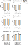
Aloe vera extract supplementation in the dose of 50 mg/kg, for 12 weeks, either by oral gavage or intraperitoneal injection similarly alleviated ovariectomy‐induced bone loss examined by pQCT. Sham and ovariectomized rats received either distilled water (SV and OV) or Aloe vera extract (AE) administration. For AE supplemented groups, rats were administered in the dose of 50 mg/kg either by oral gavage (SAG and OAG, n = 6 each) or by intraperitoneal injection (SAP and OAP). Rats were sacrificed at 12 weeks post‐supplementation. (a) experimental timeline, (b) pQCT analysis at distal metaphyseal femur and (c) representative 3D micro‐CT images of the femur. Scale bar, 1 mm. A, bone area; BMC, bone mineral content; BMD, bone mineral density; Ct, cortical; Ct.Sub, bone region included cortical and subcortical compartment; Es.Pm, endosteal perimeter; Ps.Pm, periosteal perimeter; Tb, trabecular; Th, thickness; TOT, total tissue which included both trabecular and cortical compartments. Results are expressed as means ± SE with individual values. Two‐way ANOVA followed by Tukey's multiple comparisons post hoc test was used to analyze the difference between independent sets of data and to analyze interaction between 2 factors, that is, ovariectomy effect and AE effect. *p < 0.05, **p < 0.01, ***p < 0.001 compared with SV. p < 0.05, †† p < 0.01 compared with OV.
FIGURE 6
FIGURE 6
The effect of 50 mg/kg Aloe vera extract supplementation for 12 weeks on cortical bone structure at femora mid‐shaft diaphysis examined by pQCT. Experimental design was similar to that shown in Figure 5. A, bone area; BMC, bone mineral content; BMD, bone mineral density; Ct, cortical; Es.Pm, endosteal perimeter; Ps.Pm, periosteal perimeter; Th, thickness. Results are expressed as means ± SE with individual values. Two‐way ANOVA followed by Tukey's multiple comparisons post hoc test was used to analyze the difference between independent sets of data and to analyze the interaction between 2 factors, that is, ovariectomy effect and AE effect. *p < 0.05, **p < 0.01, *** p < 0.001 compared with SV. p < 0.05 compared with OV.
FIGURE 7
FIGURE 7
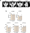
The effect of 50 mg/kg Aloe vera extract supplementation for 12 weeks on lumbar vertebrae 5 (L5) examined by ultra‐high resolution μCT. Experimental design was similar to that shown in Figure 5. (a) representative 3D micro‐CT images of the femur and (b) μCT analysis of trabecular compartment of lumbar vertebrae L5. BMC, bone mineral content; BMD, bone mineral density; BV/TV, bone volume normalized by tissue volume; Tb.Sp, trabecular separation; Tb.Th, trabecular thickness. Results are expressed as means ± SE with individual values. The differences between experimental groups were determined by one‐way ANOVA followed by Tukey post hoc test. *p < 0.05, **p < 0.01, ***p < 0.001 compared with SV. p < 0.05 compared with OV.
FIGURE 8
FIGURE 8
Oral supplementation of Aloe vera extract alleviated ovariectomy‐induced bone loss by stimulating bone formation and suppressing osteoclastic bone resorption examined by bone histomorphometry. Sham (S) and ovariectomized (O) rats were supplemented with distilled water (SV and OV) or Aloe vera extract (AE). For AE supplemented groups, rats were administered in the dose of 50 mg/kg via either oral gavage (SAG and OAG) or intraperitoneal injection (SAP and OAP). Another group of OV rats was oral administered with 100 mg/kg. Rats were sacrificed 12 weeks post‐supplementation. (a) serum level of bone formation marker, pro‐collagen type 1 and (b) serum level of bone resorption marker, tartrate‐resistant acid phosphatase. (a, b) Results are expressed as means ± SE with individual values. The differences between experimental groups were determined by one‐way ANOVA followed by Tukey post‐hoc test. * p < 0.05 compared with SV, p < 0.05 compared with OV, (c) representative images of tibia section stained Goldner's trichrome. Allows indicate trabeculae. Ma, marrow. Scale bar, 1 mm, and (d) bone microstructure and bone cell parameters. Two‐way ANOVA followed by Tukey's multiple comparisons post‐hoc test was used to analyze the difference between independent sets of data and to analyze interaction between 2 factors, i.e., ovariectomy effect and AE effect. * p < 0.05, ** p < 0.01 and *** p < 0.001.

Similar articles

References

    1. Zhao C., Shi J., Shang D., Guo M., Zhou C., and Zhao W., “Protective Effect of Phytoestrogens on Nonalcoholic Fatty Liver Disease in Postmenopausal Women,” Frontiers in Pharmacology 14 (2023): 1237845, 10.3389/fphar.2023.1237845. - DOI - PMC - PubMed
    1. Koubaa‐Ghorbel F., Chaabane M., Jdidi H., Turki M., Makni‐Ayadi F., and El Feki A., “ Salvia officinalis Mitigates Uterus and Liver Damages Induced by an Estrogen Deficiency in Ovariectomized Rats,” Journal of Food Biochemistry 45 (2021): e13542, 10.1111/jfbc.13542. - DOI - PubMed
    1. Mann S. N., Pitel K. S., Nelson‐Holte M. H., et al., “17alpha‐Estradiol Prevents Ovariectomy‐Mediated Obesity and Bone Loss,” Experimental Gerontology 142 (2020): 111113, 10.1016/j.exger.2020.111113. - DOI - PMC - PubMed
    1. Jin Y., Gao X. Y., Zhao J., et al., “Estrogen Deficiency Aggravates Fluoride‐Induced Small Intestinal Mucosa Damage and Junctional Complexes Proteins Expression Disorder in Rats,” Ecotoxicology and Environmental Safety 246 (2022): 114181, 10.1016/j.ecoenv.2022.114181. - DOI - PubMed
    1. Che C. T., George V., Ijinu T., Pushpangadan P., and Andrae‐Marobela K., “Traditional Medicine,” in Pharmacognosy (Elsevier, 2017), 15–30.

LinkOut - more resources