. 2025 Jul 17;21(7):e1013157.
doi: 10.1371/journal.ppat.1013157. eCollection 2025 Jul.

Membrane-wide screening identifies potential tissue-specific determinants of SARS-CoV-2 tropism

Affiliations

Membrane-wide screening identifies potential tissue-specific determinants of SARS-CoV-2 tropism

Ravi K Dinesh et al. PLoS Pathog. .

Abstract

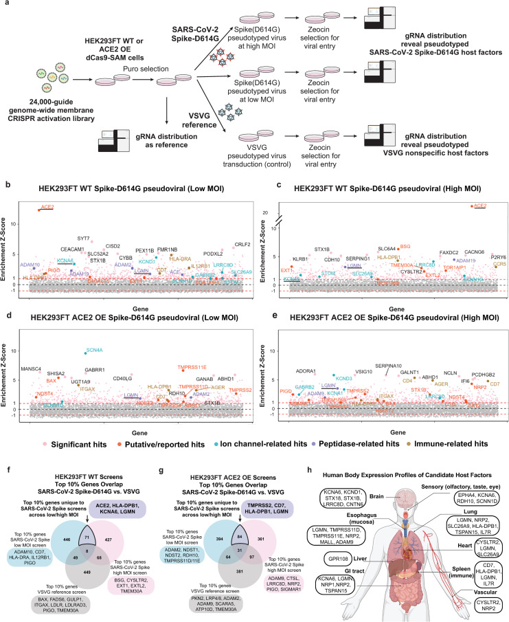
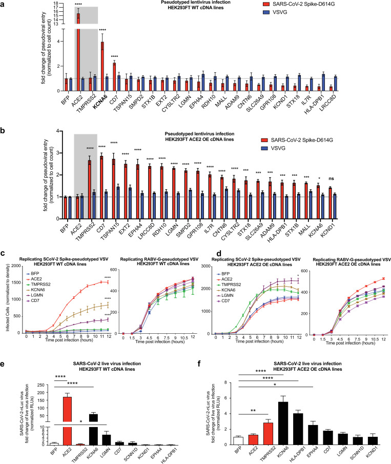
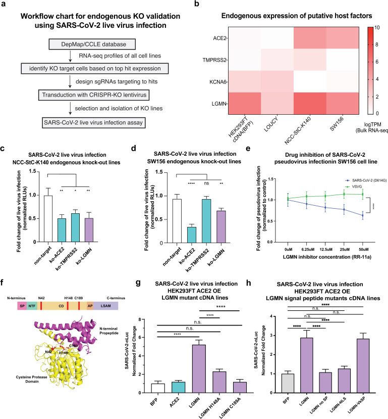
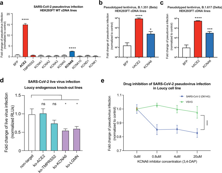
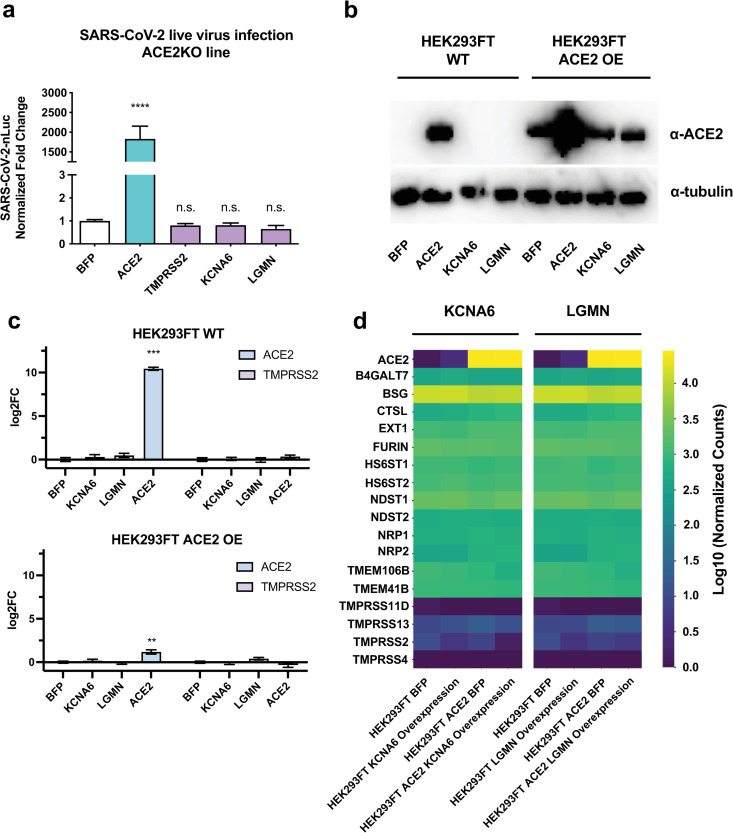
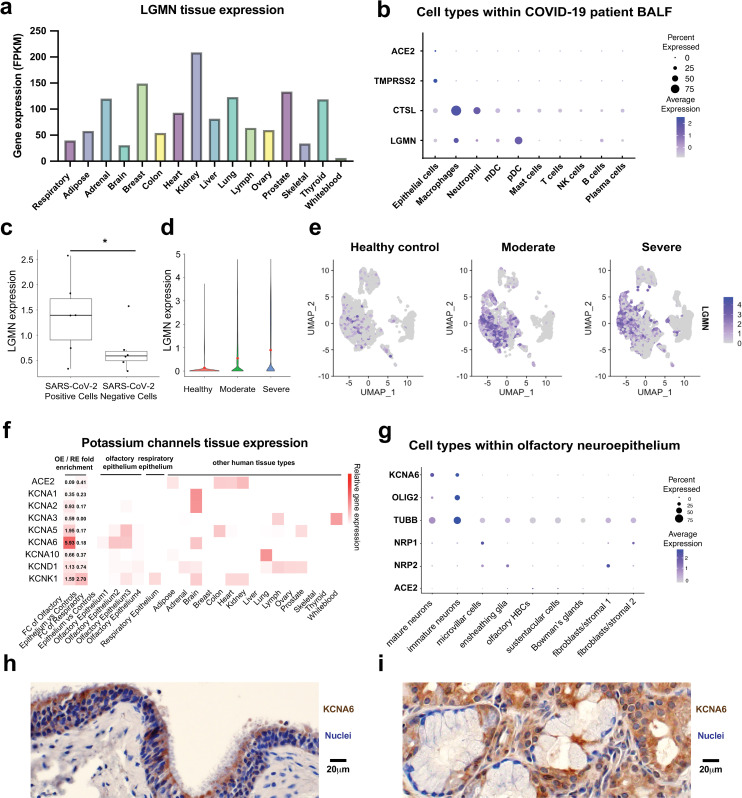
While SARS-CoV-2 primarily infects the respiratory tract, clinical evidence indicates that cells from diverse cell types and organs are also susceptible to infection. Using the CRISPR activation (CRISPRa) approach, we systematically targeted human membrane proteins in cells with and without overexpression of ACE2, thus identifying unrecognized host factors that may facilitate viral entry. Validation experiments with replication-competent SARS-CoV-2 confirmed the role of newly identified host factors, particularly the endo-lysosomal protease legumain (LGMN) and the potassium channel KCNA6, upon exogenous overexpression. In orthogonal experiments, we show that disruption of endogenous LGMN or KCNA6 decreases viral infection and that inhibitors of candidate factors can reduce viral entry. Additionally, using clinical data, we find possible associations between expression of either LGMN or KCNA6 and SARS-CoV-2 infection in human tissues. Our results identify potentially druggable host factors involved in SARS-CoV-2 entry, and demonstrate the utility of focused, membrane-wide CRISPRa screens in uncovering tissue-specific entry factors of emerging pathogens.


Conflict of interest statement

The authors have declared that no competing interests exist.

Figures

Fig 1
Fig 1. Membrane-focused CRISPRa screening identifies potential host factors involved in Spike-dependent SARS-CoV-2 virus entry.
a, Screen pipeline showing different conditions used and the downstream analyses and validation workflow. b-e, Enrichment scores of CRISPRa screens across different conditions with top hits highlighted and colored by their functional categories. Known (ACE2, TMPRSS2) and top-ranked new host factors are underlined. The enrichment cut-off score (generated using MAGeCK pipeline) was 1.0, and noted by a dashed line. f-g, Differential analysis of top 10% of hits from SARS-CoV-2 Spike and reference VSVG screens. The unique hits in SARS-CoV-2 screens help to identify putative virus-specific host factors from the screen. h, Tissue expression [29] body map to visualize top-ranking genes from the analysis of candidate host factors in panels f-g. Panels a,h were created with Biorender.
Fig 2
Fig 2. Validation of top-ranked genes using pseudoviral and replication-competent SARS-CoV-2 infection assays.
a-b, Arrayed validation of top hits in cDNA overexpressing cell lines of individual genes using SARS-CoV-2 Spike-D614G pseudotyped lentiviral assay. The control VSVG-pseudotyped lentivirus results are shown side-by-side. Results are normed to the BFP control for each respective pseudotype. Data from two independent experiments. c-d, Arrayed validation using time-lapse imaging of SARS-CoV-2 Spike-pseudotyped VSV infection in cDNA overexpression cell lines in 293FT WT cells (c) and 293FT ACE2OE (d). Companion control infection was performed using Rabies virus (RABV) G protein pseudo-typed VSV. Data from two independent experiments. e-f, Validation of top-ranked genes using SARS-CoV-2-nLuc virus infection. Data from three independent experiments. All data represent mean with SEM. Statistical analyses: for the panels a-d, using two-way ANOVA with correction for multiple comparisons during hypothesis testing, and for the panels e-f, one-way ANOVA with correction for multiple comparisons. *, p < 0.05; **, p < 0.01; ***, p < 0.001; ****, p < 0.0001.
Fig 3
Fig 3. Disruption of putative host factor LGMN decreases SARS-CoV-2 viral entry.
a, Workflow for endogenous knockout validation using SARS-CoV-2 virus infection. b, RNA-seq heat-map of gene expression in representative cell lines alongside cDNA-overexpressing reference lines. c-d, SARS-CoV-2 live virus infection of NCC-Stc-K140 (c) and SW156 (d) cells perturbed with CRISPR-based loss-of-function constructs. Three guideRNAs were used per gene and results for the three independent lines were performed via individual arrayed infections and pooled for analysis. The entire infection was repeated twice to collect replicates for all cell lines. Results are normed to the BFP control/vehicle for each respective pseudotype. e, Data showing the dosage-dependent effect on infection of SARS-CoV-2 Spike-D614G or VSV-G pseudotyped lentivirus in LGMN-expressing SW156 cells treated with RR-11a, a specific inhibitor of legumain (LGMN). f, Structure and domains of human Legumain. SP: signal peptide, NTF: N-terminal fragment, CD: catalytic domain, AP: activation peptide, LSAM: legumain stabilization and activity modulation domain. g-h, SARS-CoV-2 live virus infection of ACE2OE 293FT cells expressing LGMN or LGMN catalytic (g) and signal peptide (h) mutant cDNA. Results are normed to BFP control. Data in (g) from three independent experiments and (h) from two independent experiments. All data represent mean with SEM. Statistical analyses: for c-d,f performed via one-way ANOVA with BFP or non-target as the control condition and for e, g-h with two-way ANOVA, all with correction for multiple comparisons during hypothesis testing. *, p < 0.05; **, p < 0.01; ***, p < 0.001; ****, p < 0.0001.
Fig 4
Fig 4. KCNA6 is a putative SARS-CoV-2 host factor.
a, Infection of SARS-CoV-2 Spike-D614G pseudotyped lentivirus in 293FT cells overexpressing Kv channel members. Three independent experiments performed. b-c, Infection of cDNA overexpressing 293FT cells with lentivirus pseudotyped with either B.1.351 (Beta) or B.1.617 (Delta) Spike. n = 5. d, SARS-CoV-2 live virus infection of Loucy cells perturbed with CRISPR-based loss-of-function constructs. Three guideRNAs were used per gene and results for the three independent lines were performed via individual arrayed infections and pooled for analysis. The entire infection was repeated twice to collect replicates for all cell lines. e, Inhibition of SARS-CoV-2 viral entry with FDA-pproved compound 3,4-Diaminopyridine (Amifampridine), a potassium channel inhibitor. Data showing the dosage-dependent effect on infection of SARS-CoV-2 Spike-D614G or VSV-G pseudotyped lentivirus in KCNA6-expressing Loucy cells treated with 3,4-AP. Results are normed to the BFP control/vehicle for each respective pseudotype. Data from two independent experiments. All data represent mean with SEM. Statistical analyses: for a-d are performed via one-way ANOVA with BFP or non-target as the control condition and for f with two-way ANOVA, all with correction for multiple comparisons during hypothesis testing. *, p < 0.05; **, p < 0.01; ***, p < 0.001; ****, p < 0.0001.
Fig 5
Fig 5. KCNA6 and LGMN promote SARS-CoV-2 infection dependent on ACE2 but independent of other known SARS-CoV-2 host factors.
a, Left, SARS-CoV-2 live virus infection of ACE2KO 293T cells overexpressing cDNAs. Data from two independent experiments. b, Western Blot of ACE2 levels in WT and ACE2 OE 293FT cells overexpressing cDNAs. c, Quantitative RT-PCR analysis of ACE2 and TMPRSS2 levels upon the over-expression of indicated cDNAs. d, Whole transcriptome analysis via RNA-seq of the KCNA6 and LGMN over-expressing cell lines, compared with BFP-expressing control lines, two independent replicates were sequenced. All data represent mean with SEM. Statistical analyses in a were performed via one-way ANOVA with BFP serving as the control condition, with correction for multiple comparisons during hypothesis testing, in c with two-tailed t test using Welch’s correction with BFP as the control condition. *, p < 0.05; **, p < 0.01; ***, p < 0.001; ****, p < 0.0001.
Fig 6
Fig 6. KCNA6 and LGMN are expressed in disease-relevant cell types.
a, Expression of LGMN across human tissues [49,50]. b, Expression of ACE2, TMPRSS2 and LGMN in different cell types from BALF. c, Average expression levels of LGMN between SARS-CoV-2 positive and negative cells from severely affected patients. Each dot represents a severely affected patient. Two-sided Student’s t-test. d-e, Healthy controls (n = 4); COVID-19 patients (moderate) n = 3; COVID-19 patients (severe), n = 6. d, LGMN expression from single cell RNA-seq data. e, UMAPs depicting the expression of LGMN. f, Expression of ACE2 and KCNA family genes across human tissues. Left two columns show the fold change (FC) of expression comparing olfactory epithelium (OE) or respiratory epithelium (RE) vs. control tissues. Columns on the right show the expression levels across individual samples. g, Expression of ACE2 and KCNA6 in the single-cell RNA-seq data of olfactory neuroepithelium [51]. Sizes of the dots indicate the proportion of cells having greater-than-zero expression while the color indicates mean expression. h-i, Immunohistochemistry analysis of KCNA6 protein expression in human sinonasal tissues using archival clinical samples.

Similar articles

References

    1. Cui J, Li F, Shi Z-L. Origin and evolution of pathogenic coronaviruses. Nat Rev Microbiol. 2019;17(3):181–92. doi: 10.1038/s41579-018-0118-9 - DOI - PMC - PubMed
    1. Mechanisms of SARS-CoV-2 Transmission and Pathogenesis - PubMed. [cited 9 May 2025]. Available from: https://pubmed.ncbi.nlm.nih.gov/33132005/
    1. Clausen TM, Sandoval DR, Spliid CB, Pihl J, Perrett HR, Painter CD, et al. SARS-CoV-2 infection depends on cellular heparan sulfate and ACE2. Cell. 2020;183(4):1043–1057.e15. doi: 10.1016/j.cell.2020.09.033 - DOI - PMC - PubMed
    1. Wang R, Simoneau CR, Kulsuptrakul J, Bouhaddou M, Travisano KA, Hayashi JM, et al. Genetic screens identify host factors for SARS-CoV-2 and common cold coronaviruses. Cell. 2021;184(1):106–119.e14. doi: 10.1016/j.cell.2020.12.004 - DOI - PMC - PubMed
    1. Wei J, Alfajaro MM, DeWeirdt PC, Hanna RE, Lu-Culligan WJ, Cai WL, et al. Genome-wide CRISPR screens reveal host factors critical for SARS-CoV-2 infection. Cell. 2021;184(1):76-91.e13. doi: 10.1016/j.cell.2020.10.028 - DOI - PMC - PubMed