. 2025 Jul 22;122(29):e2423358122.
doi: 10.1073/pnas.2423358122. Epub 2025 Jul 17.

Inactivation of PI3K-C2α deregulates cell death pathways and sensitizes to endotoxic shock

Affiliations

Inactivation of PI3K-C2α deregulates cell death pathways and sensitizes to endotoxic shock

York Posor et al. Proc Natl Acad Sci U S A. .

Abstract

The organismal roles of the class II PI3K isoform PI3K-C2α remain poorly understood. Recent studies have found PI3K-C2α to promote arterial thrombosis and breast cancer metastasis, generating interest in this kinase as a drug target, with small molecule PI3K-C2α inhibitors now available. However, the consequences of systemic PI3K-C2α inactivation in the nondiseased, postnatal state are largely unknown. Here, we show that induction of genetic PI3K-C2α inactivation in adult mice is well tolerated, without adverse effects on normal physiology. Surprisingly, however, mice with inactive PI3K-C2α display strong sensitization to challenge with bacterial lipopolysaccharide (LPS), a model of endotoxic shock. This sensitization is recapitulated by vascular endothelial-specific deletion of PI3K-C2α. Furthermore, sensitization to LPS can be fully rescued by disabling extrinsic induction of cell death by combined caspase-8- and RIPK3 deficiency. These observations validate the tolerability of systemic PI3K-C2α inhibition in principle but reveal an unexpected role for PI3K-C2α in the regulation of extrinsic cell death pathways.

Keywords: PI3K; endotoxic shock; phosphoinositide; regulated cell death; vascular endothelia.


Conflict of interest statement

Competing interests statement:B.V. is a consultant for iOnctura (Geneva, Switzerland)., and shareholder of Open Orphan and Poolbeg Pharma (Dublin, Ireland).

Figures
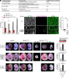
Fig. 1.
Fig. 1.
Embryonic lethal phenotype of kinase-inactive Pik3c2aD1268A/D1268A mice shows features of deregulated cell death. Homozygous inactivation of PI3K-C2α by point mutation of Asp1268 to Ala (D1268A) is lethal at mid-gestation. (A) Overview of the Pik3c2a loss-of-function models used in this study. Note that “scaffold function remaining” refers to tissues that underwent Cre-mediated recombination and designates the amount of kinase-inactive PI3K-C2α protein expressed relative to WT PI3K-C2α protein. The internally truncated PI3K-C2α protein (Fig. 2A) is less stable than the WT protein. (B) Analysis of embryo genotypes from Pik3c2a+/D1268A × Pik3c2a+/D1268A matings at day of embryonic development E9.5 to E12.5. The frequency of live embryos with homozygous inactivation of PI3K-C2α is decreased at E10.5 and E11.5. At E12.5, no live Pik3c2aD1268A/D1268A embryos were found. Figures at the Bottom of the bars show the number of embryos dissected and genotyped per group. Chi-square test comparing observed vs. expected genotype frequencies; *, P ≤ 0.05. (C and D) E10.5 yolk sacs were stained for the endothelial marker PECAM-1 and for cleaved caspase-3 as an indicator of cell death. Pik3c2aD1268A/D1268A yolk sacs show increased cleaved caspase-3 staining in endothelial cells, as quantified in (D). (C) (Scale bar, 50 µm.) (D), Two-tailed t-test with Welch’s correction. **P < 0.01. (E and F) PI3K-C2α-inactive mice were crossed to Casp8−/−;Ripk3−/−;Mlkl−/− triple KO mice (deficient for extrinsic induction of cell death) or Tnfr1−/− mice and embryos from Pik3c2a+/D1268A × Pik3c2a+/D1268A matings on the respective background were analyzed at E12.5. Each embryo is shown before (Left panels) and after removal of yolk sac and placenta (Right panels). Note that most Pik3c2aD1268A/D1268A embryos (26 out of 28 for Casp8−/−;Ripk3−/−;Mlkl−/−; 12 out of 13 for Tnfr1−/−) still showed delayed development and lethality by E12.5. However, a small minority of Pik3c2aD1268A/D1268A embryos on the Casp8−/−;Ripk3−/−;Mlkl−/− background and also on the Tnfr1−/− background showed dramatically improved development and intact vascularization at E12.5. These embryos all displayed anophthalmia, a hallmark of disrupted Hedgehog signaling. (Scale bar, 3 mm.) (F) Numbers of dissected and genotyped embryos as in (E). Figures at the bottom of the bars show the number of embryos dissected and genotyped per group.
Fig. 2.
Fig. 2.
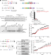
Conditional inactivation of PI3K-C2α in adult mice is well tolerated. (AE) Conditional inactivation of PI3K-C2α in mice with one constitutively point-mutated allele (D1268A) and one allele for conditional internal truncation (flox24-25). (A) Scheme depicting the flox24-25 allele for conditional internal truncation of PI3K-C2α. Exons 24 and 25 of the mouse Pik3c2a gene are flanked by loxP sites, yielding in-frame deletion upon Cre-mediated recombination. The resulting polypeptide is expected to be 175 kDa (compared to the WT protein of 187.5 kDa) and lacks the DFG-motif and activation loop in the C-lobe of the catalytic domain. UTR, untranslated region. (B) Primary embryonic fibroblasts from mice with the indicated genotypes were treated with 4-OH-tamoxifen (4-OHT) for a duration of up to 10 d and protein extracts were analyzed by immunoblotting. (C) PI3K-C2α was immunoprecipitated from embryonic fibroblasts at 3 or 5 d after 4-OHT treatment and immunoprecipitates were assayed for kinase activity against PI(4)P using γ-32P-labeled ATP followed by thin layer chromatography and phosphor imager-based quantification of the amount of synthesized PI(3, 4)P2. Note that 5 d after 4-OHT treatment, kinase activity in Pik3c2aflox24-25/D1268A;CreERT2 fibroblasts is nearly undetectable. (D) PCR-based analysis of recombination efficiency across tissues in Pik3c2aflox24-25/D1268A;CreERT2 mice subjected to tamoxifen treatment at 8 wk of age. Cre-mediated recombination shifts the size of the PCR fragment of the floxed allele from 372 bp to 409 bp. Recombination is nearly complete in many tissues including spleen, pancreas, and lymph nodes; partial in some organs such as the lung, heart, and kidney; and largely absent in the brain. (E) Body weight of mice following treatment with tamoxifen (TMX) for 2 × 5 d as indicated starting at about 8 wk of age (day 0). After treatment has ended, mice resumed gaining weight. The Pik3c2aflox24-25/D1268A;CreERT2 mice exhibited slightly reduced weight gain but showed no overt adverse phenotype. Data show body weight normalized to day 0 of treatment as mean ± s.e.m. from n = 58 (Pik3c2a+/+) or n = 46 (Pik3c2aflox24-25/D1268A) mice at the beginning of treatment. (FH) Conditional knockout by flanking exon 3 of the mouse Pik3c2a gene by loxP sites (flox3). (F) Scheme depicting gene targeting for the flox3 allele. Deletion of exon 3 causes frame shift and premature termination in exon 4. (G) Liver, spleen, and kidney from mice with the indicated genotypes and treated with tamoxifen as in (E) were collected 6 wk after treatment onset and analyzed by immunoblotting. Hsp70 and α-tubulin shown as loading control. (H) Body weight of mice following treatment with tamoxifen as in (E). Conditional knockout of PI3K-C2α led to a tendency of reduced weight gain after treatment with tamoxifen, but no other overt adverse phenotype was observed.
Fig. 3.
Fig. 3.
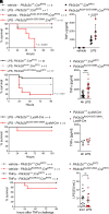
Conditional inactivation of PI3K-C2α sensitizes mice to endotoxic shock. (A and B) After global tamoxifen-induced inactivation of PI3K-C2α in adult Pik3c2aflox24-25/D1268A;CreERT2 mice (see Fig. 2 and Materials and Methods for details), mice were challenged with vehicle or a sublethal dose of lipopolysaccharide (LPS; 0.5 mg/kg LPS O111:B4 by intraperitoneal injection). (A) Kaplan–Meier curve depicting survival of mice following LPS challenge. Inactivation of PI3K-C2α strongly sensitizes mice to LPS challenge (n denotes the number of mice per group). Data were analyzed by logrank (Mantel-Cox) test. (B) TNFα concentration in blood plasma obtained from mice 4 h after vehicle/LPS challenge as in (A), determined by bead-based 13-plex flow cytometry assay (see also SI Appendix, Fig. S3). Bars show mean ± s.d. and were analyzed by the two-tailed t test with Welch’s correction. (C and D) Global tamoxifen-induced inactivation of PI3K-C2α using the flox exon 3 allele recapitulates sensitization to LPS shown after inactivation using the flox exon 24 to 25 allele (Panel A). Pik3c2a+/+;CreERT2, Pik3c2aflox3/+;CreERT2 or Pik3c2aflox3/D1268A;CreERT2 mice were challenged with LPS as described for (A and B). (C) Survival curve following LPS challenge. Data were analyzed by logrank (Mantel-Cox) test. (D) TNFα in blood plasma 4 h after LPS challenge, determined by ELISA. Data are mean ± s.d. and were analyzed by the two-tailed t test with Welch’s correction. ***P < 0.001. (E and F) Conditional inactivation of PI3K-C2α in myeloid cells does not sensitize mice to LPS challenge. Pik3c2a+/+;LysM-Cre or Pik3c2aflox24-25/D1268A;LysM-Cre mice were challenged with LPS as described for (A and B). (C) Survival curve following LPS challenge. (D) TNFα in blood plasma 4 h after LPS challenge, determined by ELISA. Bars show mean ± s.d. (G) PI3K-C2α-inactive mice are not more susceptible to challenge with TNFα. After global tamoxifen-induced inactivation of PI3K-C2α in adult Pik3c2aflox3/flox3;CreERT2 mice, mice were challenged with vehicle or a sublethal dose of TNFα (200 µg/kg murine TNFα by intravenous injection); n denotes the number of mice per group. Data were analyzed by logrank (Mantel-Cox) test, P = 0.2604. (H) LPS clearance is not affected in PI3K-C2α-inactive mice. After global tamoxifen-induced inactivation of PI3K-C2α in adult Pik3c2aflox3/D1268A;CreERT2 mice, mice were challenged with LPS as in (A and B). Blood plasma was obtained after 24 h and LPS concentration was determined by chromogenic Limulus amebocyte lysate assay.
Fig. 4.
Fig. 4.
Conditional knockout of PI3K-C2α in vascular endothelial cells recapitulates sensitization to endotoxic shock. (A) Conditional knockout in Pik3c2aflox3/flox3 mice mediated by Cre expression under control of the vascular endothelium-specific Tie2 promoter does not affect embryonic or perinatal survival of mice. Percentage genotype distribution at 14 d after birth (P14) from PI3K-C2αflox3/+;Tie2-Cre × Pik3c2aflox3/+ matings, without Tie2-Cre-negative offspring. The observed frequency is very close to the expected Mendelian distribution. Figures at the bottom of the bars show the number of mice genotyped per group. (B and C) Pik3c2a+/+;Tie2-Cre or Pik3c2aflox3/flox3;Tie2-Cre mice were challenged with a sublethal dose of lipopolysaccharide (LPS; 0.5 mg/kg LPS O111:B4 by intraperitoneal injection). (B) Kaplan–Meier curve depicting survival of mice following LPS challenge. Conditional knockout of PI3K-C2α specifically in vascular endothelial cells sensitizes mice to LPS challenge (n denotes the number of mice per group). Data were analyzed by logrank (Mantel-Cox) test. (C) TNFα in blood plasma 4 h after LPS challenge, determined by ELISA. Bars show mean ± s.d. and were analyzed by the two-tailed t test with Welch’s correction; n.s., not significant.
Fig. 5.
Fig. 5.
Knockout of Caspase-8 and RIPK3 rescues sensitization to endotoxic shock in PI3K-C2α knockout mice. (A) Knockout of TNF receptor 1 does not rescue sensitization to LPS in mice with vascular endothelial knockout of PI3K-C2α. Pik3c2a+/+;Tie2-Cre or Pik3c2aflox3/flox3;Tie2-Cre mice on a Tnfr1−/− background were challenged with a sublethal dose of lipopolysaccharide (LPS; 0.5 mg/kg LPS O111:B4 by intraperitoneal injection). Kaplan–Meier curve depicting survival of mice following LPS challenge. Note that data for mice on TNFR-1+/+ background are the same as in Fig. 4B and shown here again for direct comparison (n denotes the number of mice per group). Data were analyzed by logrank (Mantel-Cox) test, comparing mice with PI3K-C2α knockout in vascular endothelia on Tnfr1+/+ vs. Tnfr1−/− backgrounds. (B) Primary lung endothelial cells from Pik3c2a+/+;CreERT2 or Pik3c2aflox3/D1268A;CreERT2 mice were treated with 4-OHT to induce Cre-mediated recombination and cell surface receptor levels were measured by flow cytometry 5 to 8 d after recombination. Bars show mean fluorescence intensities ± s.d. of primary lung endothelial cell cultures obtained from n = 3 to 5 (Pik3c2a+/+;CreERT2) or n = 5 to 7 (Pik3c2aflox24-25/D1268A;CreERT2) mice. (C) After tamoxifen treatment at about 8 wk of age, Pik3c2a+/+;CreERT2 or Pik3c2aflox3/flox3;CreERT2 mice on a Casp8−/−;Ripk3−/− background were LPS challenged as in (A). Kaplan–Meier curve depicting survival of mice following LPS challenge. Loss of caspase-8 and RIPK3 fully protects mice against sensitization to LPS by inactivation of PI3K-C2α. Note that data for mice on Casp8+/+;Ripk3+/+ background are the same as in Fig. 3E and shown here again for direct comparison (n denotes the number of mice per group). Data were analyzed by logrank (Mantel-Cox) test, comparing mice with inactive PI3K-C2α on Casp8+/+;Ripk3+/+ vs. Casp8−/−;Ripk3−/− backgrounds. (D and E) Sensitization of mice to LPS challenge by conditional knockout of PI3K-C2α in vascular endothelia is rescued by loss of caspase-8 and RIPK3. Pik3c2a+/+;Tie2-Cre or Pik3c2aflox3/flox3;Tie2-Cre mice on a Casp8−/−;Ripk3−/− background were challenged as in (A). (D) Kaplan–Meier curve depicting survival of mice following LPS challenge. Note that data for mice on Casp8+/+;Ripk3+/+ background are the same as in Fig. 4B and shown here again for direct comparison (n denotes the number of mice per group). Data were analyzed by logrank (Mantel-Cox) test, comparing mice with PI3K-C2α knockout in vascular endothelia on Casp8+/+;Ripk3+/+ vs. Casp8−/−;Ripk3−/− backgrounds. (E) TNFα in blood plasma 4 h after LPS challenge, determined by ELISA. Bars show mean ± s.d. and were analyzed by ordinary one-way ANOVA and Sidak’s test; *P < 0.05.

References

    1. Bilanges B., Posor Y., Vanhaesebroeck B., PI3K isoforms in cell signalling and vesicle trafficking. Nat. Rev. Mol. Cell Biol. 20, 515–534 (2019). - PubMed
    1. Lo W. T., et al. , Development of selective inhibitors of phosphatidylinositol 3-kinase C2alpha. Nat. Chem. Biol. 19, 18–27 (2023). - PMC - PubMed
    1. Selvadurai M. V., et al. , Disrupting the platelet internal membrane via PI3KC2alpha inhibition impairs thrombosis independently of canonical platelet activation. Sci. Transl. Med. 12, eaar8430 (2020). - PubMed
    1. Setiabakti N. M., Tarlac V., Larsson P., Hamilton J. R., PI3KC2alpha inhibition is antithrombotic in blood from hypercholesterolemic mice. J. Thromb. Haemost. 22, 249–254 (2024). - PubMed
    1. Mountford J. K., et al. , The class II PI 3-kinase, PI3KC2alpha, links platelet internal membrane structure to shear-dependent adhesive function. Nat. Commun. 6, 6535 (2015). - PubMed

MeSH terms

Substances

LinkOut - more resources