

Post-radiation cranial fasciitis in a pediatric medulloblastoma survivor: A case report and systematic review
- PMID: 40674963
- PMCID: PMC12284691
- DOI: 10.1016/j.ijscr.2025.111695
Post-radiation cranial fasciitis in a pediatric medulloblastoma survivor: A case report and systematic review
Abstract
Introduction and importance: Cranial fasciitis (CF) is a rare, benign fibroproliferative lesion primarily affecting children. Post-radiation CF is particularly uncommon, and has been reported in only seven previous cases. Its presentation often mimics malignancy, with nonspecific preoperative findings complicating the diagnosis, therefore necessitating early intervention.
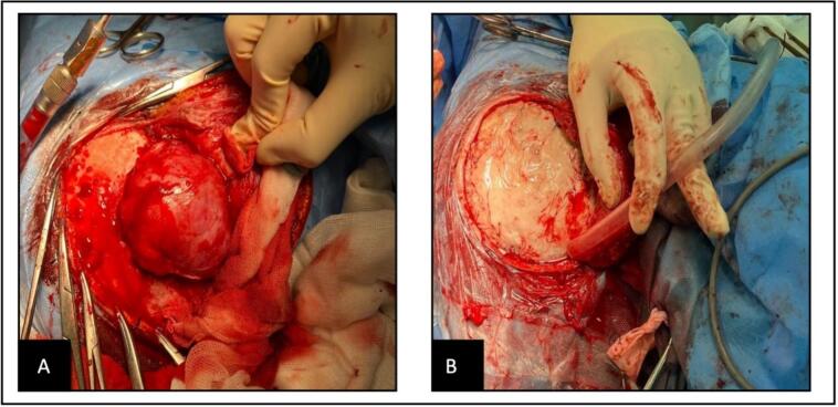
Case presentation: We report a 12-year-old male with a history of medulloblastoma treated with chemoradiotherapy four years ago, who presented with a progressive right temporo-occipital scalp mass. Imaging showed an extradural mass with calvarial bone erosion. Complete surgical excision was performed, and histopathology confirmed CF. No recurrence was observed at six-month follow-up.
Clinical discussion: Post-radiation CF is an extremely rare complication of radiotherapy. Due to its rapid growth, bony invasion, and occasional intracranial extension, it can be misdiagnosed as a radiation-induced neoplasm. Given the overlap in clinical and radiologic features with malignancies such as meningioma or sarcoma, histopathological confirmation is essential. Unlike neoplasms, CF follows a benign course, and complete surgical excision is often curative.
Conclusion: Post-radiation CF should be considered in children with prior radiotherapy presenting with scalp masses. Early diagnosis and surgical intervention are crucial for avoiding unnecessary treatments and ensuring favorable outcomes.
Keywords: Case report; Child; Cranioplasty; Fasciitis; Radiotherapy; Scalp.
Copyright © 2025 The Authors. Published by Elsevier Ltd.. All rights reserved.
Conflict of interest statement
Declaration of competing interest All authors declare that they have no conflicts of interest.
Figures


References
-
- Lecavalier M., Ogilvie L.N., Magee F., Poskitt K.J., Kozak F. Cranial fasciitis: a rare pediatric non-neoplastic lesion with 14-year follow up. Am. J. Otolaryngol. 2014;35:647–650. - PubMed
-
- Wu B., Zhu H., Liu W., Chen L. Occipital diploic cranial fasciitis after radiotherapy for a cerebellar medulloblastoma. J. Neurosurg. Pediatr. 2013;12:637–641. - PubMed
Publication types
LinkOut - more resources
Full Text Sources
