. 2025 Jul 17;16(1):6592.
doi: 10.1038/s41467-025-61541-3.

β-Hydroxybutyrate promotes cancer metastasis through β-hydroxybutyrylation-dependent stabilization of Snail

Affiliations

β-Hydroxybutyrate promotes cancer metastasis through β-hydroxybutyrylation-dependent stabilization of Snail

Wenna Jiang et al. Nat Commun. .

Abstract

β-Hydroxybutyrylation (Kbhb) modification regulates protein molecular fates in either physiology or pathology, including cancer. However, the function and regulatory mechanism of Kbhb remain completely unknown in cancer metastasis. Here, we report that β-hydroxybutyrate (BHB) is clinically associated with the progression of pancreatic cancer and functionally promotes pancreatic cancer cell metastasis. Mechanistically, BHB induces Kbhb modification of Snail at lysine 152 to enhance Snail stabilization, which is regulated by Kbhb modification enzyme CREB-binding protein (CBP), and subsequently prevents Snail degradation by blocking recognition of E3 ubiquitin ligases FBXL14. Furthermore, either targeting Snail Kbhb modification or CBP inhibitor decreases cancer metastasis and enhances the therapeutic efficacy of gemcitabine in pancreatic cancer cells. Collectively, our study reveals that Kbhb of Snail is critical to promote metastasis and provides a potential therapeutic strategy.


Conflict of interest statement

Competing interests: The authors declare no competing interests.

Figures
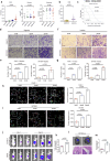
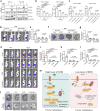
Fig. 1
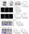
Fig. 1. BHB functions as an oncogenic metabolite in PDAC.
a Quantification of β-hydroxybutyric acid (BHB) (left), glucose (middle) and glutamine (right) level by 1H-NMR in sera from PDAC patients with various pathologic stages (n = 68, Kruskal–Wallis H Test). b Quantification of BHB in the groups of PDAC patients with (IV) or without (I-III) distant metastasis (n = 68, Mann–Whitney U Test). c Kaplan–Meier curves of OS (left) represent the percentages of surviving PDAC patients with high- and low-BHB (0.13 mmol/L for BHB was used as the cut-off value, P-values were from log-rank tests). dg Representative micrographs (d, f) and quantifications (e, g) of the indicated cells treated with different concentrations of BHB in Transwell assays. h, i Representative dynamic images (left) and quantifications (right) of displacement speed for the indicated cells treated with different concentrations of BHB in Cell Movement assays. jm Representative view images (j, l) and its quantifications (k, m) of bioluminescence for metastasis in the whole body and in liver from BALB/C nude mice treated with or without BHB (Scale bars, 50 µm) (H&E staining, M, metastatic foci; L, normal liver tissue). In (e, g, h, I, k, m), data are shown as the mean ± SD, n  =  3 (e, g, h, I) or 5 (k, m) biologically independent samples. All experimental data (di) were verified in at least three independent experiments. P-values were obtained using unpaired two-tailed Student’s t-test. Source data are provided as a Source Data file.
Fig. 2
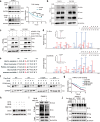
Fig. 2. BHB promotes tumor cell metastasis via Snail.
a Immunoblot analysis of the indicated EMT markers in the indicated cells treated with different concentrations of BHB. b Representative stained tissues of Snail with different concentrations of BHB (left) and BHB content were extracted from PDAC samples according to Snail staining score. (high, 4–9 staining degree, low, 1–3 staining degree, n = 50, Data are shown as the median, unpaired two-tailed Student’s t-test). cj Representative dynamic images (c) and quantifications of displacement speed (d) in scramble or Snail KD cells treated with different concentrations of BHB. Quantifications of Transwell assays in cells described above (e). Representative view images (f, h) and quantifications (g, i, j) of bioluminescence for metastasis in the whole body and in liver, gut and mesentery from xenograft mice implanted with cells described above. In (d, e, g, i, j), data are shown as the mean ± SD, n  =  3 (d, e) or 5 (g, i, j) biologically independent samples. P-values were obtained using unpaired two-tailed Student’s t-test. All experimental data were verified in at least three (a, cg) independent experiments. Source data are provided as a Source Data file.
Fig. 3
Fig. 3. Kbhb protects Snail from ubiquitination-mediated degradation.
a Immunoblot (left) and quantification (right) of Snail in PANC-1 cells treated with or without BHB, followed by CHX treatment for the indicated time. b Immunoprecipitation assays assessing Snail Kbhb modification in the indicated cells treated by BHB. c Immunoprecipitation showing Kbhb modification of cells transfected with the indicated plasmids. d HPLC-MS/MS analysis showing the MS/MS spectrum and the retention time of the in vivo-derived Snail K152 Kbhb peptide (Upper) and the synthetic standard Snail K152 kbhb peptide (Lower). e Conservation analysis of Snail K152 in the indicated species. f, g Immunoblot analysis (f) and quantification (g) of Flag in Flag-tagged-type or mutant Snail in PANC-1 cells treated with or without BHB, followed by CHX treatment for the indicated time. h, i Immunoblot analysis of Snail (h) and ubiquitin of Snail (i) in PANC-1 cells treated with or without BHB, followed by treatment with MG132 for 6 h. j Immunoprecipitation analysis of Flag ubiquitin in Flag-tagged wild-type or mutant Snail in PANC-1 cells treated with or without BHB, followed by MG132 treatment for 6 h and transfected with HA-Ub. In (a), data were shown as the mean ± SD, n  =  3 biologically independent samples (P-values using Two-way ANOVA). All experimental data were verified in (ac, f, h, i) in at least three independent experiments. Source data are provided as a Source Data file.
Fig. 4
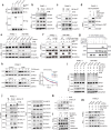
Fig. 4. CBP mediates Snail Kbhb and impairs Snail ubiquitination.
a Immunoprecipitation showing the effects of the indicated enzymes on Snail Kbhb modification in 293T cells. b, c Co-IP assays detecting the association between Snail and CBP in PANC-1 cells with or without BHB treatment. d GST pulldown assay showing the direct interaction between Snail and CBP in PANC-1 cells. e Immunoprecipitation analysis of the effect of CBP KD on Snail Kbhb in the indicated cells. f Immunoprecipitation analysis of Snail Kbhb in Myc-tagged wild-type or mutant Snail in the indicated cells transfected with vector or Flag-tagged CBP. g Purified Snail WT or K152R was incubated with or without CBP or β-hydroxybutyrate-CoA (BHB-CoA). Snail Kbhb was determined by the in vitro Kbhb assay using anti-Kbhb, anti-Snail or anti-CBP antibodies. h Immunoblot analysis (left) and quantification (right) of Snail in PANC-1 cells transfected with the indicated plasmids, followed by CHX treatment for the indicated time. i Immunoprecipitation analysis of the effect of CBP KD on Snail ubiquitin in PANC-1 cells transfected with the indicated plasmids. j Co-IP analysis of the interaction between Snail and the indicated enzymes in PANC-1 cells treated with or without BHB. k Immunoblot assay of Snail in control or shFBXL14 PANC-1 cells treated with or without BHB. l Co-IP analysis of the interaction between FBXL14 and wild-type or mutant Snail in PANC-1 cells transfected with HA-tagged FBXL14 and treated with or without BHB. m Immunoprecipitation showing the effect of CBP KD on the interaction between Snail and FBXL14 in PANC-1 cells transfected with the indicated plasmids. All experimental data were verified in at least three independent experiments. Source data are provided as a Source Data file.
Fig. 5
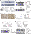
Fig. 5. Snail Kbhb promotes PDAC metastasis in a CBP-dependent manner.
ac Representative images (upper) and quantification (lower right) of Transwell assays assessing the indicated PANC-1 cells transfected with indicated plasmids. Immunoblot analysis for the indicated proteins for the abovementioned cells (lower left) (a). Representative images (left) and quantification (right) of the metastasis in the whole body (b) and in the liver (c) from BALB/C nude mice implanted with SW1990 cells transfected with firefly luciferase. d Representative section tissue images (left) and Spearman correlation analysis (right) of IHC staining for CBP and Snail from serial tissue sections (n = 50). e BHB content was extracted from PDAC samples according to CBP staining score (high, 4–9 staining degree, low, 1–3 staining degree, n = 50, Data are shown as the median, unpaired two-tailed Student’s t-test). fk Quantifications of Transwell assay (f, g) assessing CBP KD cells or control cells transfected with indicated plasmids. Representative BLI images (h, j) and quantification (i, k) of the abovementioned cells on tumor metastasis in the whole body, liver and gut from BALB/C nude mice. (Scale bars in the images of IHC staining, 100 µm). In (ac, f, g, i, k), data are shown as the mean ± SD, n  =  3 (a, f, g) or 5 (b, c, i, k) biologically independent samples. P-values were obtained using unpaired two-tailed Student’s t-test. All experimental data were verified in at least three (a, f, g) independent experiments. Source data are provided as a Source Data file.
Fig. 6
Fig. 6. Targeting Snail Kbhb enhances the efficacy of Gemcitabine for the treatment of PDAC.
a Immunoprecipitation showing the Snail Kbhb modification and the interaction between CBP and Snail in the indicated cells. b Quantifications of Transwell assays in indicated PANC-1 cells treated with different concentrations of A-485. c Quantifications of Transwell assay assessing A-485 treated in the cells transfected with indicated plasmids. dg Representative BLI images (d, f) and quantification (e, g) of the abovementioned cells on metastasis in the whole body and in the liver from xenograft mice treated with A-485. hl Representative BLI images (h, j) and quantification (i, k, l) of the A-485, GEM monotherapy or the combination on metastasis in the whole body and in the liver, gut and mesentery from xenograft mice. m Proposed model for the BHB/CBP/Snail Kbhb axis regulates cancer cell metastasis. In (b, c, e, g, I, k, i), data are shown as the mean ± SD, n  =  3 (b, c) or 5 (e, g, I, k, i) biologically independent samples. P-values were obtained using unpaired two-tailed Student’s t-test. All experimental data were verified in at least three (b, c) independent experiments. Source data are provided as a Source Data file.

References

    1. Li, J. et al. SOX9 is a critical regulator of TSPAN8-mediated metastasis in pancreatic cancer. Oncogene40, 4884–4893 (2021). - PMC - PubMed
    1. Vincent, A., Herman, J., Schulick, R., Hruban, R. H. & Goggins, M. Pancreatic cancer. Lancet378, 607–620 (2011). - PMC - PubMed
    1. Laffel, L. Ketone bodies: a review of physiology, pathophysiology and application of monitoring to diabetes. Diabetes Metab. Res. Rev.15, 412–426 (1999). - PubMed
    1. Cahill, G. F. Jr. Fuel metabolism in starvation. Annu. Rev. Nutr.26, 1–22 (2006). - PubMed
    1. Dmitrieva-Posocco, O. et al. β-Hydroxybutyrate suppresses colorectal cancer. Nature605, 160–165 (2022). - PMC - PubMed

MeSH terms