. 2025 Jul 17;15(1):25943.
doi: 10.1038/s41598-025-10854-w.

Modulating depressive-like behaviors, memory impairment, and oxidative stress in chronic stress rat model using visible light therapy

Affiliations

Modulating depressive-like behaviors, memory impairment, and oxidative stress in chronic stress rat model using visible light therapy

Elham Zahedi et al. Sci Rep. .

Abstract

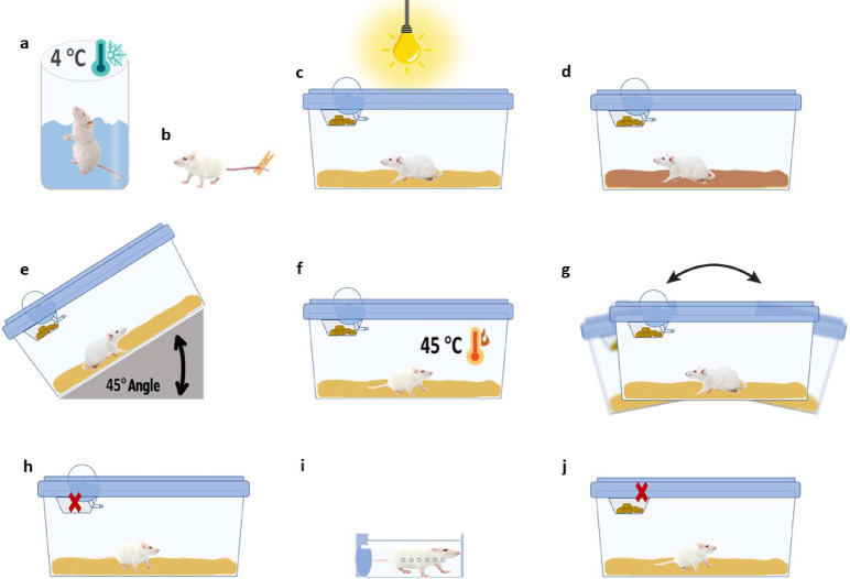
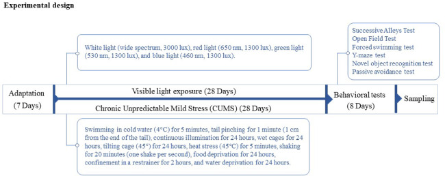
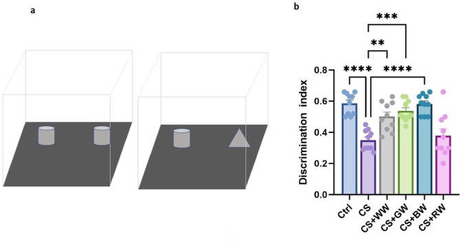
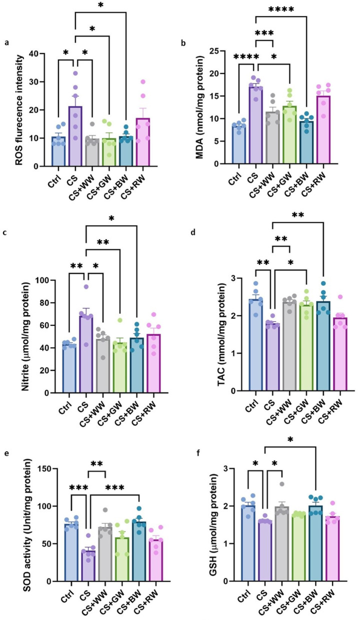
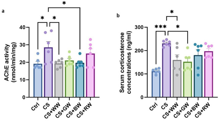
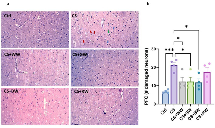
Chronic stress has been linked to significant cognitive impairments, which are exacerbated by oxidative stress and neuronal damage in brain regions such as the hippocampus and prefrontal cortex. This study investigates the therapeutic effects of different visible light wavelengths (white, red, green, and blue) on cognitive dysfunction and oxidative stress in a chronic unpredictable mild stress (CUMS)-induced rat model using stressors such as cold-water swimming, tail pinching, food and water deprivation, cage tilting, shaking, continuous illumination, wet cages, heat stress, and restraint. Sixty male Wistar rats were divided into control, CUMS, and light-exposure groups. The light-exposed groups received daily exposure to white (3000 lx), red (650 nm, 1300 lx), green (530 nm, 1300 lx), or blue (460 nm, 1300 lx) light for four hours over four weeks. Behavioral assessments were conducted to evaluate memory and depression-like behaviors. Biochemical analyses were performed to measure oxidative stress markers, acetylcholinesterase (AChE) activity, and serum corticosterone levels. Results showed that chronic stress significantly impaired cognitive function, increased oxidative stress, elevated AChE activity, and serum corticosterone level. Red light exposure had no significant effect on any of the evaluated parameters. However, Light exposure, particularly blue light, mitigated these effects by reducing reactive oxygen species (ROS), malondialdehyde (MDA), and nitrite levels, while enhancing total antioxidant capacity (TAC), superoxide dismutase (SOD) activity, and glutathione (GSH). Behavioral outcomes also improved, with increased memory performance and reduced depressive behaviors in light-treated groups. Histological analyses revealed decreased neuronal damage in the hippocampus and prefrontal cortex following light therapy. These findings suggest that visible light exposure, especially blue light, may serve as a promising non-invasive intervention for stress-induced cognitive impairments by reducing oxidative stress and neuronal damage.

Keywords: CUMS; Chronic stress; Cognitive function; Light wavelengths; Oxidative stress.


Conflict of interest statement

Declarations. Competing interests: The authors declare no competing interests.

Figures

Fig. 1
Fig. 1
A summary of the stressors used in the chronic unpredictable mild stress (CUMS) protocol. The figure illustrates the sequence of stress-inducing factors applied to the animals: (a) swimming in cold water (4 °C) for 5 min, (b) tail pinching for 1 min (1 cm from the end of the tail), (c) continuous illumination for 24 h, (d) wet cages for 24 h, (e) tilting the cage (45°) for 24 h, (f) heat stress (45 °C) for 5 min, (g) shaking for 20 min (one shake per second), (h) food deprivation for 24 h, (i) confinement in a restrainer for 2 h, and (j) water deprivation for 24 h.
Fig. 2
Fig. 2
Experimental design. The animals were first acclimatized in the animal facility for one week. Following the acclimatization period, they were subjected to chronic unpredictable mild stress for 28 days to induce the experimental model. Concurrently, the animals were assigned to groups based on exposure to different light wavelengths, with each group receiving a specific wavelength exposure for 28 days. Then, behavioral assessments were performed, followed by sample collection for the analysis of the targeted parameters.
Fig. 3
Fig. 3
Effect of visible light wavelengths on Coat State Score in chronic unpredictable mild stress (CUMS) model. Ctrl: control group, CS: CUMS model group, CS + WW: treated with white wavelength, CS + GW: treated with green wavelength, CS + BW: treated with blue wavelength, and CS + RW: treated with red wavelength. Data are expressed as the mean ± SEM (n = 10). **p < 0.01, ***p < 0.001, ****p < 0.0001.
Fig. 4
Fig. 4
Effect of visible light wavelengths on anxiety behavior in chronic unpredictable mild stress (CUMS) model. (a) This figure illustrated the successive alleys test apparatus. (b) The time spent in each zone of the apparatus as an indicator of anxiety behavior. Ctrl: control group, CS: CUMS model group, CS + WW: treated with white wavelength, CS + GW: treated with green wavelength, CS + BW: treated with blue wavelength, and CS + RW: treated with red wavelength. Data are expressed as the mean ± SEM (n = 10). *p < 0.05, **p < 0.01, ***p < 0.001, ****p < 0.0001.
Fig. 5
Fig. 5
Effect of visible light wavelengths on anxiety and depressive-like behavior in chronic unpredictable mild stress (CUMS) model. (a) This figure illustrated the open field test apparatus. (b) Number of line crossing, (c) number of center zone entry, (d) time spent for grooming, and (e) number of rearing in the open field test. (f) Immobility time in the forced swimming test. Ctrl: control group, CS: CUMS model group, CS + WW: treated with white wavelength, CS + GW: treated with green wavelength, CS + BW: treated with blue wavelength, and CS + RW: treated with red wavelength. Data are expressed as the mean ± SEM (n = 10). *p < 0.05, **p < 0.01, ***p < 0.001, ****p < 0.0001.
Fig. 6
Fig. 6
Effect of visible light wavelengths on short-term memory in chronic unpredictable mild stress (CUMS) model. (a) Schematic representation of the Y-maze apparatus. (b) Percentage of spontaneous alternation in the Y-maze, indicating working memory performance. Ctrl: control group, CS: CUMS model group, CS + WW: treated with white wavelength, CS + GW: treated with green wavelength, CS + BW: treated with blue wavelength, and CS + RW: treated with red wavelength. Data are expressed as the mean ± SEM (n = 10). *p < 0.05.
Fig. 7
Fig. 7
Effect of visible light wavelengths on recognition memory in the Novel Object Recognition Test in chronic unpredictable mild stress (CUMS) model. (a) This figure illustrating the different stages of the novel object recognition test. (b) Discrimination index, showing the time each animal spent exploring the objects during both phases of the novel object recognition test. Ctrl: control group, CS: CUMS model group, CS + WW: treated with white wavelength, CS + GW: treated with green wavelength, CS + BW: treated with blue wavelength, and CS + RW: treated with red wavelength. Data are expressed as the mean ± SEM (n = 10). **p < 0.01, ***p < 0.001, ****p < 0.0001.
Fig. 8
Fig. 8
Effect of light wavelength exposure on latency time in the Passive Avoidance test in chronic unpredictable mild stress (CUMS) model. (a) Schematic of the passive avoidance test setup. (b) Latency time for rats to enter the dark compartment in the passive avoidance test, reflecting long-term memory retention. Ctrl: control group, CS: CUMS model group, CS + WW: treated with white wavelength, CS + GW: treated with green wavelength, CS + BW: treated with blue wavelength, and CS + RW: treated with red wavelength. Data are expressed as the mean ± SEM (n = 10). *p < 0.05.
Fig. 9
Fig. 9
Effect of visible light wavelengths on oxidative stress content in chronic unpredictable mild stress (CUMS) model. (a) Reactive Oxygen Species (ROS), (b) malondialdehyde (MDA), (c) nitrite, (d) total antioxidant capacity (TAC), (e) superoxide dismutase (SOD) activity, and (f) glutathione (GSH). Ctrl: control group, CS: CUMS model group, CS + WW: treated with white wavelength, CS + GW: treated with green wavelength, CS + BW: treated with blue wavelength, and CS + RW: treated with red wavelength. Data are expressed as the mean ± SEM (n = 6). *p < 0.05, **p < 0.01, ***p < 0.001, ****p < 0.0001.
Fig. 10
Fig. 10
Effect of visible light wavelengths on brain acetylcholinesterase (AChE) activity and plasma corticosterone levels in chronic unpredictable mild stress (CUMS) model. (a) AChE activity in the brain and (b) serum corticosterone concentration were measured in the CUMS model. Ctrl: control group, CS: CUMS model group, CS + WW: treated with white wavelength, CS + GW: treated with green wavelength, CS + BW: treated with blue wavelength, and CS + RW: treated with red wavelength. Data are expressed as the mean ± SEM (n = 6). *p < 0.05, ****p < 0.0001.
Fig. 11
Fig. 11
Effect of visible light wavelengths on PFC neurons assessed by Nissl staining. (a) Morphological assessment of neurons in the prefrontal cortex (PFC) of the control group shows a normal distribution of pyramidal cells with no signs of pyknosis. Red arrows indicate pyknotic pyramidal cells, blue arrows point to degenerating pyramidal neurons, and the green arrow highlights vacuolization around a degenerated cell. (b) Quantification of damaged neurons in the PFC across different experimental groups. Ctrl: control group, CS: CUMS model group, CS + WW: treated with white wavelength, CS + GW: treated with green wavelength, CS + BW: treated with blue wavelength, and CS + RW: treated with red wavelength. Data are expressed as the mean ± SEM (n = 4). *p < 0.05, ***p < 0.001.
Fig. 12
Fig. 12
Histological analysis and neuronal quantification in the hippocampus. (a) Representative images of the CA1, CA3, and dentate gyrus (DG) regions of the hippocampus from different experimental groups. (b) Quantification of the number of neurons in the CA1 region. (c) Quantification of the number of neurons in the CA3 region. (d) Quantification of the number of neurons in the DG region. Ctrl: control group, CS: CUMS model group, CS + WW: treated with white wavelength, CS + GW: treated with green wavelength, CS + BW: treated with blue wavelength, and CS + RW: treated with red wavelength. Data are expressed as the mean ± SEM (n = 4). *p < 0.05, ***p < 0.001.

References

    1. Kültz, D. Defining biological stress and stress responses based on principles of physics. J. Exp. Zool. Ecol. Integr. Physiol.333, 350–358. 10.1002/jez.2340 (2020). - PubMed
    1. Rahman, M. M., Callaghan, C. K., Kerskens, C. M., Chattarji, S. & O’Mara, S. M. Early hippocampal volume loss as a marker of eventual memory deficits caused by repeated stress. Sci. Rep.6, 29127. 10.1038/srep29127 (2016). - PMC - PubMed
    1. Alqurashi, G. K. et al. The impact of chronic unpredictable mild Stress-Induced depression on spatial, recognition and reference memory tasks in mice: Behavioral and histological study. Behav. Sci.12, 166 (2022). - PMC - PubMed
    1. Yamamoto, S. et al. Effects of single prolonged stress and D-Cycloserine on contextual fear extinction and hippocampal NMDA receptor expression in a rat model of PTSD. Neuropsychopharmacology33, 2108–2116. 10.1038/sj.npp.1301605 (2008). - PubMed
    1. Prajapati, S. K., Singh, N., Garabadu, D. & Krishnamurthy, S. A novel stress re-stress model: Modification of re-stressor cue induces long-lasting post-traumatic stress disorder-like symptoms in rats. Int. J. Neurosci.130, 941–952. 10.1080/00207454.2019.1711078 (2020). - PubMed

MeSH terms

LinkOut - more resources