. 2025 Aug;17(8):2095-2114.
doi: 10.1038/s44321-025-00266-8. Epub 2025 Jul 17.

LIMA1-alpha staining predicts curative intent surgery response in HPV negative head and neck cancer

Affiliations

LIMA1-alpha staining predicts curative intent surgery response in HPV negative head and neck cancer

Xi Qiao et al. EMBO Mol Med. 2025 Aug.

Abstract

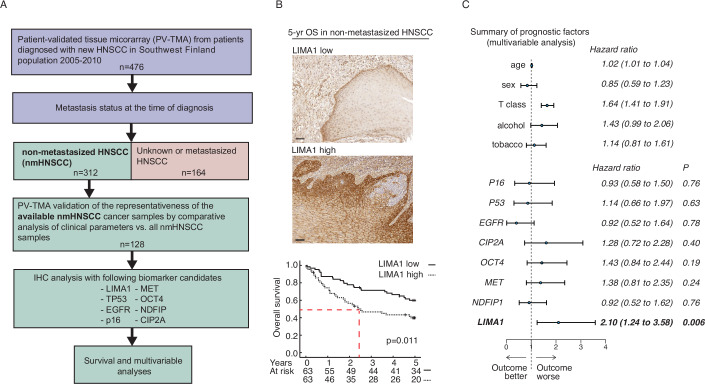
In many solid cancer types, surgery alone could be a sufficient first therapy option for a significant number of cancer patients. However, there are currently no diagnostic solutions to identify patients who could be stratified to surgery alone. To identify a biomarker predicting cancer surgery response, candidate biomarkers were studied in a non-metastatic head and neck squamous cell carcinoma (nmHNSCC) cohort well representative of the HPV-negative patient population. LIMA1 immunohistochemistry (IHC) with specificity-validated antibodies outperformed all other biomarkers in multivariable survival analyses of patients with nmHNSCC (n = 128, HR 2.10, P = 0.006). The prognostic effect was selective to LIMA1-alpha isoform IHC detection in patients who had received surgical therapy (n = 184, HR 2.39, P > 0.001). Strikingly, our real-world validation results, using two prospectively collected cohorts (n = 15 and n = 86), demonstrate that none of the LIMA1 negative patients died of HNSCC during the follow-up. Collectively, we report here the discovery of a diagnostic LIMA1-alpha IHC assay for HPV-negative HNSCC patient stratification to surgery-only therapy. Application of LIMA1 detection in routine nmHNSCC diagnostics would revolutionize the clinical management of HNSCC patients.

Keywords: Disease-Specific Survival; EPLIN; PV-TMA; Population-validated Tissue Microarray.


Conflict of interest statement

Disclosure and competing interests statement. The University of Turku has filed a patent covering the diagnostic use of LIMA1 detection on behalf of SV and JW. SV and JW are co-founders and have an ownership interest in Thestra Ltd., developing LIMA1 IHC diagnostics for clinical use.

Figures

Figure 1
Figure 1. Low LIMA1 expression in surgical nmHNSCC sample is an independent biomarker for favorable prognosis.
(A) Description of the clinical cohort that was used in the formation of an unbiased and population-validated HNSCC tissue microarray (PV-TMA) and for identification of non-metastasized (nmHNSCC) HNSCC patients (n = 128). Patient samples were immunostained with LIMA1-specific antibody. The same PV-TMA was stained additionally for TP53, EGFR, p16, CIP2A, MET, and OCT4 (Appendix Figs. S1 and S2). (B) Representative immunohistochemical stains of LIMA1 LOW and LIMA1 HIGH HNSCC samples. High LIMA1 expression strongly associated with poor 5-year overall survival in the nmHNSCC PV-TMA patient cohort. Log-rank method was used for determining the significance of the difference for survival distributions. Scale bar: 100 µm. The exact P values were as indicated. (C) Multivariable clinical prognostic model including age, high T class, and alcohol consumption (n = 312). The test demonstrated strong survival impact for LIMA1 immunohistochemistry that is independent of any other analyzed factors.
Figure 2
Figure 2. LIMA1-alpha IHC detection identifies HNCSS patients that will have favorable response to curative intent surgery.
(A) A prospective 2-year follow-up study of 15 randomly selected patients diagnosed with new HNSCC and underwent with curative intended surgery (HNSCC cohort 3). LIMA1 immunohistochemical (IHC) staining and western blot analysis (both with sc-136399) were performed on the carcinoma samples (ca). Moreover, LIMA1 western blot analyses were performed from a fresh tissue sample taken outside the tumor area (Norm). A traffic light model that considers HNSCC patient survival as well as HNSCC cancer metastasis was created as follows: patients who were alive 2 years after surgery received a green light, patients who had regional lymph node metastases, received a yellow light, patients with distant metastases received a red light and the patients who died during the follow-up were marked with a black. (B) LIMA1 positivity was significantly associated with the occurrence of nodal metastasis both at presentation and during follow-up. Two-sided Fisher’s exact test was used for the statistical analysis. The exact P values were as indicated. (C) LIMA1 positivity was remarkably associated with poor survival during follow-up. (D) Shown are representative H&E, LIMA1-alpha (sc-136399), LIMA1-beta (RB581) Low and High IHC stainings. Overall survival (OAS), disease-specific survival (DSS) and disease-free survival (DFS) were analyzed from the oral cavity HNSCC cohort 4 (n = 185; Table 2). Exact P values were as indicated. Source data are available online for this figure.
Figure 3
Figure 3. A real-world demonstration of performance of LIMA1 as a predictive HNSCC biomarker.
(A) Flowchart representation of the Finnish multicenter prospective HNSCC study protocol. Altogether, 95 patients with new HNSCC and curative intended surgery were recruited to the study. LIMA1 IHC staining was performed with two isoform-specific antibodies. Two-tier interpretation was used in the analysis of staining intensities. (B, C) Representative LIMA1 IHC stains. (D) Disease-specific survival data (DSS) with 2-tire LIMA1 staining results with sc-136399 antibody. (E, F) Representative LIMA1 IHC stainings (with RB581 antibody) and disease-specific survival (DSS) data with LIMA1-beta-specific antibody. All scale bars indicated are 100 µm.
Figure 4
Figure 4. LIMA1 promotes HNSCC in vivo invasion, metastasis, and EMT.
(A) Western blot analysis of UT-SCC-14 cells for LIMA1 silencing after siRNA transfection. (B) Measurement of primary tumor size in zebrafish embryo xenograft experiment with UT-SCC-14 cells. siCTRL, n = 8 embryos; siLIMA1, n = 9 embryos. (C) Quantification of the number of invading cells in the zebrafish embryo xenograft experiment with UT-SCC-14 cells. siCTRL, n = 8 embryos; siLIMA1, n = 9 embryos. (D) Western blot analysis of HNSCC16# cells for LIMA1 silencing after siRNA transfection. (E) Measurement of primary tumor size in the zebrafish embryo xenograft experiment with HNSCC16# cells. siCTRL, n = 11 embryos; siLIMA1, n = 15 embryos. (F) Quantification of the number of invading cells in the zebrafish embryo xenograft experiment with HNSCC16# cells. siCTRL, n = 11 embryos; siLIMA1, n = 15 embryos. (G) Western blot analysis of HNSCC17# cells for LIMA1 overexpression after doxycycline induction. (H) Measurement of primary tumor size in the zebrafish embryo xenograft experiment with HNSCC17# cells. Fluorescent-labeled HNSCC17# cells cultured in the presence or absence of doxycycline were transplanted into zebrafish embryos and embryos cultures with doxycycline to induce LIMA1 expression (DOX + ) or without doxycycline for uninduced controls (DOX + ). DOX−, n = 40 embryos; DOX + , n = 16 embryos. (I) Quantification of the number of invading cells in the zebrafish embryo xenograft experiment with HNSCC17# cells with or without doxycycline induction of LIMA1 overexpression. DOX−, n = 40 embryos; DOX + , n = 16 embryos. Non-parametric Mann–Whitney test was used for the statistical analysis of zebrafish experiments. (J) HNSCC16# cells were transfected with scrambled (siSCR) or LIMA1 (siLIMA1) siRNA and analyzed 72 h post-transfection. (K) The relative expression of vimentin represents vimentin level normalized to loading control GAPDH (n = 5). Unpaired two-tailed t test with Welch’s correction was used for statistical analysis. The exact P values were as indicated. (L) Western blot analysis of LIMA1 and vimentin protein levels in response to doxycycline-induced LIMA1 overexpression in HNSCC17# cells. Cells were treated with 1 µg/ml doxycycline for 2 weeks. Source data are available online for this figure.
Figure EV1
Figure EV1. Validation of LIMA1 antibodies.
(A) Schematic presentation of LIMA1-alpha and -beta isoforms and the antigen regions used for raising the LIMA1 antibodies used in this study. (B) Western blot characterization of LIMA1 expression (HPA023871 antibody) in 14 different HNSCC cancer cell lines and (C) in 13 triple-negative breast cancer cell lines. (D) Western full blot analysis of LIMA1 antibody (SC-136399) specificity after LIMA1 silencing with six different LIMA1 siRNA transfections. (E) Western full blot characterization of custom-made polyclonal LIMA1-beta specific antibody (RB581). UT-SCC-14 and -72 are highly LIMA1-alfa-positive HNSCC cell lines. HN09 HNSCC cancer cell line containing low LIMA1-beta expression were transduced either with LIMA1-alpha (a-induced) or LIMA1-beta (b-induced) lentiviral Tet-inducible gene expression vector. LIMA1 expression and antibody specificity upon Doxycycline treatment were ensured by western blot with LIMA1-beta specific antibody (RB581). (F, G) Western blot and immunofluorescence analyzes of HPA023871 and SC-136399 antibodies after LIMA1 siRNA silencing of patient-derived HNSCC cell lines. (H) Specificity in immunofluorescence of LIMA1-beta specific antibody (RB581) was ensured by conducting RB581 IF staining in HNSCC cell lines containing both LIMA1-alpha and -beta expression (MISB10) and with the cell line lacking LIMA1-beta expression (UT-SCC-14). Scale bars indicated were 10 µm.
Figure EV2
Figure EV2. Prognostic role for LIMA1 in TCGA HNSCC and pancreatic cancer data.
(A, B) The prognostic effect of LIMA1 was confirmed in TCGA HNSCC (P < 0.001) and operatively treated pancreatic cancer (P = 0.009) datasets.
Figure EV3
Figure EV3. Validation of LIMA1 protein inhibition by siRNA.
Depletion of LIMA1 in three different patient-derived HNSCC cell lines (UT-SCC14, UT-SCC60B and UT-SCC45).
Figure EV4
Figure EV4. LIMA1 promotes epithelial mesenchymal transition.
(A) Gene set enrichment analysis (GSEA) for genes co-expressing with LIMA1 in TCGA HNSCC data set. The normalized enrichment scores (NES) for HALLMARK gene sets with FDR < 0.05 are shown. (B) Enrichment plot for HALLMARK EMT gene set (P < 0.0001) from GSEA analysis. Default statistics of GSEA was used (Subramanian et al, 2005).

References

    1. Bozec A, Culié D, Poissonnet G, Dassonville O (2019) Current role of primary surgical treatment in patients with head and neck squamous cell carcinoma. Curr Opin Oncol 31:138–145 - PubMed
    1. Burtness B, Harrington KJ, Greil R, Soulières D, Tahara M, de Castro G, Psyrri A, Basté N, Neupane P, Bratland Å (2019) Pembrolizumab alone or with chemotherapy versus cetuximab with chemotherapy for recurrent or metastatic squamous cell carcinoma of the head and neck (KEYNOTE-048): a randomised, open-label, phase 3 study. Lancet 394:1915–1928 - PubMed
    1. Chow LQM (2020) Head and neck cancer. N Engl J Med 382:60–72 - PubMed
    1. Collins RJ, Jiang WG, Hargest R, Mason MD, Sanders AJ (2015) EPLIN: a fundamental actin regulator in cancer metastasis? Cancer Metastasis Rev 34:753–764 - PMC - PubMed
    1. Gillison ML, Trotti AM, Harris J, Eisbruch A, Harari PM, Adelstein DJ, Jordan RCK, Zhao W, Sturgis EM, Burtness B (2019) Radiotherapy plus cetuximab or cisplatin in human papillomavirus-positive oropharyngeal cancer (NRG Oncology RTOG 1016): a randomised, multicentre, non-inferiority trial. Lancet 393:40–50 - PMC - PubMed

MeSH terms

Substances

LinkOut - more resources