

Development of a multispectral imaging apparatus for cost-effective fundus disease detection
- PMID: 40677385
- PMCID: PMC12265482
- DOI: 10.1364/BOE.563950
Development of a multispectral imaging apparatus for cost-effective fundus disease detection
Abstract
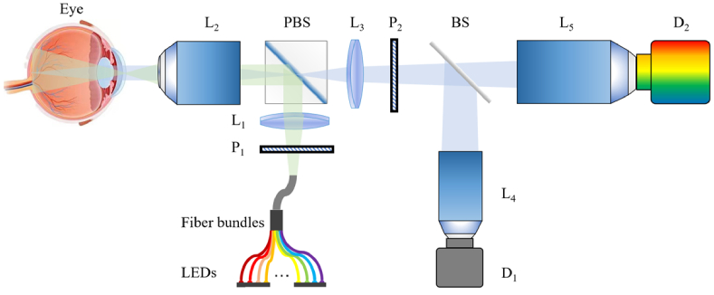
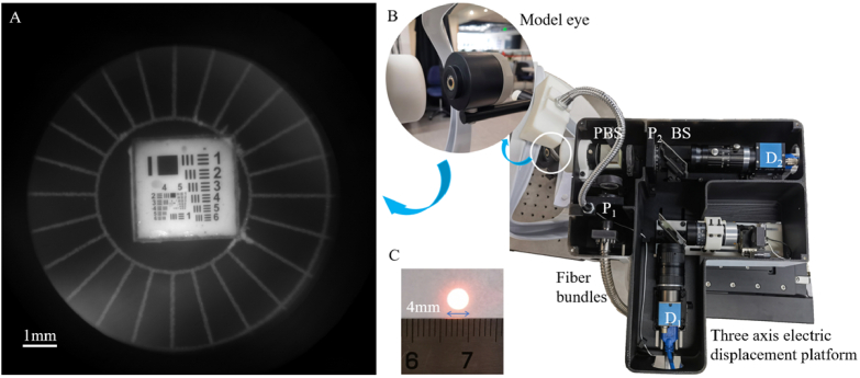
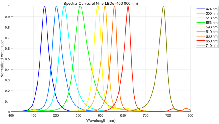
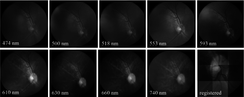
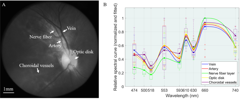
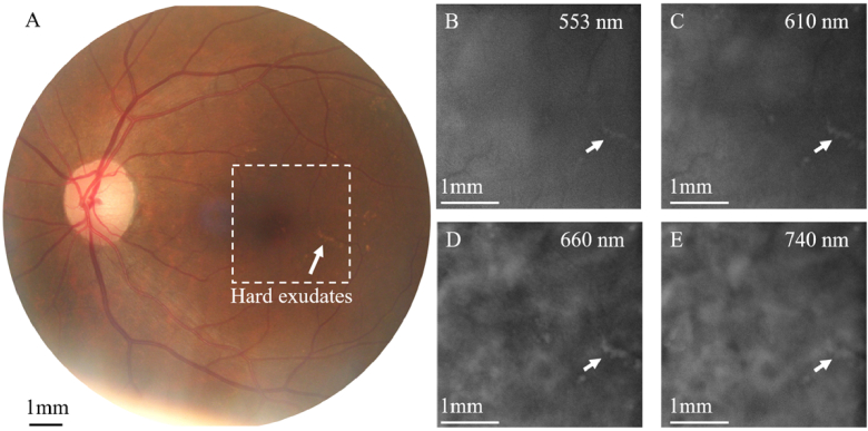
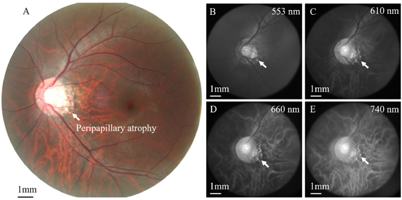
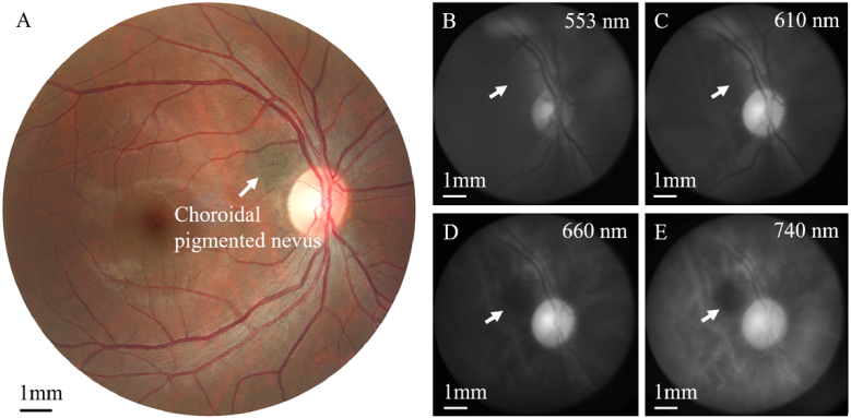
Fundus spectral imaging (FSI) integrates fundus photography with spectral techniques, providing both spatial and spectral information for retinal imaging. Whereas existing FSI systems have demonstrated advantages in structural and functional imaging, their widespread adoption is hindered by high costs and complex optical designs. To address these challenges, we propose a low-cost multispectral fundus camera with a simplified optical design, built from off-the-shelf optics, 3D-printed parts, and equipped with fiber-bundle-coupled multi-wavelength LED illumination source (470-740 nm). Additionally, the proposed multispectral imaging apparatus incorporates a coaxial non-separated polarization-based reflection suppression technique, using orthogonal polarizers to suppress corneal reflections without pupil-plane separation. To the best of our knowledge, this is the first application of such an architecture in the context of FSI. Experimental results demonstrate that the developed system achieves high-quality FSI under low-cost conditions, validating its feasibility as a practical solution. Clinical validation validates its diagnostic capability for diabetic retinopathy, choroidal pigmented nevus, and, notably, the first reported spectral imaging of peripapillary atrophy. The system achieves performance comparable to conventional color fundus photography while enabling superior diagnosis of deep fundus conditions such as choroidal lesions, offering a cost-effective and practical FSI solution for broader deployment in resource-limited settings.
© 2025 Optica Publishing Group.
Conflict of interest statement
The authors declare no conflicts of interest.
Figures
Similar articles
-
Artificial intelligence for diagnosing exudative age-related macular degeneration.Cochrane Database Syst Rev. 2024 Oct 17;10(10):CD015522. doi: 10.1002/14651858.CD015522.pub2. Cochrane Database Syst Rev. 2024. PMID: 39417312
-
Short-Term Memory Impairment.2024 Jun 8. In: StatPearls [Internet]. Treasure Island (FL): StatPearls Publishing; 2025 Jan–. 2024 Jun 8. In: StatPearls [Internet]. Treasure Island (FL): StatPearls Publishing; 2025 Jan–. PMID: 31424720 Free Books & Documents.
-
Optical coherence tomography (OCT) for detection of macular oedema in patients with diabetic retinopathy.Cochrane Database Syst Rev. 2011 Jul 6;(7):CD008081. doi: 10.1002/14651858.CD008081.pub2. Cochrane Database Syst Rev. 2011. Update in: Cochrane Database Syst Rev. 2015 Jan 07;1:CD008081. doi: 10.1002/14651858.CD008081.pub3. PMID: 21735421 Updated.
-
Signs and symptoms to determine if a patient presenting in primary care or hospital outpatient settings has COVID-19.Cochrane Database Syst Rev. 2022 May 20;5(5):CD013665. doi: 10.1002/14651858.CD013665.pub3. Cochrane Database Syst Rev. 2022. PMID: 35593186 Free PMC article.
-
Optical coherence tomography (OCT) for detection of macular oedema in patients with diabetic retinopathy.Cochrane Database Syst Rev. 2015 Jan 7;1(1):CD008081. doi: 10.1002/14651858.CD008081.pub3. Cochrane Database Syst Rev. 2015. PMID: 25564068 Free PMC article.
References
-
- Banu K. S., Lerma M., Ahmed S. U., et al. , “Hyperspectral microscopy-applications of hyperspectral imaging techniques in different fields of science: a review of recent advances,” Appl. Spectrosc. Rev. 59(7), 935–958 (2024). 10.1080/05704928.2023.2270035 - DOI
LinkOut - more resources
Full Text Sources