. 2025 Jun 20:33:102006.
doi: 10.1016/j.mtbio.2025.102006. eCollection 2025 Aug.

Porous granular hydrogel scaffolds biofabricated from dual-crosslinked hydrogel microparticles for breast tissue engineering

Affiliations

Porous granular hydrogel scaffolds biofabricated from dual-crosslinked hydrogel microparticles for breast tissue engineering

Yaqi Guo et al. Mater Today Bio. .

Abstract

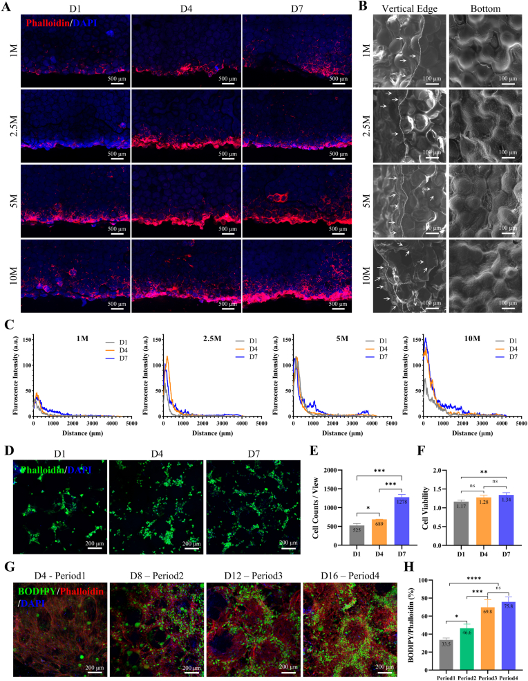
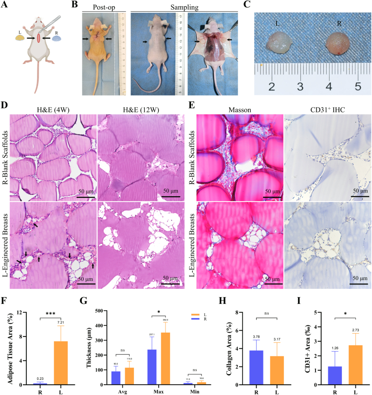
Hydrogel scaffolds play a crucial role in tissue engineering; however, traditional bulk hydrogel scaffolds (BHS) often suffer from insufficiently sized pores (nanoscales), impeding cellular infiltration, development, and expansion. This limitation affects oxygen and nutrient exchange efficiency, in which case it relies extensively on liquid permeation and bulk hydrogels swelling. In contrast, hydrogel microparticles (HMPs) have proven to be both printable and injectable, allowing the development of modular thick constructs with interconnected pores. This study introduces a novel method of fabricating porous granular hydrogel scaffolds (GHS) by printing thermo-crosslinked gelatin methacryloyl (GelMA) HMPs granular hydrogels before chemical crosslinking (dual-crosslinking). The scaffolds exhibit an average pore fraction ranging from 14 % to 23 % and an average pore size varying from 4923 μm2 to 8185 μm2 (with equivalent circular diameter of 80-102 μm). In vitro experiments demonstrated the effective infiltration, adhesion, proliferation, and adipogenic differentiation of human adipose-derived stem cells (hADSCs) within the scaffold pores. Additionally, in vivo observations confirmed the presence of differentiated adipose cells within the central pores after 4 weeks. These results collectively suggest the proposed microspheres printing technique holds significant promise for fabricating microporous scaffolds and further applications in tissue engineering.

Keywords: Breast tissue engineering; Extrusion printing; Granular hydrogel scaffolds; Hydrogel microparticles; hADSCs.


Conflict of interest statement

The authors declare that they have no known competing financial interests or personal relationships that could have appeared to influence the work reported in this paper.

Figures

Image 1
Graphical abstract
Fig. 1
Fig. 1
Schematic representation of preparation of GelMA hydrogel microparticles (HMPs) and GelMA granular hydrogel scaffolds (GHS) through extrusion printing. Human adipose-derived stem cells (hADSCs) were isolated and expanded from human adipose tissue and then seeded on GelMA GHS to generate tissue-engineered breasts, which were then implanted subcutaneously in nude mice for in vivo adipose regeneration experiments.
Fig. 2
Fig. 2
Preparation and Characteristics of GelMA Granular Hydrogel. (A) Coaxial needle used to generate GelMA HMPs. (B) Schematic procedure of preparing GelMA granular hydrogels, including collection on an ice plate, (i-iv) washing with PBS and (v-vi) jamming with a vacuum filter. (C) Diameter of GelMA HMPs with different flow rate ratio and inner channel diameter of coaxial needle. (D) Frequency distribution histogram of GelMA-HMPs diameter at flow rate ratio of 6:60 (Small), 6:24 (Medium) and 6:10 (Large). (E–G) Rheological characteristics of GelMA granular hydrogels with GelMA precursor concentration of 5 % w/v (green), 7.5 % w/v (orange) and 10 % w/v (blue). (E) Shear moduli (G′, filled symbols) and loss moduli (G″, open symbols) of GelMA granular hydrogels as a function of oscillation strain γ (0.1 %–1000 %). (F) Shear viscosity with increasing shear rate (0.1–100 s−1) demonstrates the shear-thinning property of GelMA granular hydrogels. (G) Evaluation of self-recovery property of 7.5 % w/v GelMA granular hydrogels under alternating shear strain (1 % and 200 %). (H) GelMA HMPs (i) in paraffin oil, (ii) in PBS, (iii) after jamming and (iv) after printing and (I) the diameter of GelMA HMPs. Bar = 500 μm, n ≥ 50,∗p < 0.01,∗∗∗∗p < 0.00001.
Fig. 3
Fig. 3
Construction of Scaffold by Microparticles Printing. (A) A schematic graph of microparticles printing process, including GelMA granular hydrogels being extruded onto the plate under room temperature and then UV crosslinked at 365 nm. (B) GelMA granular hydrogels extruded through the nozzle. (C) Representative image of a semi-spherical scaffold with a diameter of 10 mm constructed by microparticles printing. (D) Images of the letters “WHUH” printed and UV crosslinked and picking up the letter “H”. (E) Images of GelMA granular hydrogels before and after extrusion under a range of nozzle temperature (8, 12, 16, 20 and 24 °C) using an 18 G nozzle and their maximum extrusion length before a fracture. (F) 3D models and representative images of printed scaffolds with 4–10 slice layers using 7.5 % w/v GelMA granular hydrogel and their actual height. (G) Images of printed scaffolds with a range of extrusion speed (1.00, 1.25, 1.50, 1.75 and 2.00 mm3/s) and their Feret's diameter. n ≥ 3,ns = none sense, ∗p < 0.1, ∗∗p < 0.01, ∗∗∗∗p < 0.0001.
Fig. 4
Fig. 4
Characteristics of Printed Scaffold. (A) Images of a printed scaffold (i) before clamping, (ii) after clamping, (iii) before pressing and (iv) after pressing. (B) Photographs of the 7.5 % w/v GelMA GHS under compressing test to measure the Young's moduli. (C) Young's moduli (kPa) of 7.5 % w/v GelMA bulk hydrogel scaffolds (blue) and GelMA HMPs scaffolds (orange) before (dash column) and after (solid column) swelling in PBS for 24 h n ≥ 3. (D) Stereo microscope images and SEM images of freeze-dried printed scaffolds, (E)laser microscope images, height distribution heatmap and 3D reconstruction heatmap of printed scaffolds using 7.5 % w/v GelMA granular hydrogel with the microparticles diameter at 200, 300 and 400 nm. (F) Permeability test by adding 100 μL red dye solution and the infiltration depth at different timepoint. (G) Solution retention volume test by adding dropwise 10 μL red dye solution until residue liquid was seen on the glass slide. The black arrow shows the residue liquid. (H) 3D confocal projection of printed scaffolds using 7.5 % w/v GelMA granular hydrogel with the microparticles diameter at 200, 300 and 400 nm. Pores was images by incubating the scaffolds with 0.2 mg/mL high-molecular weight fluorescein isothiocyanate (FITC)-labeled dextran (70 kDa). Pore fraction and, number of pores and pore size were assessed by detecting the pore spaces in 2D slices using ImageJ. n ≥ 3, ns = none sense, ∗p < 0.1, ∗∗p < 0.01, ∗∗∗p < 0.001 and ∗∗∗∗p < 0.0001.
Fig. 5
Fig. 5
Composition and Culturation of Tissue Engineered Breast in vitro. (A) Cross-sectional views showing the cell distribution and infiltration depth phalloidin of the tissue engineered breast by seeding cells onto the printed scaffolds undersurface with a cell suspension of 1, 2.5, 5 and 10 million/mL after 1, 4 and 7 days. (B) SEM images showing the vertical edge and undersurface of the tissue engineered breast on day 7. White dashed lines represent the edges of the longitudinal section with the bottom surface. White arrows indicated the cells. (C) Fluoresce intensity of phalloidin as a function of the distance to the undersurface. (D) After co-incubation for 1, 4 and 7 days, the number of cells per view(E) and CCK-8 assay (F) showed the proliferation activity of cells on scaffolds. (G–H) Cell differentiation test showing the relative area of BODIPY to phalloidin at the end of period 1, 2, 3 and 4. n ≥ 3, ns = none sense, ∗p < 0.1, ∗∗p < 0.01, ∗∗∗p < 0.001 and ∗∗∗∗p < 0.0001.
Fig. 6
Fig. 6
In vivo Study. (A) A schematic of implantation and a representative photo after implantation. A small incision of 20 mm was made on the dorsal subcutaneous pockets of 8 weeks female BALB/c Nude Crlj mice. Tissue engineered breast was implanted into the left pocket while the blank printed scaffold was implanted into the right. (B) The representative photographs of retrieving the implants after 4 weeks. The black arrows showed the implants. (C) The photograph of implants after 4 weeks. (D) H&E staining of blank printed scaffolds (R) and tissue engineered breast (L) after 4 weeks and 12 weeks in vivo. The black arrows showed the neovascularization with red blood cells inside. (E) Images of Masson's trichrome staining and immunohistochemistry staining of blank printed scaffolds (R) and tissue engineered breast (L) after 4 weeks in vivo. (F) The relative adipose tissue area (%) by assessing the vacuole area to the total tissue area in scaffold pores of H&E staining images in Image Pro Plus. (G) The average (Avg), maximum (Max) and minimum (Min) thickness of fibrous capsule of implants by assessing the vertical section images of H&E staining in Image Pro Plus. (H) The relative collagen area (%) by assessing the blue stained area to the total tissue area in scaffold pores of Masson's trichrome staining images in Image Pro Plus. (I) The relative CD31+ cells area (‰) by assessing the positive stained area to the total tissue area in scaffold pores of immunohistochemistry staining images in Image Pro Plus.
Fig. 7
Fig. 7
Immunofluorescence Staining. IF analysis for (A) BODIPY/CD31/DAPI and (B) CD86/CD206/DAPI staining of blank printed scaffolds (R) and tissue engineered breast (L) after 4 weeks of implantation. BODIPY for lipids, CD31 for vascular endothelial cells, CD86 for M1 macrophages and CD206 for M2 macrophages.

Similar articles

References

    1. Sung H., Ferlay J., Siegel R.L., Laversanne M., Soerjomataram I., Jemal A., Bray F. Global cancer statistics 2020: GLOBOCAN estimates of incidence and mortality worldwide for 36 cancers in 185 countries. CA Cancer J. Clin. 2021;71:209–249. doi: 10.3322/caac.21660. - DOI - PubMed
    1. Zehra S., Doyle F., Barry M., Walsh S., Kell M.R. Health-related quality of life following breast reconstruction compared to total mastectomy and breast-conserving surgery among breast cancer survivors: a systematic review and meta-analysis. Breast Cancer. 2020;27:534–566. doi: 10.1007/s12282-020-01076-1. - DOI - PubMed
    1. Fanakidou I., Zyga S., Alikari V., Tsironi M., Stathoulis J., Theofilou P. Mental health, loneliness, and illness perception outcomes in quality of life among young breast cancer patients after mastectomy: the role of breast reconstruction. Qual. Life Res. 2018;27:539–543. doi: 10.1007/s11136-017-1735-x. - DOI - PubMed
    1. Browne J.P., Jeevan R., Gulliver-Clarke C., Pereira J., Caddy C.M., van der Meulen J. The association between complications and quality of life after mastectomy and breast reconstruction for breast cancer. Cancer. 2017;123:3460–3467. doi: 10.1002/cncr.30788. - DOI - PubMed
    1. Israeli R., Funk S., Reaven N.L. Comparative analysis of 18-month outcomes and costs of breast reconstruction flap procedures. Plast. Reconstr. Surg. 2014;133:471–479. doi: 10.1097/PRS.0000000000000064. - DOI - PubMed

LinkOut - more resources