. 2025 Jan 30:2025:4711663.
doi: 10.1155/humu/4711663. eCollection 2025.

Whole Genome Sequencing of "Mutation-Negative" Individuals With Cornelia de Lange Syndrome

Affiliations

Whole Genome Sequencing of "Mutation-Negative" Individuals With Cornelia de Lange Syndrome

Morad Ansari et al. Hum Mutat. .

Abstract

This study was aimed at assessing the diagnostic utility of whole genome sequence analysis in a well-characterised research cohort of individuals referred with a clinical suspicion of Cornelia de Lange syndrome (CdLS) in whom prior genetic testing had not identified a causative variant. Short-read whole genome sequencing was performed on 195 individuals from 105 families, 108 of whom were affected. 100/108 of the affected individuals had prior relevant genetic testing, with no pathogenic variant being identified. The study group comprised 42 trios in which both parental samples were available for testing (42 affected individuals and 126 unaffected parents), 61 singletons (unrelated affected individuals), and two families with more than one affected individual. The results showed that 32 unrelated probands from 105 families (30.5%) had likely causative coding region-disrupting variants. Four loci were identified in > 1 proband: NIPBL (10), ANKRD11 (6), EP300 (3), and EHMT1 (2). Single variants were detected in the remaining genes (EBF3, KMT2A, MED13L, NLGN3, NR2F1, PHIP, PUF60, SET, SETD5, SMC1A, and TBL1XR1). Possibly causative variants in noncoding regions of NIPBL were identified in four individuals. Single de novo variants were identified in five genes not previously reported to be associated with any developmental disorder: ARID3A, PIK3C3, MCM7, MIS18BP1, and WDR18. The clustering of de novo noncoding variants implicates a single upstream open reading frame (uORF) and a small region in Intron 21 in NIPBL regulation. Causative variants in genes encoding chromatin-associated proteins, with no defined influence on cohesin function, appear to result in CdLS-like clinical features. This study demonstrates the clinical utility of whole genome sequencing as a diagnostic test in individuals presenting with CdLS or CdLS-like phenotypes.


Conflict of interest statement

The authors declare no conflicts of interest.

Figures

Figure 1
Figure 1
Pathogenic or likely pathogenic variants in known developmental disorder loci. This figure shows cartoons of 13 different proteins encoded by the loci in which causative heterozygous variants have been identified in this study. Each of these loci are known cause of developmental disorders. The proteins in bold script have a direct role in mediating the normal function of cohesin. (a) Four proteins in which variants in > 1 unrelated affected individual have been identified. The position and type of the variants are indicated using the key below this panel. (b) Proteins, mutation type, and position of the variants that have been identified in a single proband. The domain name is indicated when a missense variant lies within the domain.
Figure 2
Figure 2
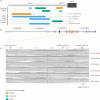
Causative structural variants. (a) Cartoon of the genomic structure of NIPBL coloured bars indicating the position of the structural variants shown in (b) and (c). (b) IGV plot of Proband 4197 and their parents showing a region of Chromosome 5. The green lines on the proband IGV plot indicate an inverted segment of chromosome with the blue lines representing a possible duplicated region (the coverage graph does not support this increased copy number). The inversion is predicted to encompass NIPBL Exons 42 and 43 and disrupt the open reading frame. (c) IGV plot of Proband 4497 and their parents. A heterozygous de novo deleted region is indicated by the drop in coverage in the proband and the grey lines on the IGV plot indicating paired end reads that cover the deletion breakpoints. This deletion encompasses Exons 46 and 47 which encode the most C-terminal region of NIPBL. (d) The IGV plot of Proband 4482 and their parents indicate a de novo deletion encompassing the whole SET gene. (e) The read plot of Proband 4353 and their parents indicating a de novo intragenic deletion involving the PUF60 gene (plotted using samplot [https://github.com/ryanlayer/samplot]).
Figure 3
Figure 3
De novo variants affecting uORF structure and clustering in Intron 21 of NIPBL. (a) Cartoon of the position of the predicted uORFs in the 5⁣′UTR encoded by Exon 1 and Exon 2 of NIPBL, indicating the strength of the Kozak translational start sequence shown in yellow, blue, and green for strong, moderate, and weak, respectively. The positions of the de novo variants in Probands 4079 and 4709 and their predicted effects are also shown. (b) Cartoon of the NIPBL genomic structure derived from the UCSC Genome Browser indicating the position of the noncoding variants detailed in (a) and (c). (c) IGV snapshot of the ~1 kb interval containing the de novo, deep intronic variants identified in Intron 21. Three de novo variants (arrowed above the IGV plots) were identified in Proband 4722 and a single variant (arrowed below the IGV plot) in Proband 4427.
Figure 4
Figure 4
De novo protein-coding variants in genes not known to be associated with developmental disorders. This figure shows cartoons of five different proteins encoded by the loci in which de novo variants have been identified in this study. None of these loci are known causes of developmental disorders. The position and type of the variants are indicated using the key used in Figure 1. The domain name is indicated when a missense variant lies within the domain.

References

    1. Kline A. D., Moss J. F., Selicorni A., et al. Diagnosis and management of Cornelia de Lange syndrome: first international consensus statement. Nature Reviews Genetics . 2018;19(10):649–666. doi: 10.1038/s41576-018-0031-0. - DOI - PMC - PubMed
    1. Krantz I. D., McCallum J., DeScipio C., et al. Cornelia de Lange syndrome is caused by mutations in NIPBL, the human homolog of Drosophila melanogaster Nipped-B. Nature Genetics . 2004;36(6):631–635. doi: 10.1038/ng1364. - DOI - PMC - PubMed
    1. Tonkin E. T., Wang T. J., Lisgo S., Bamshad M. J., Strachan T. _NIPBL_ , encoding a homolog of fungal Scc2-type sister chromatid cohesion proteins and fly Nipped-B, is mutated in Cornelia de Lange syndrome. Nature Genetics . 2004;36(6):636–641. doi: 10.1038/ng1363. - DOI - PubMed
    1. Huisman S. A., Redeker E. J., Maas S. M., Mannens M. M., Hennekam R. C. High rate of mosaicism in individuals with Cornelia de Lange syndrome. Journal of Medical Genetics . 2013;50(5):339–344. doi: 10.1136/jmedgenet-2012-101477. - DOI - PubMed
    1. Ansari M., Poke G., Ferry Q., et al. Genetic heterogeneity in Cornelia de Lange syndrome (CdLS) and CdLS-like phenotypes with observed and predicted levels of mosaicism. Journal of Medical Genetics . 2014;51(10):659–668. doi: 10.1136/jmedgenet-2014-102573. - DOI - PMC - PubMed

LinkOut - more resources