. 2025 Jul 17:8:0777.
doi: 10.34133/research.0777. eCollection 2025.

Cell Shock Absorption via Stress Relaxation Hydrogel Microspheres for Alleviating Endoplasmic Reticulum Stress in Chondrocytes

Affiliations

Cell Shock Absorption via Stress Relaxation Hydrogel Microspheres for Alleviating Endoplasmic Reticulum Stress in Chondrocytes

Ding Zhao et al. Research (Wash D C). .

Abstract

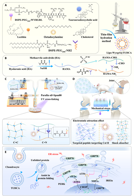
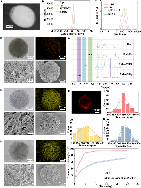
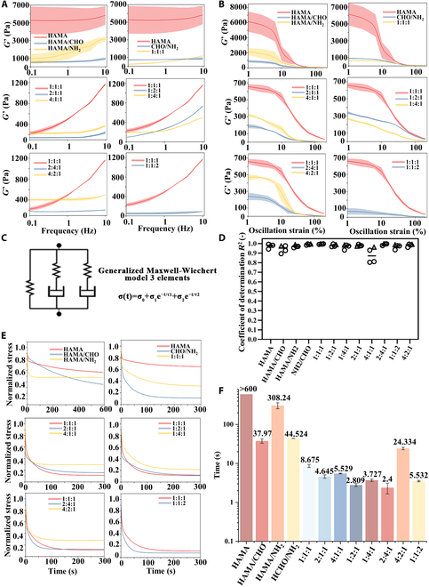
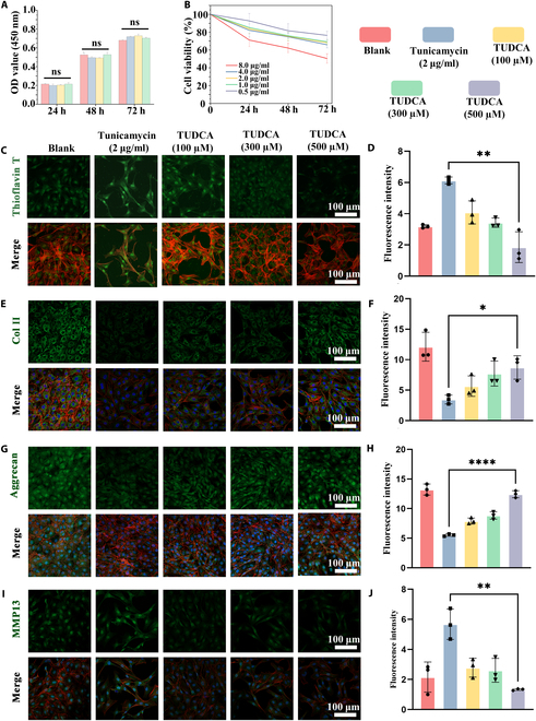
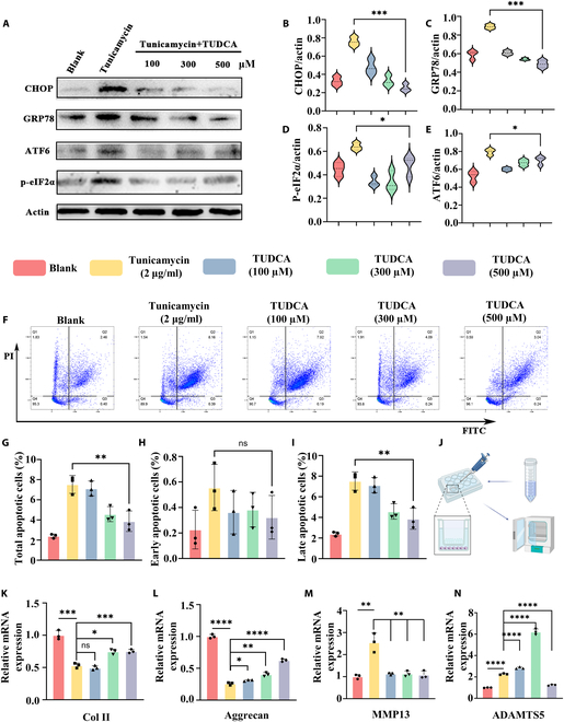
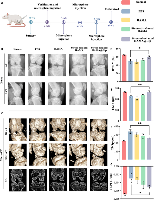
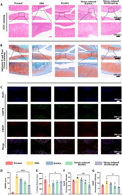
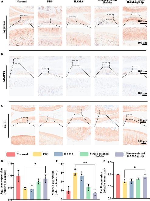
Chronic mechanical vibrations and endoplasmic reticulum (ER) stress are major contributors to osteoarthritis (OA) progression. This study proposes a novel "cellular shock absorption" approach by developing viscoelastic hydrogel microspheres with tunable stress relaxation properties. By modulating the chemical bonds in the hydrogel network through oxidation and hydrazine coupling reaction, hydrogel microspheres capable of absorbing shock and reducing mechanical stimulus-induced ER stress in chondrocytes are created. Cationic liposomes, modified with the cartilage-targeting peptide Wyrgrl and loaded with tauroursodeoxycholic acid (TUDCA), are encapsulated within these hydrogel microspheres. The microspheres not only dissipate intra-articular impact forces, reducing vibration and pressure transmission, but also provide sustained release of TUDCA, alleviating ER stress and slowing OA progression. In vitro studies showed that the stress relaxation time constant (τ) of the microspheres was tuned to 23.81 s, closely resembling the mechanical properties of the cartilage matrix. This property, combined with targeted TUDCA delivery, reduced Grp78 and CHOP expression, alleviating ER stress and inhibiting chondrocyte apoptosis. In vivo, the microspheres preserved joint cartilage structure, suppressed ER stress responses, and substantially delayed OA progression. This strategy presents a promising approach to mitigating cartilage damage and delaying OA by reducing mechanical stress and alleviating ER stress.


Conflict of interest statement

Competing interests: The authors declare that they have no competing interests.

Figures

Fig. 1.
Fig. 1.
Schematic design and experimental flow diagram. (A) Preparation of cationic liposomes targeting cartilage. (B) Chemical modification of MA-grafted HAMA by oxidation and OPA/N-nucleophile condensation reactions. (C) Fabrication of stress-relaxed HAMA@Lip by multiphase microfluidics and UV crosslinking. (D) Targeting approach of stress-relaxed HAMA@Lip and mechanism of mechanical stress relaxation effect. (E) TUDCA alleviates apoptosis by aiding protein folding and inhibiting UPR.
Fig. 2.
Fig. 2.
Construction and characterization of cationic liposomes and stress-relaxed HAMA@Lip. (A) TEM image of liposomes. (B and C) Particle size and zeta potential analysis of various liposome formulations, including unloaded liposomes (Lipo), WYRGRL-modified liposomes (Lipo-WYRGRL), TUDCA-loaded liposomes (Lipo-WYRGRL@TUDCA), and DiR-labeled liposomes (Lipo-WYRGRL@DiR), as determined by DLS. (D to F) Optical microscopy, phosphorus elemental mapping analysis, and SEM images of microspheres from different groups (HAMA microspheres, stress-relaxed HAMA, and stress-relaxed HAMA@Lip). (G) Proton nuclear magnetic resonance (1H NMR) spectra and substitution rates of various chemically modified HA products (HAMA, HAMA-CHO, and HAMA-NH₂). (H) Confocal microscopy images showing uniform loading of Lipo-WYRGRL@DiR on stress-relaxed HAMA. (I to K) Particle size distribution of microspheres from different groups (HAMA microspheres, stress-relaxed HAMA, and stress-relaxed HAMA@Lip). (L) Cumulative drug release profiles of Lipo-WYRGRL@TUDCA and stress-relaxed HAMA@Lip.
Fig. 3.
Fig. 3.
Mechanical characterization of stress-relaxed hydrogels with varying compositions. (A) Frequency sweep (n = 3) curves of hydrogels with different compositions at 37 °C under a fixed strain of 0.1% over a frequency range of 0.1 to 10 Hz; shaded areas represent the standard deviation within each group. (B) Amplitude sweep (n = 3) curves of hydrogels with different compositions at 37 °C under a fixed frequency of 1 Hz over a strain range of 1% to 100%; shaded areas represent the standard deviation within each group. (C) Experimental data fitted using the generalized 3-element Maxwell–Wiechert model. (D) The Maxwell–Wiechert model accurately fits the experimental data, with the goodness of fit determined by the coefficient of determination (R2). Hollow circles represent individual data points for each group (n = 3), and gray triangles indicate the mean values for each dataset. (E) Stress relaxation fitting curves of hydrogels with different compositions at 37 °C. The fitted data for each group were derived from independently prepared hydrogels (n = 3). (F) Stress relaxation constants of hydrogels with different compositions (n = 3) over an observation period of 600 s. Error bars are not shown for the HAMA group as the relaxation times exceeded 600 s.
Fig. 4.
Fig. 4.
Stressed-relaxed HAMA@Lip exhibits good biocompatibility and alleviates chondrocyte ERS and OA phenotypes. (A) CCK-8 assay results for chondrocytes treated with blank, Lip, stress-relaxed HAMA, and stress-relaxed HAMA@Lip on days 1, 2, and 3 (Blank, Lipo-Wyrgrl@TUDCA; Stress-relaxed HAMA; Stress-relaxed HAMA@Lipo). (B) Cell growth curves under different concentrations of tunicamycin. (C and D) ThT staining results and corresponding fluorescence quantification for different treatment groups (n = 3). (E and F) Col II staining results and corresponding fluorescence quantification for different treatment groups (n = 3). (G and H) Aggrecan staining results and corresponding fluorescence quantification for different treatment groups (n = 3). (I and J) MMP13 staining results and corresponding fluorescence quantification for different treatment groups (n = 3). One-way ANOVA with Tukey’s post hoc test. ns: no significance, *P < 0.05, **P < 0.01.
Fig. 5.
Fig. 5.
Stressed-relaxed HAMA@Lip promotes protein folding, alleviates ERS, and reduces apoptosis. (A) Western blot analysis of CHOP, GRP78, ATF6, p-eIF2α, and actin expression levels across different treatment groups. (B to E) Quantitative analysis of CHOP, GRP78, ATF6, and p-eIF2α expression levels normalized to actin (n = 3). (F) Flow cytometry analysis of early and late apoptosis in different treatment groups. (G to I) Quantitative statistics of total apoptotic cells, early apoptosis, and late apoptosis proportions in different treatment groups (n = 3). (J) Schematic illustration of coculture of Stressed-relaxed HAMA@Lip with chondrocytes. (K to N) Expression levels of OA-related genes in chondrocytes after coculture (n = 3). One-way ANOVA with Tukey’s post hoc test. ns: no significance, *P < 0.05, **P < 0.01, ***P < 0.001, **** P < 0.0001.
Fig. 6.
Fig. 6.
Radiological evaluation of in vivo therapeutic effects of Stressed-relaxed HAMA@Lip. (A) Schematic diagram and timeline of the animal experiment. (B) X-ray images showing the knee joint space and osteophyte formation in both anterior-posterior (AP) and lateral (LAT) views. (C) Micro-CT images illustrating the knee joint and subchondral bone in both 2D and 3D views. (D to G) Quantitative analysis of subchondral bone using bone volume fraction (BV/TV), trabecular thickness (Tb.Th), trabecular separation (Tb.Sp), and trabecular pattern factor (Tb.Pf) (n = 3). One-way ANOVA with Tukey’s post hoc test. *P < 0.05, **P < 0.01.
Fig. 7.
Fig. 7.
Stressed-relaxed HAMA@Lip alleviates ERS and preserves cartilage structure in OA. (A) Representative H&E staining images of rat knee joints from different groups. (B). Representative Safranin O–Fast Green staining images of rat knee joints from different groups. (C) Representative immunofluorescence images of CHOP and GRP78 in rat joints from different groups. (D) OARSI scoring of OA progression in rats from different groups (n = 3); detailed scoring criteria are provided in the Supplementary Materials. (E) Quantitative analysis of articular cartilage thickness in rats from different groups (n = 3). (F and G) Quantitative analysis of fluorescence intensity for CHOP and GRP78 in rat joints across different groups (n = 3). One-way ANOVA with Tukey’s post hoc test. *P < 0.05, **P < 0.01, ***P < 0.001.
Fig. 8.
Fig. 8.
Stressed-relaxed HAMA@Lip inhibits the progression of OA in rats. (A) Representative immunohistochemical staining images of aggrecan in rat joints from different groups. (B) Representative immunohistochemical staining images of MMP13 in rat joints from different groups. (C) Representative immunohistochemical staining images of Col II in rat joints from different groups. (D) Quantitative analysis of aggrecan expression in rat joints normalized to the normal group (n = 3). (E) Quantitative analysis of MMP13 expression in rat joints normalized to the normal group (n = 3). (F) Quantitative analysis of Col II expression in rat joints normalized to the normal group (n = 3). One-way ANOVA with Tukey’s post hoc test. *P < 0.05, **P < 0.01.

Similar articles

References

    1. Saraswathibhatla A, Indana D, Chaudhuri O. Cell-extracellular matrix mechanotransduction in 3D. Nat Rev Mol Cell Biol. 2023;24(7):495–516. - PMC - PubMed
    1. Seetharaman S, Vianay B, Roca V, Farrugia AJ, de Pascalis C, Boëda B, Dingli F, Loew D, Vassilopoulos S, Bershadsky A, et al. Microtubules tune mechanosensitive cell responses. Nat Mater. 2022;21(3):366–377. - PubMed
    1. Ma S, Dubin AE, Romero LO, Loud M, Salazar A, Chu S, Klier N, Masri S, Zhang Y, Wang Y, et al. Excessive mechanotransduction in sensory neurons causes joint contractures. Science. 2023;379(6628):201–206. - PMC - PubMed
    1. Yang X, Bi D, Czajkowski M, Merkel M, Manning ML, Marchetti MC. Correlating cell shape and cellular stress in motile confluent tissues. Proc Natl Acad Sci USA. 2017;114(48):12663–12668. - PMC - PubMed
    1. Mao Y, Wickström SA. Mechanical state transitions in the regulation of tissue form and function. Nat Rev Mol Cell Biol. 2024;25(8):654–670. - PubMed

LinkOut - more resources