Rectus sheath hematoma causing cecal perforation: a case report
- PMID: 40678159
- PMCID: PMC12266957
- DOI: 10.1093/jscr/rjaf550
Rectus sheath hematoma causing cecal perforation: a case report
Abstract
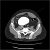
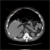
Rectus sheath hematoma is a rare condition caused by bleeding from the epigastric arteries, with an incidence of 1.2-1.5 cases per year. We present a 49-year-old male with a smoking history and recent corona virus disease 2019 (COVID-19) infection who presented with symptoms of an upper respiratory infection and suspected venous thromboembolism. Imaging revealed bilateral pulmonary emboli and a left rectus sheath hematoma, which was initially managed conservatively. However, the patient's condition worsened with a significant drop in hemoglobin and development of encephalopathy. Imaging showed an enlarging hematoma, leading to transfusion and selective embolization. On day 8, the patient developed generalized abdominal pain, and imaging confirmed a hollow viscus perforation. An exploratory laparotomy revealed cecal perforation due to mass effect from the hematoma. An ileocecectomy was performed.
Keywords: abdominal pain; anticoagulation; case report; cecal perforation; rectus sheath hematoma.
© The Author(s) 2025. Published by Oxford University Press and JSCR Publishing Ltd.
Conflict of interest statement
None declared.
Figures


References
Publication types
LinkOut - more resources
Full Text Sources

