Comment
. 2025 Jul 16;11(4):e200265.
doi: 10.1212/NXG.0000000000200265. eCollection 2025 Aug.

Muscle Biopsy Findings in Valosin-Containing Protein Multisystem Proteinopathy

Marianela Schiava  1 Yolande Parkhurst  2 Matthew Henderson  2 Tuomo Polvikoski  1 Manouela V Valtcheva  3 Ichizo Nishino  4 Michio Inoue  4 Yukako Nishimori  4 Yoshihiko Saito  4 Tanya Stojkovic  5 Rocio N Villar-Quiles  5 Norma Beatriz Romero  5 Teresinha Evangelista  5 Edoardo Malfatti  6 Sarah Souvannanorath  6 Elena Pegoraro  7 Pietro Riguzzi  7 Mauro Monforte  8 Sara Bortolani  8 Eleonora Torchia  8 Mario Sabatelli  8 Giorgio Tasca  1 Volker Straub  1 Chiara Marini-Bettolo  1 Michela Guglieri  1 Hakan Cetin  9 Ellen Gelpi  9 Sigrid Klotz  9 Jan L De Bleecker  10 Alicia Alonso-Jimenez  11 Jonathan Baets  11 Willem De Ridder  11 Peter De Jonghe  11 Kristl G Claeys  12   13 Dietmar Rudolf Thal  14 Jorge A Bevilacqua  15 Sushan Luo  16   17 Wenhua Zhu  16   17 Jie Lin  16   17 George Papadimas  18 Constantinos Papadopoulos  18 Eleni Zamba-Papanicolaou  18 Sophia Xirou  18 Endre Pal  19 Carmelo Rodolico  20 Anna Kostera-Pruszczyk  21 Biruta Kierdaszuk  21 Anna Kaminska  21 Nuria Muelas  22 Juan Jesus Vilchez  22 Cristina Domínguez-González  23   24 Aurelio Hernandez-Lain  25 Jorge Alonso-Perez  26 Velina Nedkova-Hristova  27 Carlos Aledo  25 Anders Oldfors  28 Umesh A Badrising  29 Hani Kushlaf  30 Thomas E Lloyd  31 Chiseko Ikenaga  31 Lindsay N Alfano  32 Colin C Quinn  33 David Walk  34 Matthias Vorgerd  35 Conrad Weihl  3 Montse Olivé  36 Jordi Diaz-Manera  1 VCP International Study Group
Collaborators, Affiliations
Comment

Muscle Biopsy Findings in Valosin-Containing Protein Multisystem Proteinopathy

Marianela Schiava et al. Neurol Genet. .

Abstract

Background and objectives: Valosin Containing Protein-associated multisystem proteinopathy (VCP-MSP) is a progressive, autosomal dominant disorder caused by pathogenic variants in the VCP gene, resulting in a heterogeneous clinical presentation. Muscle biopsy findings are characteristic but not pathognomonic. This study aimed to comprehensively analyse VCP-related myopathology and explore correlations with clinical phenotypes, genetic variants, and disease progression.

Methods: Muscle biopsy images and data were collected retrospectively from adults (≥18 years) with pathogenic or likely pathogenic VCP variants enrolled in the VCP Multicentre International Study. Biopsy data were standardized using the "Common Data Elements for Muscle Biopsy Reporting." Variations in biopsy findings were analysed by biopsy site, time from disease onset, the four most common VCP variants, and clinical phenotypes.

Result: A total of 112 muscle biopsies were included. Most individuals were male (66.0%). The mean age at biopsy was 53.3 years (SD 10.0), with a mean disease duration of 6.5 years (SD 4.5). The most frequent VCP variant was c.464G>A (p.Arg155His) (18.8%). The top clinical phenotypes were isolated myopathy (37.5%), myopathy with Paget disease of bone (17.9%), and myopathy with motor neuron involvement (13.4%). The vastus lateralis was the most common biopsy site (34.8%), and 91% were open biopsies. Histopathologic findings included atrophic fibres (87.5%), rimmed vacuoles (72.3%), endomysial fibrosis (58.0%), and protein aggregates (51.8%), primarily p62 (60.3%) and VCP (36.2%). Degeneration niches with fibrofatty replacement and atrophic fibres were seen in 33.3% of biopsies without frequency differences by clinical phenotypes. There were no differences in biopsy findings among the 4 most common VCP gene variants, except for the absence of degeneration niches in muscle biopsies of 12 patients with c.277C>T (p.Arg93Cys). MRI data from 30 patients showed fat pockets corresponding to these niches and STIR hyperintensity correlated with inflammatory infiltrates in 42.9%. Concordance between clinical phenotype, biopsy, and neurophysiology was observed in only 49.4% of cases, indicating significant heterogeneity.

Discussion: VCP-MSP muscle biopsies consistently show myopathic or mixed patterns with rimmed vacuoles and p62/VCP-positive inclusions, regardless of clinical phenotype, age, or progression. Some lack vacuoles, challenging diagnosis. Discrepancies between clinical, neurophysiology, and biopsy findings should prompt consideration of VCP-MSP to improve detection and management.


Conflict of interest statement

The authors report no relevant disclosures. Go to Neurology.org/NG for full disclosures.

Figures

Figure 1
Figure 1. Frequency of Findings on Muscle Biopsies of Patients With VCP-MSP Disease
(A) Frequency of general patterns identified in muscle biopsies of the study. (B) Frequency of histologic abnormalities observed in the muscle biopsies included.
Figure 2
Figure 2. Examples of Histologic Findings Observed in Muscle Biopsies of Patients With VCP-MSP Disease
(A) Hematoxylin and eosin staining showing angulated atrophic fibers, rounded fibers, and prominent variability in fiber size. (B and C) Rimmed vacuoles identified on hematoxylin and eosin staining (B) and trichrome Gomori staining (C). (D) Hematoxylin and eosin staining showing a necrotic fiber undergoing phagocytosis. The necrotic fiber is in the center; it has lost its normal polygonal shape and appears eosinophilic (pink) because of protein denaturation. Individual nuclei are not identifiable because they may have disappeared or fragmented. Numerous macrophages, appearing as cells with foamy cytoplasm and dark nuclei, surround and invade the necrotic fiber. (E) Hematoxylin and eosin staining showing a muscle fiber with protein aggregates seen as eosinophilic inclusions in the sarcoplasm. These protein aggregates stained positive for the following proteins on immunohistochemistry: desmin (F), myotilin (G), VCP (H), and p62 (I). (J) TDP-43 aggregates in the sarcoplasm of a muscle fiber identified on immunohistochemistry. (K) Amyloid structures identified with Congo red staining. (L) Ubiquitin-positive inclusions and (M) AB-crystallin–positive protein aggregates identified on immunohistochemistry. Calibration bar: 20 × 50 μm for images A and B and 40 × 20 μm for the rest.
Figure 3
Figure 3. Examples of Degenerative Niches Identified in Muscle Biopsies of Patients With VCP-MSP Myopathy
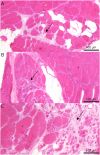
(A, B, and C) Three examples of degenerative niches (arrows) characterized by areas of the muscle containing atrophic fibers embedded in fibrotic and fat tissue closely located to areas of the muscle with clearly lower involvement (asterisks). Calibration bar: 10 × 100 μm.
Figure 4
Figure 4. Natural History of Pathologic Progression in Muscle Biopsies of Patients With VCP-MSP Myopathy
(A) Example of a very mild case where only few atrophic scattered fibers (asterisk) were identified. VCP (A′) and p62 (A″) inclusions were identified in isolated fibers. (B) Mild case showing a group of small atrophic fibers (circle) with p62 inclusions (B′). (C) More advanced case displaying several groups of atrophic fibers closely located (arrow) and vacuolated fibers (asterisk). (D) Degenerative foci containing atrophic fibers staining positive for caveolin (D′) embedded in expanded fibrotic tissue surrounded by an area of normal muscle. (E) More advanced case with a large degenerative foci. (F) Hematoxylin and eosin staining at low resolution where 2 degenerative niches were identified (G and H) with several fibres containing p62-positive inclusions (G′ and H′) while the rest of muscle biopsy was almost nonaffected. (I) Severe case where the muscle biopsy was almost completely replaced by fat and fibrotic tissue (J), except an area (square) showing milder anomalies (K). (L) End-stage case where muscle architecture was lost, and extremely atrophic fibers were observed staining positive for embryonic myosin heavy chain (L′) embedded in expanded fibrotic tissue.
Figure 5
Figure 5. Correlations Between Muscle Findings, Phenotype, and EMG Changes
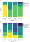
(A) Frequency of general muscle biopsy patterns in patients with VCP-MSP with 4 different phenotypes. (B) Level of agreement between the phenotypic description, EMG, and muscle biopsy results. MND = motor neuron disease; n = number of patients.

Comment on

References

    1. Meyer H, Weihl CC. The VCP/p97 system at a glance: connecting cellular function to disease pathogenesis. J Cell Sci. 2014;127(Pt 18):3877-3883. doi: 10.1242/jcs.093831 - DOI - PMC - PubMed
    1. Meyer H, Bug M, Bremer S. Emerging functions of the VCP/p97 AAA-ATPase in the ubiquitin system. Nat Cell Biol. 2012;14(2):117-123. doi: 10.1038/ncb2407 - DOI - PubMed
    1. Dalal S, Hanson PI. Membrane traffic: what drives the AAA motor? Cell. 2001;104(1):5-8. doi: 10.1016/S0092-8674(01)00186-6 - DOI - PubMed
    1. Pontifex CS, Zaman M, Fanganiello RD, Shutt TE, Pfeffer G. Valosin-containing protein (VCP): a review of its diverse molecular functions and clinical phenotypes. Int J Mol Sci. 2024;25(11):5633. doi: 10.3390/ijms25115633 - DOI - PMC - PubMed
    1. Pye VE, Dreveny I, Briggs LC, et al. Going through the motions: the ATPase cycle of p97. J Struct Biol. 2006;156(1):12-28. doi: 10.1016/j.jsb.2006.03.003 - DOI - PubMed