. 2025 Jun 24:10:100349.
doi: 10.1016/j.ijpx.2025.100349. eCollection 2025 Dec.

Plant-derived extracellular vesicles as a natural drug delivery platform for glioblastoma therapy: A dual role in preserving endothelial integrity while modulating the tumor microenvironment

Affiliations

Plant-derived extracellular vesicles as a natural drug delivery platform for glioblastoma therapy: A dual role in preserving endothelial integrity while modulating the tumor microenvironment

Lishan Cui et al. Int J Pharm X. .

Abstract

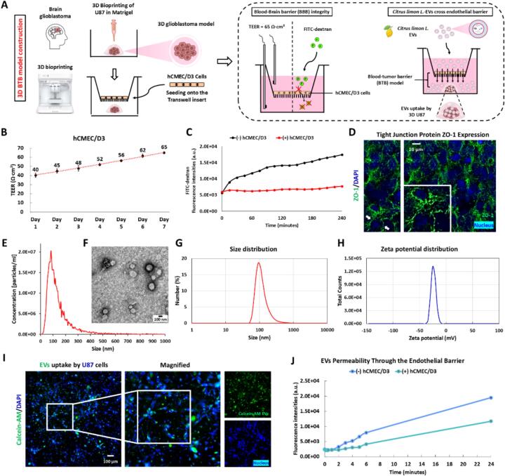
Glioblastoma (GBM) is the most aggressive primary brain tumor, with limited treatment options due to the restrictive blood-brain barrier (BBB) and the heterogeneity of the blood-tumor barrier (BTB). Temozolomide (TMZ), the standard chemotherapy, suffers from poor BBB permeability, rapid degradation, and systemic toxicity. Plant-derived extracellular vesicles (PDEVs) have emerged as promising natural nanocarriers, offering biocompatibility, stability, and the ability to cross biological barriers. This study investigates the use of extracellular vesicles from Citrus limon L. (LDEs) to encapsulate and deliver TMZ (EVs@TMZ) for GBM treatment. LDEs were isolated, characterized, and loaded with TMZ via ultrasonication. Encapsulation efficiency, stability, and physicochemical properties were assessed using UV-Vis and FTIR spectroscopy. A 3D BTB model was developed using bioprinted U87 glioblastoma cells in Matrigel, co-cultured with hCMEC/D3 endothelial cells to replicate the tumor microenvironment. Barrier integrity was evaluated through TEER and FITC-dextran assays. Uptake, cytotoxicity, and tumor invasion were assessed in this model, along with oxidative stress and VEGF-A secretion. LDEs effectively encapsulated TMZ, enhancing drug stability under physiological conditions. EVs@TMZ crossed the endothelial barrier while preserving barrier integrity and reducing TMZ-induced ROS production. In the 3D glioblastoma model, EVs@TMZ showed strong cytotoxic effects on tumor cells while minimizing endothelial toxicity and oxidative stress. Moreover, VEGF-A secretion was suppressed, disrupting pro-tumorigenic pathways. These findings highlight Citrus-derived EVs as biocompatible, efficient carriers for TMZ delivery, offering a promising approach to overcome current challenges in GBM therapy and supporting further development of PDEVs for brain tumor treatment.

Keywords: 3D bioprinting; Blood-tumor barrier; Drug delivery; Glioblastoma; Plant-derived extracellular vesicles; VEGF-A.


Conflict of interest statement

The authors declare that the research was conducted in the absence of any commercial or financial relationships that could be construed as a potential conflict of interest.

Figures

Unlabelled Image
Graphical abstract
Fig. 1
Fig. 1
Construction of a 3D BTB model and assessment of the barrier-crossing ability of Citrus limon L.-derived EVs. (A) Schematic representation of the 3D BTB model construction and EVs permeability assessment. (B) TEER measurements assessing endothelial barrier formation in hCMEC/D3 cells over seven days. (C) FITC-dextran permeability in the absence (−) or presence (+) of hCMEC/D3 cells. (D) Immunofluorescence staining of the tight junction protein ZO-1 (green) in hCMEC/D3 cells. Nuclei are stained with DAPI (blue). (E) Nanoparticle tracking analysis (NTA) of size distribution and concentration of EVs. (F) Representative transmission electron microscopy (TEM) image of EVs (Scale bar = 100 nm). (G) Size and (H) zeta potential distribution of Citrus limon L.-derived EVs. (I) Fluorescence images showing the uptake of Calcein-AM-labeled EVs (green) by U87 glioblastoma cells. Nuclei are stained with DAPI (blue). scale bar = 100 μm. The magnified images depict high-magnification views of the white boxed areas. (J) Fluorescence intensities of endothelial barrier-crossed EVs were measured using a Cytation 3 Cell Imaging Multi-Mode Reader (BioTek, Winooski, VT, USA). Error bars represent the standard deviation (or standard error) of the mean; however, they are shorter than the height of the symbols and therefore not visible in the figure. (For interpretation of the references to colour in this figure legend, the reader is referred to the web version of this article.)
Fig. 2
Fig. 2
EVs Uptake by 3D U87 glioblastoma cells and the cytotoxic effects of TMZ in the 3D model. (A) Representative images of 3D U87 cells incubated with (+) or without (−) EVs at 37 °C for 24 h. EVs were labeled with calcein-AM (shown in green), actin filaments were labeled with rhodamine phalloidin, and nuclei were counterstained with 4′,6-diamidino-2-phenylindole (DAPI) (scale bar = 10 μm). The magnified images depict high-magnification views of the white boxed areas. (B) Fluorescence intensity profiles of cellular components under (−) EVs and (+) EVs conditions. (C) Representative stitched microscopic images of 3D U87 after 48 h of TMZ treatment ranging from 0 to 2.5 mM (Scale bar = 1 mm). (D) Dose-dependent cytotoxicity of TMZ on 3D U87 model. (E) IC50 of TMZ in 3D U87 model. The IC50 value is fitted from sigmoidal dose-response curve to quantify drug potency. Bars, mean ± SEM. Statistical significance was evaluated using one-way ANOVA followed by Tukey's post-hoc tests (*p < 0.05; **p < 0.01; ***p < 0.001). (For interpretation of the references to colour in this figure legend, the reader is referred to the web version of this article.)
Fig. 3
Fig. 3
pH-dependent spectral properties and degradation kinetics of TMZ. (A) UV–visible absorption spectrum of TMZ in DMSO with a characteristic peak at 328 nm. (B) Calibration curve of TMZ in DMSO at 328 nm. (C) Absorption spectra of TMZ in PBS at different pH values (7.4, 6.8, 5.5, and 2.5). (D) Time-dependent degradation of TMZ under different pH conditions measured at 328 nm. (E-H) UV–Vis absorption spectra of TMZ in PBS at pH 7.4 (E), pH 6.8 (F), pH 5.5 (G), and pH 2.5 (H) over 48 h. At pH 7.4 and 6.8, a progressive decrease in absorbance at 328 nm and the emergence of an isosbestic point indicate hydrolytic degradation. UV–vis spectra were measured by a BioTek Cytation 3 Cell Imaging Multimode Reader.
Fig. 4
Fig. 4
Encapsulation and Release of TMZ in Citrus limon L.-Derived EVs. (A) Schematic diagram of TMZ encapsulation in EVs (EVs@TMZ) by ultrasound, separation of free TMZ and EVs@TMZ by ultrafiltration, and release of TMZ after Triton X-100 lysis. (B) UV–Vis absorption spectrum of TMZ in PBS at pH 5.5, with a characteristic peak at 328 nm. (C) Calibration curve of TMZ absorbance at 328 nm in PBS (pH 5.5). (D) UV–Vis absorption spectra of free TMZ, EVs@TMZ, and total TMZ. (E) Encapsulation efficiency of TMZ in EVs, showing the percentage of free TMZ and EVs@TMZ relative to total TMZ. (F) UV–vis spectra of TMZ before and after Triton X-100 lysis of EVs. (G) Quantification of TMZ after EVs lysis. (H–J) Fourier-transform infrared (FTIR) spectra of free TMZ (H), EVs (I), and EVs@TMZ (J) with characteristic functional groups and potential interactions between TMZ and EVs.
Fig. 5
Fig. 5
Effects of Citrus limon L.-derived EVs on U87 and hCMEC/D3 Cells. (A, D) Representative live/dead staining images of U87 and hCMEC/D3 cells, respectively, following 48-h treatment with EVs (80 μg/mL). Live cells are stained green (Calcein AM), while dead cells are stained red (PI). (B, E) Cell viability of U87 and hCMEC/D3 cells treated with increasing concentrations of EVs (10, 20, 40, and 80 μg/mL). Data are expressed as the percentage of viable cells relative to the control (mean ± SEM, n = 4). (C, F) Intracellular ROS levels in U87 and hCMEC/D3 cells were normalized to the number of viable cells. (G) Wound healing assay showing hCMEC/D3 cell migration at 0-, 4-, and 24-h post-scratch, (+) with or (−) without EVs. (H) Quantification of migrating cells and hCMEC/D3 cells after scratch wounding. Statistical significance was determined using one-way or two-way ANOVA with Tukey's post hoc test (*p < 0.05; **p < 0.01; ***p < 0.001; ****p < 0.0001). (For interpretation of the references to colour in this figure legend, the reader is referred to the web version of this article.)
Fig. 6
Fig. 6
Evaluation of EVs@TMZ uptake, cytotoxicity, glioblastoma spheroid progression, and VEGF-A secretion in a 3D BTB model. (A) Schematic illustration of EVs@TMZ uptake by U87 in a 3D BTB co-culture model with hCMEC/D3 cells. (B) Confocal microscopy images of 3D U87 cells incubated with EVs@TMZ at 37 °C for 24 h. EVs@TMZ were labeled with calcein-AM (shown in green), actin filaments were labeled with rhodamine phalloidin, and nuclei were counterstained with 4′,6-diamidino-2-phenylindole (DAPI) (scale bar = 10 μm). The magnified images depict high-magnification views of the white boxed areas. (C) 3D Z-stack image of spatial distribution and uptake of EVs@TMZ in U87 cells. (D) Progressive BTB crossing of EVs@TMZ over 24 h. (E) Cytotoxic effects and (F) ROS levels in hCMEC/D3 cells after 48 h of untreated or treatment with TMZ and EVs@TMZ. (G) Representative stitched microscopic images of 3D U87 in the untreated (Ctrl) group at 24 and 48 h, with arrows indicating aggregates and migration. (H) Representative fluorescence images of 3D U87 after 48 h under three conditions: untreated (Ctrl), TMZ-treated, and EVs@TMZ-treated. (I-L) Quantification of 3D U87, including viable cells (I), ROS levels (J), migrated cells (K), and core density (L). (M) VEGF-A levels in U87, U87 co-cultured with hCMEC/D3 (U87 + hCMEC/D3), TMZ-treated, and EVs@TMZ-treated. Statistical significance was determined using one-way ANOVA with Tukey's post hoc test (*p < 0.05; **p < 0.01; ***p < 0.001; ****p < 0.0001). (For interpretation of the references to colour in this figure legend, the reader is referred to the web version of this article.)

References

    1. Alzahrani F.A., Khan M.I., Kameli N., Alsahafi E., Riza Y.M. Plant-derived extracellular vesicles and their exciting potential as the future of next-generation drug delivery. Biomolecules. 2023;13 - PMC - PubMed
    1. Amarandi R.-M., Ibanescu A., Carasevici E., Marin L., Dragoi B. Liposomal-based formulations: a path from basic research to temozolomide delivery inside glioblastoma tissue. Pharmaceutics. 2022;14:308. - PMC - PubMed
    1. Auffinger B., Tobias A.L., Han Y., Lee G., Guo D., Dey M., Lesniak M.S., Ahmed A.U. Conversion of differentiated cancer cells into cancer stem-like cells in a glioblastoma model after primary chemotherapy. Cell Death & Differentiation. 2014;21:1119–1131. - PMC - PubMed
    1. Berumen Sánchez G., Bunn K.E., Pua H.H., Rafat M. Extracellular vesicles: mediators of intercellular communication in tissue injury and disease. Cell Communication and Signaling. 2021;19:104. - PMC - PubMed
    1. Cai Q., Li X., Xiong H., Fan H., Gao X., Vemireddy V., Margolis R., Li J., Ge X., Giannotta M., Hoyt K., Maher E., Bachoo R., Qin Z. Optical blood-brain-tumor barrier modulation expands therapeutic options for glioblastoma treatment. Nat. Commun. 2023;14:4934. - PMC - PubMed

LinkOut - more resources