

Can artificial intelligence in spine imaging affect current practice? Practical developments and their clinical status
- PMID: 40678684
- PMCID: PMC12269973
- DOI: 10.1016/j.xnsj.2025.100621
Can artificial intelligence in spine imaging affect current practice? Practical developments and their clinical status
Abstract
Background: As artificial intelligence (AI) increases its footprint in spine imaging, gauging the clinical relevance of new developments poses an increasingly difficult challenge, especially given the majority of developments reflect experimental or early work. With this summary of available AI tools, focusing on those in clinical use, the benefits of AI in spine imaging are explained for radiologists and surgeons to understand the current state of and potentially the decision to adopt AI in clinical practice.
Methods: Through a narrative review of publications relating to "artificial intelligence" and "spine imaging" in the PubMed database, this article provides an update on AI applications in spine imaging being utilized in current clinical practice.
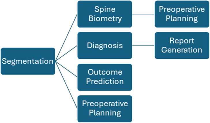
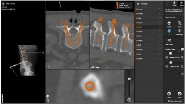
Results: Current applications of AI in spine imaging range from deep learning image reconstruction and denoising, spine segmentation and biometry, radiological report generation, surgical outcomes prediction, surgical planning, to intraoperative assistance. Developments in deep learning reconstruction (DLR) are most mature and demonstrate improvements to imaging speed and interpretability compared to non-AI alternatives. While clinical implementations exist in other use cases, their performance remains either an area of active investigation or comparable to the level of a human.
Conclusions: Uses of AI in spine imaging span multiple applications with early clinical implementation in most areas, suggesting a promising future ahead.
Keywords: Artificial intelligence; Augmented reality; Image reconstruction; Radiology; Segmentation; Spine imaging; Surgical planning.
© 2025 The Authors. Published by Elsevier Inc. on behalf of North American Spine Society.
Conflict of interest statement
The authors declare that they have no known competing financial interests or personal relationships that could have appeared to influence the work reported in this paper.
Figures


Similar articles
-
A Systematic Review and Bibliometric Analysis of Applications of Artificial Intelligence and Machine Learning in Vascular Surgery.Ann Vasc Surg. 2022 Sep;85:395-405. doi: 10.1016/j.avsg.2022.03.019. Epub 2022 Mar 24. Ann Vasc Surg. 2022. PMID: 35339595
-
Research status, hotspots and perspectives of artificial intelligence applied to pain management: a bibliometric and visual analysis.Updates Surg. 2025 Jun 28. doi: 10.1007/s13304-025-02296-w. Online ahead of print. Updates Surg. 2025. PMID: 40580377
-
The impact of artificial intelligence on the endoscopic assessment of inflammatory bowel disease-related neoplasia.Therap Adv Gastroenterol. 2025 Jun 23;18:17562848251348574. doi: 10.1177/17562848251348574. eCollection 2025. Therap Adv Gastroenterol. 2025. PMID: 40556746 Free PMC article. Review.
-
Artificial intelligence for detecting keratoconus.Cochrane Database Syst Rev. 2023 Nov 15;11(11):CD014911. doi: 10.1002/14651858.CD014911.pub2. Cochrane Database Syst Rev. 2023. PMID: 37965960 Free PMC article.
-
Current Radiology workforce perspective on the integration of artificial intelligence in clinical practice: A systematic review.J Med Imaging Radiat Sci. 2025 Jan;56(1):101769. doi: 10.1016/j.jmir.2024.101769. Epub 2024 Oct 21. J Med Imaging Radiat Sci. 2025. PMID: 39437624
References
-
- Bash S., Johnson B., Gibbs W., Zhang T., Shankaranarayanan A., Tanenbaum L.N. Deep learning image processing enables 40% faster spinal MR scans which match or exceed quality of standard of care: a prospective multicenter multireader study. Clin Neuroradiol. 2022;32 doi: 10.1007/s00062-021-01121-2. - DOI - PMC - PubMed
LinkOut - more resources
Full Text Sources
Research Materials
