. 2025 Aug 11;11(8):4758-4772.
doi: 10.1021/acsbiomaterials.5c00828. Epub 2025 Jul 18.

Spontaneous Crimping of Gelatin Methacryloyl Nanofibrils Induced by Limited Hydration

Affiliations

Spontaneous Crimping of Gelatin Methacryloyl Nanofibrils Induced by Limited Hydration

Chien-Wei Wu et al. ACS Biomater Sci Eng. .

Abstract

The crimped appearance of native collagen fibrils in youthful tissues serves as a mechanical buffer and phenotype determinant for resident cells. In vitro platforms emulating these native crimped networks facilitate the study of cell-matrix dynamics in various pathophysiological contexts. However, creating fibrillar networks with sizes and crimping matching native tissues using collagen-derived substrates remains challenging. We present an innovative approach to produce spontaneous, tunable crimping of electrospun, aligned gelatin methacryloyl nanofibrils using limited hydration. The diameter of the synthesized fibrils approximated that of native fibrils. Beyond individual fibril crimping, the network exhibited large-scale, periodic crimping with wavelengths matching native collagen networks. Tensile stress tests revealed that crimping reduced network stiffness but enhanced stretchability, consistent with native tissues. Additionally, crimping promoted cell translocation into the network. Fibroblasts cultured on crimped fibrils showed smaller cell areas, higher vinculin and α-tubulin expression, and lower α-smooth muscle actin levels compared to those on straight fibrils. This novel method not only replicates the native fibril characteristics using collagen-derived materials, but also offers a valuable tool for advancing our understanding of cell-matrix interactions, with significant implications for tissue engineering and regenerative medicine.

Keywords: 3D cell culture; Collagen derivatives; GelMA; fibril crimping; tissue scaffold.


Figures

1
1
Schematic diagrams for synthesis of crimped GelMA fibrils. (a) Structural formula representation for GelMA synthesis and photo-cross-linking. (b) Setup for generating random and parallel-aligned GelMA fibrils using electrospinning. (c) Procedure to induce crimping in the electrospun fibrils. To produce crimped GelMA fibrils, the fibril network was detached from the aluminum foil, soaked in 91%-pure ethanol solutions, photo-cross-linked, and dried using CPD for subsequent cell culture. Fibril networks that remained attached to the foil and were treated with pure ethanol solution maintained a straight morphology.
2
2
Spontaneous fibril network shrinkage upon limited hydration. (a) Sequential photos of as-spun fibril networks soaked in ethanol solutions of 91%, 93%, 95%, 97%, and pure ethanol, taken at varying intervals over 25 min (square scale: 1 cm). Arrows indicate the direction of fibril alignment. (b) Temporal dynamics of network shrinkage represented by the normalized projected area of the fibril network versus soaking time in ethanol solutions of varying concentrations. (n = 3) The adjacent zoom-in plot highlights the first 3 min of temporal dynamics marked by a dotted box.
3
3
Spontaneous crimping of GelMA fibrils upon limited hydration. SEM images of fibrils soaked in ethanol at concentrations of (a) 91%, (b) 93%, (c) 95%, (d) 97%, and (e) pure ethanol, along with (f) untreated straight fibrils. Images (g) and (h) show the larger-scale, periodic crimping pattern in fibrils soaked in 95% ethanol, with dotted lines marking the edges of the crimp pattern. The arrow indicates the crimping wavelength, defined as the distance between adjacent edges. (i) Crimping degrees and (j) diameters of fibrils treated under various conditions. Comparisons with straight fibrils are marked with *, and comparisons with fibrils soaked in 95% ethanol are marked with #. P-values less than 0.05, 0.01, 0.001, and 0.0001 are indicated by *, **, ***, and ****, respectively. The notations NS­(*) and NS(#) indicate that the comparison with straight fibrils and fibrils soaked in 95% ethanol, respectively, was not statistically significant. (n = 20–65).
4
4
Time-dependent crimping and diameter changes of fibrils under limited hydration. SEM images of fibrils immediately after electrospinning (a) and following immersion in 95% ethanol for (b) 20 s, (c) 40 s, (d) 60 s, (e) 120 s, (f) 300 s, (g) 600 s, and (h) 1500 s. (i) Quantitative analysis of fibril diameters and crimping degrees as a function of soaking time (n = 50–70).
5
5
Representative images and stress–strain profiles for tensile stress analysis of fibril network. (a) Photograph of a network clamped for stretching. (b) Stress–strain curves recorded during the four stretching cycles: three for preconditioning and one until failure. The yield point marks the transition from elastic to plastic deformation. Young’s modulus was derived from the initial linear elastic region, and ultimate strength from maximum stress during failure. Stress–strain behavior within the initial 1.5% strain is highlighted (dotted box). (c) SEM images of crimped fibrils: (1) in the unstretched state, (2) after preconditioning, and (3) after stretched-to-failure. (d) Schematic representation of a network stretched to failure, with SEM images taken sequentially from (1) broken end, (2) near broken end, (3) intermediate portion, to (4) clamped end of a stretched-to-failure network.
6
6
Mechanical properties of various fibril networks. Stress–strain relationship for (a) parallel-aligned and (b) randomly oriented dried fibrils. Statistical comparison for (c) yield stress, (d) ultimate stress, (e) Young’s modulus, (f) yield strain, and (g) ultimate strain between dried fibril networks prepared in various conditions (n = 3–5). The marks * denote comparisons within parallel-aligned fibrils, and # denote comparisons within randomly oriented fibrils. P-values lower than 0.05, 0.01, 0.001, and 0.0001 are marked as */#, **/##, ***/###, and ****/####, respectively. (h) Stress–strain relationship for hydrated fibrils (n = 4).
7
7
Impact of fibril topology on cell morphology. (a) Representative fluorescence images of NIH-3T3 fibroblasts cultured on various fibril networks and Petri dishes, with F-actin stained using phalloidin (magenta) and nuclei stained using DAPI (blue). (b) Distribution of fibroblast orientation on fibril networks of different topologies, shown by the deviation angles of individual cell orientation from the mean cell orientation. (c) Aspect ratio and (d) area of fibroblasts cultured on various materials, determined from F-actin fluorescence. (e) Aspect ratio and (f) area of cell nuclei, calculated using nuclear fluorescence. P-values less than 0.05, 0.01, 0.001, and 0.0001 are denoted as *, **, ***, and ****, respectively, (n = 44–76).
8
8
Cellular integration within fibril networks. SEM images of (a) 3T3 fibroblasts and (b) bone marrow-derived mesenchymal stem cells (BMSCs) cultured on straight and crimped fibrils. Statistic comparison of the crimping degree of the fibrils adjacent to adhered (c) fibroblasts and (d) BMSCs between networks with straight and crimped fibrils. (e) Representative SEM image of crimped fibrils soaked in cell culture media without cells for 48 h. (f) Statistic compassion of the crimping degrees of crimped fibrils in networks with and without cell culture. (g) Schematic diagram and SEM images of the cross-section of a crimped fibril mat cultured with fibroblasts for 48 h, showing cell migration to a depth of approximately 200 μm from the mat surface toward the central region. Arrows denote the cut ends of the fibrils at the cross section. (h) Immunofluorescence images of fibroblasts cultured on crimped fibrils, overlaid with bright-field images. (blue for DAPI, magenta for phalloidin, green for vinculin) (i) SEM images of fibroblasts cultured on straight and crimped fibrils for 7 days. Cells had a rounder shape on crimped fibrils, similar to those observed after 48 h culture. P-values lower than 0.0001 are marked as ****. (n = 50 in panels c, d, f).
9
9
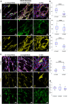
Impact of fibril topology on protein expression. (a) Immunofluorescence images of NIH-3T3 fibroblasts cultured on Petri dishes, straight fibrils, or crimped fibrils for 48 h, stained for F-actin (magenta), nucleus (blue), α-tubulin (yellow), and vinculin (green). Statistical comparison of (b) fluorescence intensity per cell for vinculin expression, (c) vinculin expression area per cell, and (d) fluorescence intensity per cell for α-tubulin expression. (e) Immunofluorescence images of fibroblasts cultured on Petri dishes or fibrillar structures for 48 h, stained for F-actin (magenta), nucleus (blue), α-SMA (yellow), and YAP1 (green). Statistical comparison of fluorescence intensity for (f) α-SMA and (g) YAP1 expression. Note that α-SMA data for cells cultured on Petri dishes were not included in the comparison due to different fluorescence settings used for image acquisition. P-values less than 0.0001 are denoted as **** (n = 26–36 cells in panels b, c; n = 44–47 cells in panels e, f).
10
10
Proposed mechanisms of ethanol concentration-dependent topology changes in GelMA fibrils induced by limited hydration. (a) Non-cross-linked GelMA chains confined within as-spun fibrils. (b) Fibrils become buckled due to interactions with hydrogen bonds provided by water and ethanol molecules when immersed in high-concentration ethanol. (c) Increased water content promotes more hydrogen-bonding network inside the fibrils, resulting in fibril thickening and aggregation. (d) Fibrils completely dissolve when the ethanol concentration drops below 50%.

Similar articles

References

    1. Franchi M., Raspanti M., Dell’orbo C., Quaranta M., De Pasquale V., Ottani V., Ruggeri A.. Different crimp patterns in collagen fibrils relate to the subfibrillar arrangement. Connect. Tissue Res. 2008;49(2):85–91. doi: 10.1080/03008200801913635. - DOI - PubMed
    1. Gathercole L., Keller A.. Crimp morphology in the fibre-forming collagens. Matrix. 1991;11(3):214–234. doi: 10.1016/S0934-8832(11)80161-7. - DOI - PubMed
    2. Gruber H. E., Hanley E. N. Jr.. Observations on morphologic changes in the aging and degenerating human disc: secondary collagen alterations. BMC Musculoskeletal Disord. 2002;3(1):9. doi: 10.1186/1471-2474-3-9. - DOI - PMC - PubMed
    1. Diamant J., Keller A., Baer E., Litt M., Arridge R. G. C.. Collagen; ultrastructure and its relation to mechanical properties as a function of ageing. Proc. R Soc. Lond. B Biol. Sci. 1972;180(1060):293–315. doi: 10.1098/rspb.1972.0019. - DOI - PubMed
    1. Hansen K. A., Weiss J. A., Barton J. K.. Recruitment of tendon crimp with applied tensile strain. J. Biomech. Eng. 2002;124(1):72–77. doi: 10.1115/1.1427698. - DOI - PubMed
    2. Rigby B. J., Hirai N., Spikes J. D., Eyring H.. The mechanical properties of rat tail tendon. J. Gen. Physiol. 1959;43(2):265–283. doi: 10.1085/jgp.43.2.265. - DOI - PMC - PubMed
    1. Hurschler C., Loitz-Ramage B., Vanderby R. Jr.. A structurally based stress-stretch relationship for tendon and ligament. J. Biomech. Eng. 1997;119:392–399. doi: 10.1115/1.2798284. - DOI - PubMed

LinkOut - more resources