. 2025 Aug 26;44(8):116007.
doi: 10.1016/j.celrep.2025.116007. Epub 2025 Jul 17.

Epistasis in the receptor-binding domain of contemporary H3N2 viruses that reverted to bind sialylated di-LacNAc repeats

Affiliations

Epistasis in the receptor-binding domain of contemporary H3N2 viruses that reverted to bind sialylated di-LacNAc repeats

Ruonan Liang et al. Cell Rep. .

Abstract

Since their introduction into humans, H3N2 influenza A viruses have evolved continuously to escape immunity through antigenic drift, driven by mutations in and around the receptor-binding site. Recently, these changes resulted in viruses that recognize elongated glycans, which are less abundant in the human respiratory tract, complicating vaccine strain propagation. This study employed ELISA, glycan arrays, tissue staining, flow cytometry, and hemagglutinin (HA) assays to demonstrate the molecular determinants of recent H3N2 viruses that regained recognition of shorter glycans. Mutations Y159N/T160I in contemporary strains replace Y159/T160, weakening receptor binding. However, this is compensated by Y195F in the 190-helix. These findings highlight epistasis across critical residues in the HA receptor-binding site, including the 130-loop, 150-loop, and 190-helix. Interestingly, a positive correlation exists between binding to an asymmetrical N-glycan and binding to human and ferret respiratory tract tissues. These results elucidate the epistatic nature of receptor-binding specificity during influenza A virus H3N2 evolution.

Keywords: CP: Microbiology; H3N2; N-glycan; epistasis; hemagglutinin; influenza; receptor binding; sialic acid.


Conflict of interest statement

Declaration of interests The authors declare no competing interests.

Figures

Figure 1.
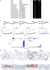
Figure 1.. Y159 and T160 are responsible for glycan-binding specificity toward LacNAc
(A) Alignment of amino acids 159 and 160 between A/CH/13 and A/SG/16. (B) Glycan structures AE and binding avidities of A/CH/13 HAs (WT and mutations) and A/SG/16 to these structures was measured by ELISA. Error bars represent the standard deviation of a duplicate measurement, which represent three biological independent assays. (C) Tissue staining of A/CH/13 HAs (WT and mutations) and A/SG/16 WT to ferret lung and human trachea. (D) Flow cytometry analysis of A/CH/13 HAs (WT and mutations) and A/SG/16 WT to hCK and hCK-B3GNT2 cells. Error bars represent the standard deviation of triplicate measurements, which represent three biological independent assays. (E) Hemagglutination assay of A/CH/13 HAs (WT and mutations) and A/SG/16 WT to WT and glycan-remodified chicken erythrocytes. Error bars represent the standard deviation of triplicate measurements, which represent three biological independent assay.
Figure 2.
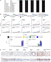
Figure 2.. The binding specificities of A/SG/16 HAs (WT and mutations)
(A) Alignment of residues at 159 and 160 of 3C.2a.1 and its descendants. (B) Binding avidities of A/SG/16 HAs (WT and mutations) measured by ELISA. Error bars represent the standard deviation of the duplicate measurements, which represent three biological independent assays. (C) Glycan array analysis of A/SG/16 WT and Y159N/T160I. Error bars represent the standard deviation of four replicates on the array and represent two biological independent assays. (D) The tissue staining of the A/SG/16 HAs (WT and mutations) to ferret lung and human trachea.
Figure 3.
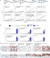
Figure 3.. Y195F rescues abrogated binding due to Y159N/T160I
(A) Alignment of the residues in 130-loop and 190-helix of H3N2 3C.2a1 viruses. (B) Binding avidities of A/SG/16 and A/SG/16 Y159N/T160I mutant H3 proteins. Error bars represent the standard deviation of the duplicate measurements, which represent three biological independent assays. (C) Glycan array analysis of A/SG/16 Y195F and Y159N/T160I/Y195F. Error bars represent the standard deviation of four replicates and represent two biological independent assays. (D) Tissue staining of A/SG/16 HAs (WT and mutations).
Figure 4.
Figure 4.. The effect of residues 131, 186, 190, 193, and 195 on receptor binding
(A) Binding avidities of different mutational combinations. Error bars represent the standard deviation of the duplicate measurement, which represent three biological independent assays. (B) Glycan array analysis of H3 A/SG16 Y195F mutants. Error bars represent the standard deviation of four replicates and represent two biological independent assays. (C) Tissue staining of the same HA proteins.
Figure 5.
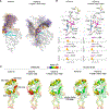
Figure 5.. The Y159N, T160I, and Y195F mutations significantly reconfigure receptor-binding site flexibility
(A) Overlay of 120 frames sampled with an even stride from three 400 ns MD replicas (1.2 μs total simulation time) of A/SG/16 and A/SG/16 Y159N/T160I/Y195F HA monomeric RBS models bound to compound D. D is shown as colored lines and the N158 glycan as white lines. The 130-loop and 150-loop are highlighted in cyan and orange, respectively. (B) Distribution of hydrogen bond distances between the sialic acid of D and the side chains of Y98 and of S136 and S137 along the same MD simulations. Relevant distances are shown as orange dashed lines. C97, C139, L154, Y/N159, T/I160, H183, and Y/F195 are shown as sticks. (C) Protein backbone root-mean-square fluctuations (RMSFs) along the MD simulations (1.2 μs) of unbound HA monomeric RBS models of A/SG/16, A/SG/16 Y159N/T160I, and A/SG/16 Y159N/T160I/Y195F and the same models of A/SG/16 and A/SG/16 Y159N/T160I/Y195F bound to D. The 130-loop, 150-loop, 190-helix, and 220-loop are highlighted. Cα atoms of Y98, S136, and S137 are shown as spheres. D and the N158 glycan are not shown for clarity.
Figure 6.
Figure 6.. The Y195F mutation allosterically rescues A/SG/16 Y159N/T160I receptor binding
(A) Selected frames from three 400 ns MD replicas (1.2 μs total simulation time) of the A/SG/16 HA monomeric RBS model illustrate the allosteric network linking Y195 to Y98 and the 130-loop containing S136 and S137, the alternative hydrogen bond interactions of Y195, and the occasional shift in Y98 rotameric preference, resulting in a conformation not competent for sialic acid binding (3% of the simulation). The gray arrow indicates the extent of the allosteric network that enables communication between RBS elements. The Y98 conformational change is marked with a red arrow. C97, Y98, S136, S137, C139, L154, Y159, T160, H183, and Y195 are shown as sticks. (B) Left: a selected frame from three 400 ns MD replicas (1.2 μs total simulation time) of the Y159N/T160I HA monomeric RBS model, illustrating the alternative backbone-flipped conformation in which the side chain of S136 forms a hydrogen bond with the backbone carbonyl of C97, making it unavailable for sialic acid binding (39% of the simulation). The backbone conformational change is marked with a red arrow. C97, Y98, S136, S137, C139, L154, N159, I160, H183, and Y195 are shown as sticks. Right: distribution of the hydrogen bond distance between the hydroxyl group of S136 and the backbone carbonyl of C97 in the MD simulations of the unbound forms of A/SG/16, Y159N/T160I, and Y159N/T160I/Y195F, as well as the D-bound forms of A/SG/16 and Y159N/T160I/Y195F. A vertical line at 3 Å marks the threshold used to quantify the population of the alternative conformation. (C) A selected frame from three 400 ns MD replicas (1.2 μs total simulation time) of the Y159N/T160I/Y195F HA monomeric RBS model showing that H183 is in close contact with Y98. C97, Y98, S136, S137, C139, L154, N159, I160, H183, and F195 are shown as sticks.

Update of

References

    1. Karakus U, Mena I, Kottur J, El Zahed SS, Seoane R, Yildiz S, Chen L, Plancarte M, Lindsay L, Halpin R, et al. (2024). H19 influenza A virus exhibits species-specific MHC class II receptor usage. Cell Host Microbe 32, 1089–1102. 10.1016/j.chom.2024.05.018. - DOI - PMC - PubMed
    1. Fouchier RAM, Munster V, Wallensten A, Bestebroer TM, Herfst S, Smith D, Rimmelzwaan GF, Olsen B, and Osterhaus ADME (2005). Characterization of a novel influenza A virus hemagglutinin subtype (H16) obtained from black-headed gulls. J. Virol. 79, 2814–2822. 10.1128/jvi.79.5.2814-2822.2005. - DOI - PMC - PubMed
    1. West J, Röder J, Matrosovich T, Beicht J, Baumann J, Mounogou Kouassi N, Doedt J, Bovin N, Zamperin G, Gastaldelli M, et al. (2021). Characterization of changes in the hemagglutinin that accompanied the emergence of H3N2/1968 pandemic influenza viruses. PLoS Pathog. 17, e1009566. 10.1371/journal.ppat.1009566. - DOI - PMC - PubMed
    1. Rogers GN, and Paulson JC (1983). Receptor determinants of human and animal influenza virus isolates: differences in receptor specificity of the H3 hemagglutinin based on species of origin. Virology 127, 361–373. 10.1016/0042-6822(83)90150-2. - DOI - PubMed
    1. Koel BF, Burke DF, Bestebroer TM, van der Vliet S, Zondag GCM, Vervaet G, Skepner E, Lewis NS, Spronken MIJ, Russell CA, et al. (2013). Substitutions near the receptor binding site determine major antigenic change during influenza virus evolution. Science 342, 976–979. 10.1126/science.1244730. - DOI - PubMed

MeSH terms

Substances

LinkOut - more resources