. 2025 Jul;123(2):e70356.
doi: 10.1111/tpj.70356.

Barley resistance and susceptibility to fungal cell entry involve the interplay of ROP signaling with phosphatidylinositol-monophosphates

Affiliations

Barley resistance and susceptibility to fungal cell entry involve the interplay of ROP signaling with phosphatidylinositol-monophosphates

Lukas Sebastian Weiß et al. Plant J. 2025 Jul.

Abstract

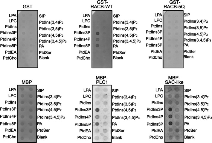
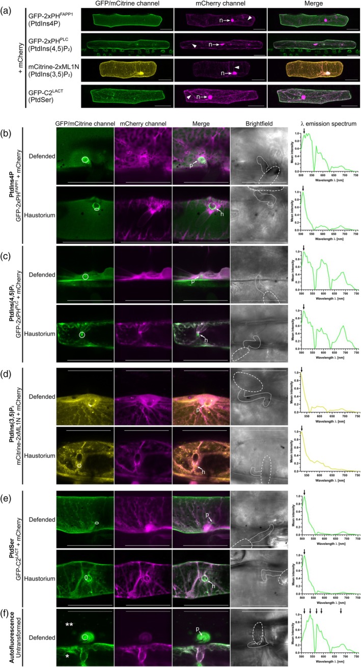
Rho-of-plant small GTPases (ROPs) are regulators of plant polar growth and of plant-pathogen interactions. The barley ROP, RACB, is involved in susceptibility toward infection by the barley powdery mildew fungus Blumeria hordei (Bh) but little is known about the cellular pathways that connect RACB signaling to disease susceptibility. Here we identify novel RACB interaction partners of plant or fungal origin by untargeted co-immunoprecipitation of constitutively active (CA) RACB tagged by green fluorescent protein from Bh-infected barley epidermal layers and subsequent analysis by liquid chromatography-coupled mass spectrometry. Three of the immunoprecipitated proteins, a plant phosphoinositide phosphatase, a plant phosphoinositide phospholipase, and a putative Bh-effector protein, are involved in the barley-Bh-pathosystem and support disease resistance or susceptibility, respectively. RACB and its plant interactors bind to overlapping anionic phospholipid species in vitro, and in the case of RACB, this lipid interaction is mediated by its carboxy-terminal polybasic region (PBR). Fluorescent markers for anionic phospholipids show altered subcellular distribution in barley cells during Bh attack and under expression of a RACB-binding fungal effector. Phosphatidylinositol 4-phosphate, phosphatidylinositol 3,5-bisphosphate, and phosphatidylserine show a distinct enrichment at the haustorial neck region, suggesting a connection to subcellular targeting of RACB at this site. The interplay of ROPs with anionic phospholipids and phospholipid-metabolizing enzymes may thus enable the subcellular enrichment of components pivotal for success or failure of fungal penetration.

Keywords: ROP GTPAse; effector; haustorium; phosphatidylinositol‐monophosphate; phosphoinositide phosphatase; phosphoinositide phospholipase; polarity; polybasic domain; susceptibility.


Conflict of interest statement

The authors declare no conflict of interest.

Figures

Figure 1
Figure 1
Transgenic barley plants overexpressing plasma membrane‐localized eGFP‐RACB‐CA are super‐susceptible to Bh infection. (a) eGFP and tagged fusion proteins show stable expression in first barley leaves and can be enriched via α‐GFP IP. eGFP‐tagged proteins were detected by α‐GFP Western blotting. IP fractions: Input (I), Unbound (U), Wash (W) and enriched proteins (IP). (b) Full‐length eGFP‐RACB‐CA localizes to the PM of barley epidermal cells, while eGFP‐RACB‐CA‐ΔCSIL and free eGFP are visible in the cytosol (arrowhead: cytosolic strands) and nucleus (n, arrows). CLSM investigated subcellular localization. Scale bar: 50 μm. Images are maximum projections of at least 29 Z‐steps of 2 μm increment optical sections. Image brightness was uniformly enhanced post‐scanning for better visibility. (c) Overexpression of full‐length eGFP‐RACB‐CA renders barley plants more susceptible to Bh invasion, whereas eGFP‐RACB‐CA‐ΔCSIL and free eGFP show similar levels of lower susceptibility. For each construct, three primary leaves of one plant line were tested. Lines were: BG654 E2 for eGFP‐RACB‐CA, BG655 E1 for eGFP‐RACB‐CA‐ΔCSIL and BG656 E1 for eGFP. Each data point represents the susceptibility of one leaf that was analyzed at 100 interaction sites. Crossbar depicts average susceptibility relative to the eGFP control. Statistical analysis was performed using a one‐way ANOVA with Tukey's honestly significant difference test.
Figure 2
Figure 2
RACB‐CA interacts with 9o9, PLC1, and SAC‐like in FRET‐FLIM experiments. (a) Co‐expression of N‐ or C‐terminally mCherry‐tagged 9o9 decreased the fluorescence lifetime of GFP‐RACB‐CA, which indicates direct protein–protein interaction. (b) N‐terminally tagged mCherry‐SAC‐like but not mCherry‐PLC1 significantly reduced the fluorescence lifetime of GFP‐RACB‐CA. (c) C‐terminally tagged PLC1‐mCherry but not SAC‐like‐mCherry decreased the lifetime of GFP‐RACB‐CA. GST‐mCherry and CRIB46‐mCherry were used as negative and positive controls, respectively (Schultheiss et al., ; Trutzenberg et al., 2022). FRET‐FLIM measurements were conducted at the cell periphery of epidermal cells of transiently transformed Nicotiana benthamiana plants. Co‐expression of fusion proteins was confirmed before measurements. All measurements were collected in three independent biological replicates. The number of observations (n) per construct is shown below each column. The crossbar and error bars show the average and standard deviation. Statistical differences were calculated with Wilcoxon‐Rank‐Sum tests with Bonferroni correction. ns P > 0.05, **P < 0.01, ***P < 0.001. −/−: FRET‐donor‐only control.
Figure 3
Figure 3
Gene expression of Bh 9o9 increases during Bh invasion. The expression levels of Bh 9o9 (a) and barley PLC1 (b) and SAC‐like (c) were measured during the early phase of Bh colonization at the indicated timepoints. Transcript of 9o9 was detectable in spores and increased during infection. Expression levels of PLC1 and SAC‐like only slightly differed during infection but showed partially significant differences compared with their non‐infected controls (as indicated by the P‐values). Bars show reference gene‐normalized fold changes in infected leaves, which were calculated according to using normalized gene expression in spores (a) or corresponding non‐infected leaves (b, c) as a baseline. β‐TUB2 was chosen as a reference gene for Bh, while UBC2 was taken as housekeeping genes for barley (Schnepf et al., ; Sherwood & Somerville, 1990). Bars show average fold changes in normalized gene expression with standard deviation. Data was collected over three independent biological replicates. Statistical differences were calculated separately for each timepoint and gene, comparing the normalized expression of PLC1 and SAC‐like in infected leaves to that of corresponding non‐infected leaves. Differences were assessed using t‐tests with Holm‐Sidak correction for multiple testing and only indicated in the graph when significant, *P < 0.05.
Figure 4
Figure 4
Single‐cell overexpression or silencing of 9o9, PLC1, and SAC‐like influence in the barley‐Bh pathosystem. The susceptibility of barley epidermal cells toward penetration by Bh was investigated after transient overexpression (a, c) or RNAi‐mediated silencing (b, d) of 9o9, PLC1, and SAC‐like at 40 hpi. The respective empty vectors were used as controls. Overexpression of 9o9 and silencing of PLC1 and SAC‐like led to increased susceptibility, whereas silencing of 9o9 and overexpression of PLC1 and SAC‐like showed no influence. RNAi‐silencing specificity was confirmed using the si‐Fi RNAi‐off‐target prediction tool (Lück et al., 2019). Each datapoint shows the Bh penetration efficiency of a single experiment relative to its averaged empty vector control. Crossbars display the average susceptibility from 10 (a), five (b, d) and seven (c) independent biological experiments. Statistical differences were calculated with two‐tailed Student's t‐tests comparing an overexpression or silencing construct with its respective empty vector control: ns P > 0.05, *P < 0.05, **P < 0.01. OEX: overexpression.
Figure 5
Figure 5
RACB and its candidate interactors associate with PtdIns‐monophosphates in vitro. RACB and its candidate interactors show lipid‐binding in in vitro protein–lipid‐overlay experiments. Recombinant proteins were purified from E. coli using affinity chromatography. Recombinant proteins were incubated with lipid‐spotted membranes and detected via α‐GST or α‐MBP antibodies. Colored dots indicate association with the respective lipid species. Free GST and MBP were used as non‐lipid‐binding controls. blank: no lipid spotted. LPA, lysophosphatidic acid; LPC, lysophosphatidylcholine; SIP, sphingosine 1‐phosphate.
Figure 6
Figure 6
RACB‐CA localization and function depend on positively charged amino acid residues in its PBR. (a) The subcellular localization of GFP‐tagged RACB‐CA or its derivate RACB‐CA‐5Q together with its mCherry‐tagged putative downstream scaffold protein RIC171 were investigated via CLSM after transient transformation of epidermal cells. Size bars represent 50 μm. (b) Yeast two‐hybrid assay of binding domain (Vossen et al.) tagged RACB‐variants (WT, wild‐type; CA, constitutively activated; DN, dominant negative) with activation domain (AD) tagged RIC171 in yeast on protein interaction non‐selective complete supplement medium (CSM) plates lacking leucine and tryptophan (‐L ‐W) or protein interaction selective plates lacking leucine, tryptophan, histidine, and adenine (‐L ‐W ‐H ‐Ade), or protein interaction selective plates supplemented with 0.5 mm 3‐aminotriazole (‐L ‐W ‐H + 0.5 mm 3‐AT). Interaction indicative growth is visible for combinations between RIC171 and each of RACB‐WT, RACB‐WT‐5Q, RACB‐CA, and RACB‐CA‐5Q. Interaction between T‐antigen and p53 served as a positive control for the yeast two‐hybrid experiment. (c) Interaction of GFP‐tagged versions of RACB‐CA with mCherry‐RIC171 in FRET‐FLIM experiments. Barley epidermal cells were transiently transformed via particle bombardment with expression constructs for either GFP‐RACB‐CA or GFP‐RACB‐CA‐5Q as FRET‐donors and mCherry‐RIC171 as a FRET acceptor. Free mCherry served as no‐interaction controls. FRET‐FLIM measurements were conducted at the cell periphery of the equatorial plane of barley epidermal cells 2 days after transformation. Stars display statistically significant differences between the samples (Tukey test, ns P > 0.05, **P < 0.01, ***P < 0.001, **** P < 0.0001 based on an ANOVA [significant at α = 0.05]). Measured cells were collected in at least three independent biological replicates. (d) The susceptibility of barley epidermal cells toward penetration by Bh was investigated after transient overexpression of RACB‐CA or RACB‐CA‐5Q or an empty vector control, respectively. Each data point shows the Bh penetration efficiency of a single experiment relative to its averaged control. Crossbars display the average susceptibility from four independent biological experiments. Statistical differences were calculated with Tukey test, ns P > 0.05, **P < 0.01, ***P < 0.001 based on an ANOVA (significant at α = 0.05). Control = empty vector transformation.
Figure 7
Figure 7
Subcellular localization of PtdIns4P, PtdIns(4,5)P2, PtdIns(3,5)P2, and PtdSer in unstressed and Bh‐infected barley epidermal cells. (a) The subcellular localization of four anionic phospholipid species was investigated in barley via CLSM after transient transformation of epidermal cells with lipid‐species‐specific genetically encoded biomarkers (Hirano et al., ; Platre et al., ; Simon et al., 2014). All four species showed at least partial localization to the PM. Free mCherry was co‐transformed as a marker highlighting localization in the cytosol (arrowhead: cytosolic strands) and nucleus (n, arrows). All images show Z‐stack maximum intensity projections composed of at least 16 XY‐optical sections captured in 1.5 μm Z‐steps. Scale bar: 50 μm. Representative images from at least two independent biological replicates are shown. Image brightness was uniformly enhanced post‐scanning for better visibility. (b–f) Images of infected cells were taken between 16 and 20 hpi. High‐magnification images of non‐penetrated (“Defended”) and Bh‐colonized (“Haustorium”) cells are shown. Arrows point to unsuccessful penetration attempts (p) or haustorial entry points (h). PtdIns4P (b) and PtdSer (e) were enriched at the haustorial neck region of Bh‐colonized cells. Signal from PtdIns(4,5)P2 (c) was primarily visible in cytosol and nucleus, but was hard to evaluate due to very low fluorescence levels. PtdIns(3,5)P2 (d) was slightly enriched at the cell wall apposition of non‐penetrated cells. The circles in the GFP/mCitrine‐images show regions‐of‐interest that were λ‐scanned to evaluate fluorescence emission spectra. λ‐scanning was performed in 5 nm detection steps after excitation with a 488 nm (GFP) or 514 nm (mCitrine) laser source. Only in non‐penetrated GFP‐2xPHFAPP1‐transformed cells and all GFP‐2xPHPLC‐transformed cells, a strong presence of non‐GFP‐signals could be detected that was likely autofluorescence emitted by phenolic compounds released during plant defense responses. In all other cells, spectra matched that of GFP or YFP (fluorophore emission maxima are highlighted by arrows;). For reference, (f) shows both autofluorescence in an untransformed, Bh‐attacked cell (**) and, side by side, a single transformed cell (*) imaged with the settings for PtdIns(4,5)P2. λ‐scanning was performed in the indicated region of interest (circle) to characterize autofluorescence emitted during high laser excitation levels and detector gain. Note the lack of the typical GFP peak around 510 nm and the peaks/shoulders from autofluorescence at about 540, 570, 590, and 690 nm. Free mCherry was co‐transformed as a marker for cytosolic and nuclear fluorescence. All images except brightfield pictures show Z‐stack maximum intensity projections of at least 4 XY‐optical sections captured in 1.5 μm Z‐steps. Brightfield images show single XY‐optical sections, in which spores are outlined with dashed lines and appressoria are highlighted with dotted lines. Scale bar: 50 μm. Representative images from at least five events in each of at least two independent biological replicates are shown. All images were taken with individual laser excitation strength and detector gain for higher image clarity. Image brightness was uniformly enhanced post‐scanning for better visibility.
Figure 8
Figure 8
Effect of 9o9 on the subcellular localization of PtdIns(3,5)P₂ marker in Bh‐infected barley epidermal cells. (a) The subcellular localization of the anionic phospholipid PtdIns(3,5)P₂ was analyzed in barley epidermal cells via confocal laser scanning microscopy following transient co‐transformation with Bh 9o9 and the PtdIns(3,5)P₂‐specific biosensor Citrine‐2x‐ML1N (Hirano et al., 2017). Free mCherry was co‐transformed as a cytosolic marker. Images of infected cells were taken between 16 and 20 hpi. High‐magnification images of Bh‐colonized cells are shown; haustorial entry points are indicated with (h) and the asterisk (*) indicates the haustorial body. All images show Z‐stack maximum intensity projections composed of at least 16 XY‐optical sections captured in 1.5 μm Z‐steps. Brightfield images represent single sections of the transmission channel, with the haustorium out of focus. Scale bar: 50 μm. Image brightness was uniformly enhanced post‐acquisition for better visualization. Representative images illustrate three categories of PtdIns(3,5)P₂ accumulation at haustorial entry sides/neck region: strong accumulation (upper panel), medium accumulation (middle panel), and no accumulation (lower panel). (b) Counting of PtdIns(3,5)P₂ accumulation patterns observed in each of 18 cells expressing 9o9 or control cells co‐transformed with the empty vector. In 9o9‐expressing cells, PtdIns(3,5)P₂ accumulation at haustorial entry points was seen in the majority of cells. In most control cells, PtdIns(3,5)P₂ accumulation was absent or weak.

References

    1. Akamatsu, A. , Wong, H.L. , Fujiwara, M. , Okuda, J. , Nishide, K. , Uno, K. et al. (2013) An OsCEBiP/OsCERK1‐OsRacGEF1‐OsRac1 module is an essential early component of chitin‐induced rice immunity. Cell Host & Microbe, 13, 465–476. - PubMed
    1. Bak, G. , Lee, E.J. , Lee, Y. , Kato, M. , Segami, S. , Sze, H. et al. (2013) Rapid structural changes and acidification of guard cell vacuoles during stomatal closure require phosphatidylinositol 3,5‐bisphosphate. Plant Cell, 25, 2202–2216. - PMC - PubMed
    1. Berken, A. (2006) ROPs in the spotlight of plant signal transduction. Cellular and Molecular Life Sciences, 63, 2446–2459. - PMC - PubMed
    1. Caldelari, D. , Sternberg, H. , Rodríguez‐Concepción, M. , Gruissem, W. & Yalovsky, S. (2001) Efficient prenylation by a plant Geranylgeranyltransferase‐I requires a functional CaaL box motif and a proximal polybasic domain. Plant Physiology, 126, 1416–1429. - PMC - PubMed
    1. Choi, S. , Prokchorchik, M. , Lee, H. , Gupta, R. , Lee, Y. , Chung, E.H. et al. (2021) Direct acetylation of a conserved threonine of RIN4 by the bacterial effector HopZ5 or AvrBsT activates RPM1‐dependent immunity in Arabidopsis. Molecular Plant, 14, 1951–1960. - PubMed

Substances

LinkOut - more resources