. 2025 Jul 18;8(1):1069.
doi: 10.1038/s42003-025-08360-w.

Myc overexpression improves recovery from myocardial infarction associated with cardiomyocyte hyperplasia in the mouse heart

Affiliations

Myc overexpression improves recovery from myocardial infarction associated with cardiomyocyte hyperplasia in the mouse heart

Cristina Villa Del Campo et al. Commun Biol. .

Abstract

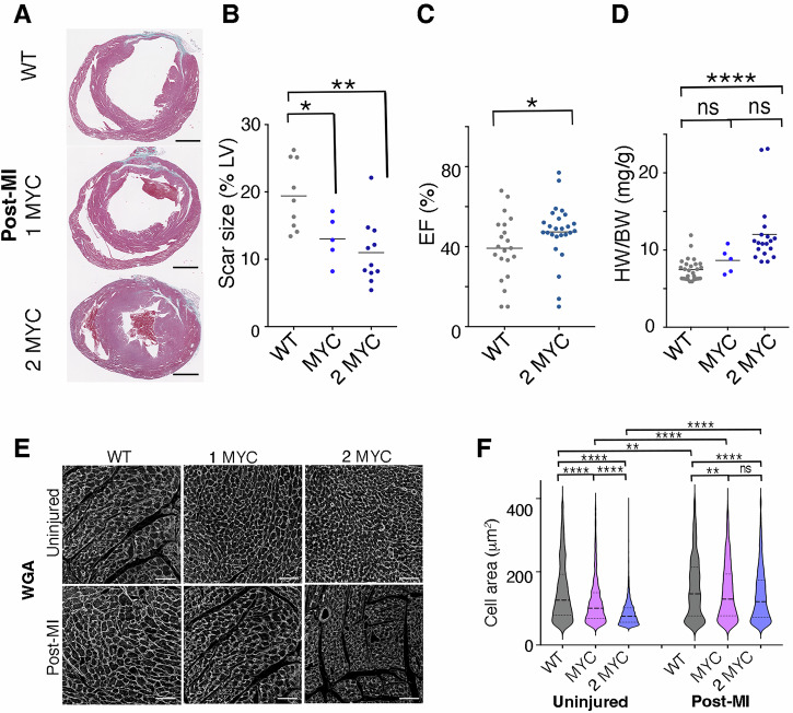
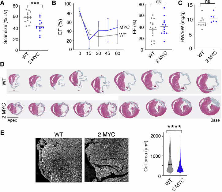
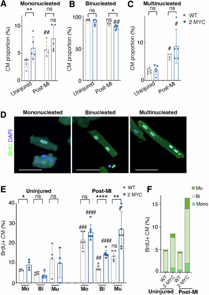
Adult mammalian hearts lack the ability to regenerate after a myocardial infarction, which leads to heart failure progression. Therefore, finding ways to promote regeneration is a major goal in the development of cardiovascular therapies. In this study, we focus on the role of Myc moderate overexpression in response to acute ischemic injury. We have previously shown that moderate Myc overexpression is not detrimental to cardiac function and promotes cell competition and a hyperplastic phenotype. Here, we describe that Myc overexpression in non-regenerative postnatal hearts promotes functional improvements and reduced scar formation after myocardial infarction. This response correlates with a hyperplastic phenotype in postnatal and adult hearts, characterized by bigger hearts, smaller cardiomyocyte size and increased BrdU incorporation without ploidy increase. Moreover, we show that Myc strongly enhances diploid cardiomyocyte proliferation and the generation of new binucleated cardiomyocytes. Thus, Myc promotes functional and morphological improvements in the heart following acute ischemia and this response correlates with promotion of hyperplasia versus hypertrophy.


Conflict of interest statement

Competing interests: The authors declare no competing interests

Figures

Fig. 1
Fig. 1. Moderate Myc overexpression in postnatal cardiomyocytes is phenotypically silent in homeostasis and promotes cardiomyocyte cell cycle activity with no increase in ploidy.
A Histological heart sections stained with hematoxylin/eosin of WT mice and mice overexpressing two alleles of Myc (2MYC) at 28 days. B Percent EF at 28 days of age. C Heart to body weight ratio (mg/g) at 28 days. Data are presented as individual hearts and mean. D Representative confocal images of isolated P1 cardiomyocytes stained for α-actinin and PH3 for WT and MYC overexpressing hearts (left). Percent of PH3 positive cardiomyocytes. E Representative confocal images of adult isolated cardiomyocytes stained for BrdU (left). BrdU+ cardiomyocyte proportion in each cardiomyocyte population in uninjured adult hearts (10–12 weeks) (right). (Mo: mononucleated, Bi: binucleated, Mu: Multinucleated). F Representative confocal images of isolated adult cardiomyocytes stained for actinin and DAPI for nuclear volume segmentation. G Average nuclear volume per heart shown in μm3 for WT and Myc-overexpressing hearts in mononucleated and binucleated cardiomyocytes (n > 300 nuclei/heart). Data presented as individual hearts, mean and error bars (when shown) represent SD. Scale bars: 1 mm in A; 70 μm in (D); 60 μm in (E); 200 μm in (F). *p < 0.05 **p < 0.01 ***p < 0.001 ****p < 0.0005.
Fig. 2
Fig. 2. Moderate Myc overexpression promotes a repair response after ischemic injury involving hyperplasia of the cardiomyocyte population.
A Histological sections stained with Masson’s trichrome showing scarring (blue) and healthy muscle (pink) after ischemic injury performed at P7 and analyzed 21 days after surgery (P28). B Scar size quantification as % of LV area 21 days post ischemic injury. C Percent EF 21 days post injury at P7(P28). D Heart weight to body weight ratio 21 days post ischemic injury at P7(P28). E Histological sections stained with wheat germ agglutinin (WGA-gray) of P28 non-infarcted hearts (Control) and post ischemic injury hearts at 21 days after the LAD at P7 (P28) (Post-MI) in wild type (WT) and Myc overexpressing mice (1MYC and 2 MYC). F Quantification of cardiomyocyte cross-sectional area distribution in P28 non-infarcted (Uninjured) and 21 days post injury (Post-MI) wild type (WT) and Myc-overexpressing (1MYC and 2 MYC) hearts. Data presented in B as distribution of individual cardiomyocyte area (n > 1000 cells/condition) and mean. Data presented as individual hearts and mean. Scale bars: 50 μm. *p < 0.05 **p < 0.01 ***p < 0.001 ****p < 0.0005.
Fig. 3
Fig. 3. Myc mild overexpression promotes a regenerative response with no hypertrophy in adult hearts following ischemic injury.
A Scar size as % of LV 60 days post ischemic injury. B Left: Percent EF post ischemic injury in adult mice showing evolution over time (days). Right: Percent EF 60 days post ischemic injury in adult mice (final time point). C Heart weight-to-body weight ratio 60 days post ischemic injury. D Histological sections stained with Masson’s Trichrome showing scarring 60 days post ischemic injury from apex to base in WT and Myc overexpressing (2MYC) hearts. E Histological sections of hearts stained with wheat germ agglutinin (WGA-gray) post ischemic injury 60 days after LAD in wild type (WT) and Myc overexpressing hearts (1MYC and 2 MYC) (left). Quantification of cardiomyocyte cross-sectional area distribution post ischemic injury 60 days after LAD ligation in wild type (WT) and Myc-overexpressing hearts (2 MYC) (right). Data presented as individual hearts, mean and error bars (when shown) represent SD. Data presented in (E) as distribution of individual cardiomyocyte area (n > 1000 cells/condition) and mean. Scale bars: 2 mm. *p < 0.05 **p < 0.01 ***p < 0.001 ****p < 0.0005.
Fig. 4
Fig. 4. Myc mild overexpression promotes no major alteration in cardiomyocyte populations and activates cell cycle in cardiomyocytes after ischemic injury.
AC Quantification of cardiomyocyte population proportions in P28 uninjured hearts (Uninjured) and hearts that suffered ischemic injury at P7 analyzed at P28 (Post-MI) for mononucleated (A), binucleated (B) and multinucleated (C) CMs in WT or Myc-overexpressing hearts. D Representative confocal images of mononucleated, binucleated and multinucleated cardiomyocytes isolated by Langendorff digestion positively stained for BrdU (Green) and DAPI (blue). E Percentage of BrdU+ cardiomyocytes according to nucleation in uninjured hearts at P28 (uninjured) and hearts analyzed 21 days post ischemic injury at P7 (P28) (Post-MI) from WT and Myc-overexpressing mice. F Percentage of BrdU+ cardiomyocytes with respect to the total cardiomyocyte population in uninjured hearts at P28 (uninjured) and hearts analyzed 21 days post ischemic injury at P7 (P28) (Post-MI) from WT and Myc-overexpressing hearts. (Mo: mononucleated, Bi: binucleated, Mu: Multinucleated). Data presented as individual hearts, mean and error bars (when shown) represent SD. (n = 500–1000 CM scored/heart). Scale bars: 50 μm *p < 0.05 **p < 0.01 ***p < 0.001 ****p < 0.0005 (* represents p-value when comparing WT and MYC for each condition and # represents p-value when comparing each genotype with its own value in control and Post-MI conditions).

References

    1. Jessup, M. et al. 2009 focused update: ACCF/AHA Guidelines for the Diagnosis and Management of Heart Failure in Adults: a report of the American College of Cardiology Foundation/American Heart Association Task Force on Practice Guidelines: developed in collaboration with the International Society for Heart and Lung Transplantation. Circulation,119, 1977–2016 (2009). - PubMed
    1. Soonpaa, M. H. et al. Cardiomyocyte DNA synthesis and binucleation during murine development. Am. J. Physiol.2712, H2183–H2189 (1996). - PubMed
    1. Senyo, S. E. et al. Mammalian heart renewal by pre-existing cardiomyocytes. Nature493, 433–436 (2013). - PMC - PubMed
    1. Porrello, E. R. et al. MiR-15 family regulates postnatal mitotic arrest of cardiomyocytes. Circ. Res.109, 670–679 (2011). - PMC - PubMed
    1. Porrello, E. R. et al. Transient regenerative potential of the neonatal mouse heart. Science331, 1078–1080 (2011). - PMC - PubMed

Substances