

Experimental study of intravoxel incoherent motion diffusion imaging combined with ultrasound renal resistance index in contrast-induced nephropathy
- PMID: 40682007
- PMCID: PMC12273328
- DOI: 10.1186/s12882-025-04329-3
Experimental study of intravoxel incoherent motion diffusion imaging combined with ultrasound renal resistance index in contrast-induced nephropathy
Abstract
Objective: This study explored the application of Intravoxel Incoherent Motion (IVIM) diffusion imaging combined with Ultrasound Renal Resistance Index (RRI) for monitoring the pathophysiological changes associated with early contrast-induced nephropathy (CIN).
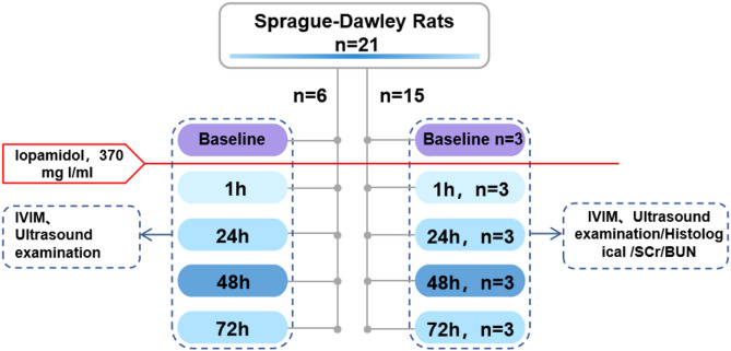
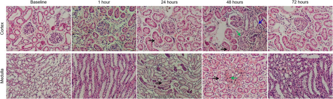
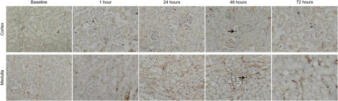
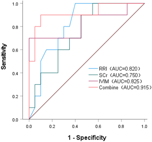
Methods: In this study, forty-two male Sprague-Dawley (SD) rats were equally divided into two groups: a contrast media (CM) group and a control group, each containing 21 animals. The CM group was administered a tail vein injection of ioversol (370 mg I/ml, 1.5 ml/kg), while the control group received a saline solution in a similar volume. Assessments using IVIM-MRI and Doppler ultrasound were performed 24 h before and at 1, 24, 48, and 72 h post-injection. These assessments aimed to evaluate the true diffusion coefficient (D), pseudo-diffusion coefficient (D*), perfusion fraction (f), apparent diffusion coefficient (ADC), and RRI. Concurrently, three rats from each group were sacrificed at these time points for renal histopathology, hypoxia-inducible factor-1α (HIF-1α) expression analysis, and the quantification of serum creatinine (SCr) and blood urea nitrogen (BUN) levels. Receiver operating characteristic (ROC) curves were plotted, and the area under the curve (AUC) was analyzed to evaluate the diagnostic performance of IVIM and RRI in predicting CIN.
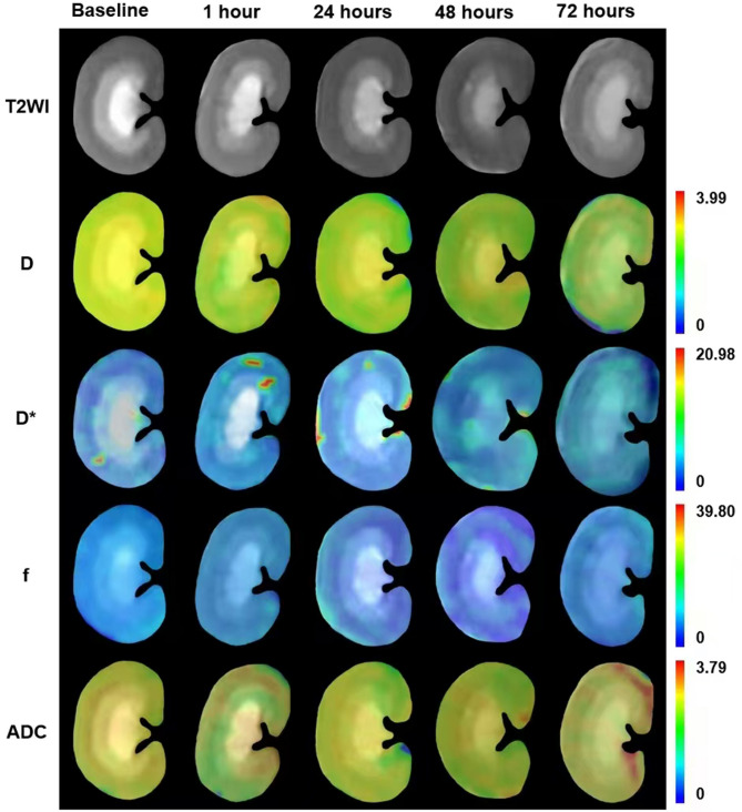
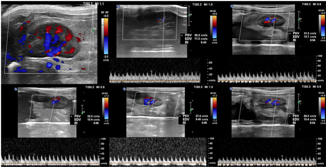
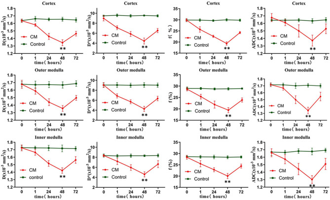
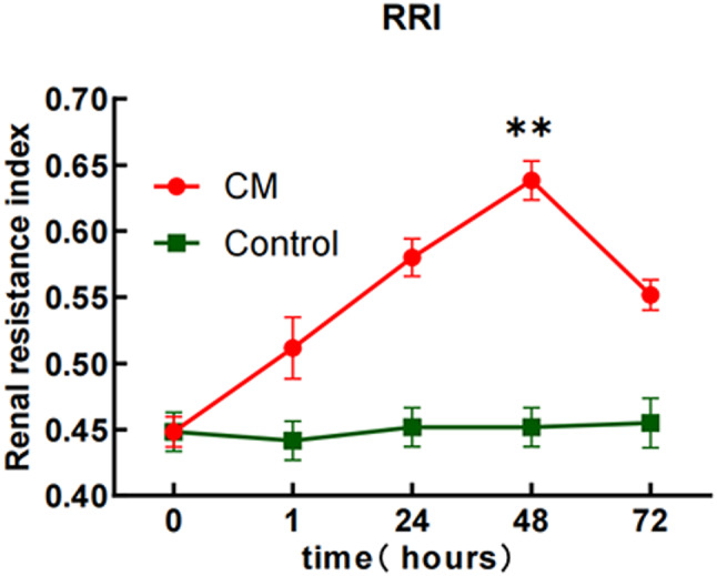
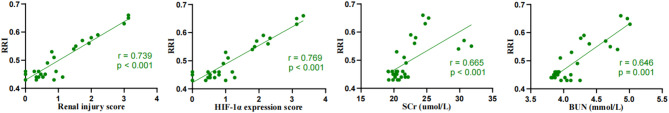
Results: Post-ioversol administration, significant declines were noted in the D, D*, f, ADC across the renal cortex (CO), outer medulla (OM), and inner medulla (IM) from 1 to 48 h (P < 0.05), with the lowest values observed at 48 h. These parameters began to recover after 72 h. Conversely, RRI values escalated from 1 to 48 h, peaking at 48 h (P < 0.05), and then diminished gradually after 72 h. The control group showed no significant changes in these parameters. Furthermore, a negative correlation was observed between RRI, histopathological grades, HIF-1α expression levels, and the levels of SCr and BUN. In contrast, RRI exhibited a positive correlation with these pathological scores and the levels of SCr and BUN. ROC curve analysis revealed that the combined predictive performance of IVIM and RRI was superior to that of individual parameters.
Conclusion: The synergistic application of IVIM and RRI techniques offers a non-invasive approach for early detection of renal damage after ioversol exposure and is a potent method for observing the pathophysiological shifts associated with early-stage CIN.
Keywords: Acute kidney injury; Contrast-induced nephropathy; Intravoxel incoherent motion; Renal resistance index.
© 2025. The Author(s).
Conflict of interest statement
Declarations. Ethics approval and consent to participate: No human samples or clinical data were included in this study, hence consent to participate is not applicable. All animal procedures were conducted according to the National Institute of Health Guide for the Care and Use of Laboratory Animals and approved by the University of Beihua Ethics Committee (approval number 2024112604). Consent for publication: Not applicable. Competing interests: The authors declare no competing interests.
Figures

References
-
- Nijssen EC, Rennenberg R, Nelemans P, van Ommen V, Wildberger JE. Post-Contrast acute kidney injury and intravenous prophylactic hydration: an update. Kontrastmittelinduzierte nephropathie: aktueller stand Präventiver Maßnahmen. Rofo. 2021;193(2):151–9. - PubMed
-
- Mamoulakis C, Tsarouhas K, Fragkiadoulaki I, et al. Contrast-induced nephropathy: basic concepts, pathophysiological implications and prevention strategies. Pharmacol Ther. 2017;180:99–112. - PubMed
-
- Boccalandro F, Shreyder K, Harmon L, Dhindsa M, Fahim T, Sheikh S. Five-Year Follow-Up of patients with Radio-Contrast-Induced acute renal injury: can intravenous sodium bicarbonate improve Long-Term outcomes?? Cardiovasc Revasc Med. 2021;31:61–8. - PubMed
MeSH terms
Substances
LinkOut - more resources
Full Text Sources
Medical
